Advertisement

Main menu

  • Home
  • Content
    • Current Issue
    • Accepted Manuscripts
    • Article Preview
    • Past Issue Archive
    • Video Articles
    • AJNR Case Collection
    • Case of the Week Archive
    • Case of the Month Archive
    • Classic Case Archive
  • Special Collections
    • AJNR Awards
    • Low-Field MRI
    • Alzheimer Disease
    • ASNR Foundation Special Collection
    • Photon-Counting CT
    • View All
  • Multimedia
    • AJNR Podcasts
    • AJNR SCANtastic
    • Trainee Corner
    • MRI Safety Corner
    • Imaging Protocols
  • For Authors
    • Submit a Manuscript
    • Submit a Video Article
    • Submit an eLetter to the Editor/Response
    • Manuscript Submission Guidelines
    • Statistical Tips
    • Fast Publishing of Accepted Manuscripts
    • Graphical Abstract Preparation
    • Imaging Protocol Submission
    • Author Policies
  • About Us
    • About AJNR
    • Editorial Board
    • Editorial Board Alumni
  • More
    • Become a Reviewer/Academy of Reviewers
    • Subscribers
    • Permissions
    • Alerts
    • Feedback
    • Advertisers
    • ASNR Home

User menu

  • Alerts
  • Log in

Search

  • Advanced search
American Journal of Neuroradiology

American Journal of Neuroradiology

ASHNR American Society of Functional Neuroradiology ASHNR American Society of Pediatric Neuroradiology ASSR
  • Alerts
  • Log in

Advanced Search

  • Home
  • Content
    • Current Issue
    • Accepted Manuscripts
    • Article Preview
    • Past Issue Archive
    • Video Articles
    • AJNR Case Collection
    • Case of the Week Archive
    • Case of the Month Archive
    • Classic Case Archive
  • Special Collections
    • AJNR Awards
    • Low-Field MRI
    • Alzheimer Disease
    • ASNR Foundation Special Collection
    • Photon-Counting CT
    • View All
  • Multimedia
    • AJNR Podcasts
    • AJNR SCANtastic
    • Trainee Corner
    • MRI Safety Corner
    • Imaging Protocols
  • For Authors
    • Submit a Manuscript
    • Submit a Video Article
    • Submit an eLetter to the Editor/Response
    • Manuscript Submission Guidelines
    • Statistical Tips
    • Fast Publishing of Accepted Manuscripts
    • Graphical Abstract Preparation
    • Imaging Protocol Submission
    • Author Policies
  • About Us
    • About AJNR
    • Editorial Board
    • Editorial Board Alumni
  • More
    • Become a Reviewer/Academy of Reviewers
    • Subscribers
    • Permissions
    • Alerts
    • Feedback
    • Advertisers
    • ASNR Home
  • Follow AJNR on Twitter
  • Visit AJNR on Facebook
  • Follow AJNR on Instagram
  • Join AJNR on LinkedIn
  • RSS Feeds

AJNR Awards, New Junior Editors, and more. Read the latest AJNR updates

Research ArticleSpine Imaging and Spine Image-Guided InterventionsT

Percutaneous Transosseous Translaminar Approach for Thecal Sac Access in Advanced Ankylosing Spondylitis with Instrumented Posterior Spinal Fusion

B.P. Liu, A. Aghaei Lasboo, M. Rozenfeld, T.A. Hijaz, S.F. Futterer and M.T. Walker
American Journal of Neuroradiology January 2010, 31 (1) 193-195; DOI: https://doi.org/10.3174/ajnr.A1701
B.P. Liu
  • Find this author on Google Scholar
  • Find this author on PubMed
  • Search for this author on this site
A. Aghaei Lasboo
  • Find this author on Google Scholar
  • Find this author on PubMed
  • Search for this author on this site
M. Rozenfeld
  • Find this author on Google Scholar
  • Find this author on PubMed
  • Search for this author on this site
T.A. Hijaz
  • Find this author on Google Scholar
  • Find this author on PubMed
  • Search for this author on this site
S.F. Futterer
  • Find this author on Google Scholar
  • Find this author on PubMed
  • Search for this author on this site
M.T. Walker
  • Find this author on Google Scholar
  • Find this author on PubMed
  • Search for this author on this site
  • Article
  • Figures & Data
  • Info & Metrics
  • Responses
  • References
  • PDF
Loading

Article Figures & Data

Figures

  • Fig 1.
    • Download figure
    • Open in new tab
    • Download powerpoint
    Fig 1.

    Lateral projection scoliosis radiograph demonstrates instrumented posterior spinal fusion from the occiput to the pelvis. There is an accentuated upper thoracic kyphosis and positive sagittal imbalance.

  • Fig 2.
    • Download figure
    • Open in new tab
    • Download powerpoint
    Fig 2.

    Sagittal fast spin-echo T2-weighted image of the thoracic spine demonstrates syringohydromyelia of the thoracic spinal cord with expansion of the cord (arrows). The syrinx extends from T2-T11. Portions of the spinal canal are obscured by artifacts.
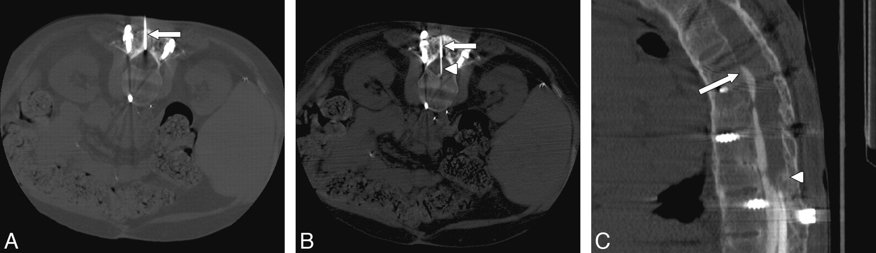
  • Fig 3.
    • Download figure
    • Open in new tab
    • Download powerpoint
    Fig 3.

    A, Axial CT scan through the L2–3 level with the patient prone shows the cannula (arrow) traversing a 2.5-cm thickness of bone graft, lamina, and ossified ligamentum flavum. B, Axial CT scan demonstrates placement of the 22-gauge spinal needle (arrowhead) through the cannula (arrow) and into the SAS. The needle tip was ventral, and though we did not puncture the ventral dura, that is a theoretic risk. In future attempts, we plan to measure the distance to the middle of the thecal sac and use a depth gauge or collar to identify our intended depth. We will then secure that depth with a clamp on the spinal needle so that it cannot inadvertently advance with gravity. C, Sagittal reconstruction of the CT myelogram demonstrates subarachnoid compartmentalization, adhesions, and a compete myelographic block ventrally at T8 (arrow) and dorsally at T12 (arrowhead).

PreviousNext
Back to top

In this issue

American Journal of Neuroradiology: 31 (1)
American Journal of Neuroradiology
Vol. 31, Issue 1
1 Jan 2010
  • Table of Contents
  • Index by author
Advertisement
Print
Download PDF
Email Article

Thank you for your interest in spreading the word on American Journal of Neuroradiology.

NOTE: We only request your email address so that the person you are recommending the page to knows that you wanted them to see it, and that it is not junk mail. We do not capture any email address.

Enter multiple addresses on separate lines or separate them with commas.
Percutaneous Transosseous Translaminar Approach for Thecal Sac Access in Advanced Ankylosing Spondylitis with Instrumented Posterior Spinal Fusion
(Your Name) has sent you a message from American Journal of Neuroradiology
(Your Name) thought you would like to see the American Journal of Neuroradiology web site.
CAPTCHA
This question is for testing whether or not you are a human visitor and to prevent automated spam submissions.
Cite this article
B.P. Liu, A. Aghaei Lasboo, M. Rozenfeld, T.A. Hijaz, S.F. Futterer, M.T. Walker
Percutaneous Transosseous Translaminar Approach for Thecal Sac Access in Advanced Ankylosing Spondylitis with Instrumented Posterior Spinal Fusion
American Journal of Neuroradiology Jan 2010, 31 (1) 193-195; DOI: 10.3174/ajnr.A1701

Citation Manager Formats

  • BibTeX
  • Bookends
  • EasyBib
  • EndNote (tagged)
  • EndNote 8 (xml)
  • Medlars
  • Mendeley
  • Papers
  • RefWorks Tagged
  • Ref Manager
  • RIS
  • Zotero
0 Responses
Respond to this article
Share
Bookmark this article
Percutaneous Transosseous Translaminar Approach for Thecal Sac Access in Advanced Ankylosing Spondylitis with Instrumented Posterior Spinal Fusion
B.P. Liu, A. Aghaei Lasboo, M. Rozenfeld, T.A. Hijaz, S.F. Futterer, M.T. Walker
American Journal of Neuroradiology Jan 2010, 31 (1) 193-195; DOI: 10.3174/ajnr.A1701
del.icio.us logo Twitter logo Facebook logo Mendeley logo
  • Tweet Widget
  • Facebook Like
  • Google Plus One
Purchase

Jump to section

  • Article
    • Abstract
    • Technical Report
    • Discussion
    • References
  • Figures & Data
  • Info & Metrics
  • Responses
  • References
  • PDF

Related Articles

  • No related articles found.
  • PubMed
  • Google Scholar

Cited By...

  • No citing articles found.
  • Crossref
  • Google Scholar

This article has not yet been cited by articles in journals that are participating in Crossref Cited-by Linking.

More in this TOC Section

  • Management Outcomes For VO Spine Biopsy
  • Characteristics of SIH Type I Culprit Lesions
  • Advanced Imaging of Type 2 Spinal CSF Leaks
Show more Spine Imaging and Spine Image-Guided Interventions

Similar Articles

Advertisement

Indexed Content

  • Current Issue
  • Accepted Manuscripts
  • Article Preview
  • Past Issues
  • Editorials
  • Editor's Choice
  • Fellows' Journal Club
  • Letters to the Editor
  • Video Articles

Cases

  • Case Collection
  • Archive - Case of the Week
  • Archive - Case of the Month
  • Archive - Classic Case

More from AJNR

  • Trainee Corner
  • Imaging Protocols
  • MRI Safety Corner
  • Book Reviews

Multimedia

  • AJNR Podcasts
  • AJNR Scantastics

Resources

  • Turnaround Time
  • Submit a Manuscript
  • Submit a Video Article
  • Submit an eLetter to the Editor/Response
  • Manuscript Submission Guidelines
  • Statistical Tips
  • Fast Publishing of Accepted Manuscripts
  • Graphical Abstract Preparation
  • Imaging Protocol Submission
  • Evidence-Based Medicine Level Guide
  • Publishing Checklists
  • Author Policies
  • Become a Reviewer/Academy of Reviewers
  • News and Updates

About Us

  • About AJNR
  • Editorial Board
  • Editorial Board Alumni
  • Alerts
  • Permissions
  • Not an AJNR Subscriber? Join Now
  • Advertise with Us
  • Librarian Resources
  • Feedback
  • Terms and Conditions
  • AJNR Editorial Board Alumni

American Society of Neuroradiology

  • Not an ASNR Member? Join Now

© 2025 by the American Society of Neuroradiology All rights, including for text and data mining, AI training, and similar technologies, are reserved.
Print ISSN: 0195-6108 Online ISSN: 1936-959X

Powered by HighWire