
Article Figures & Data
Figures
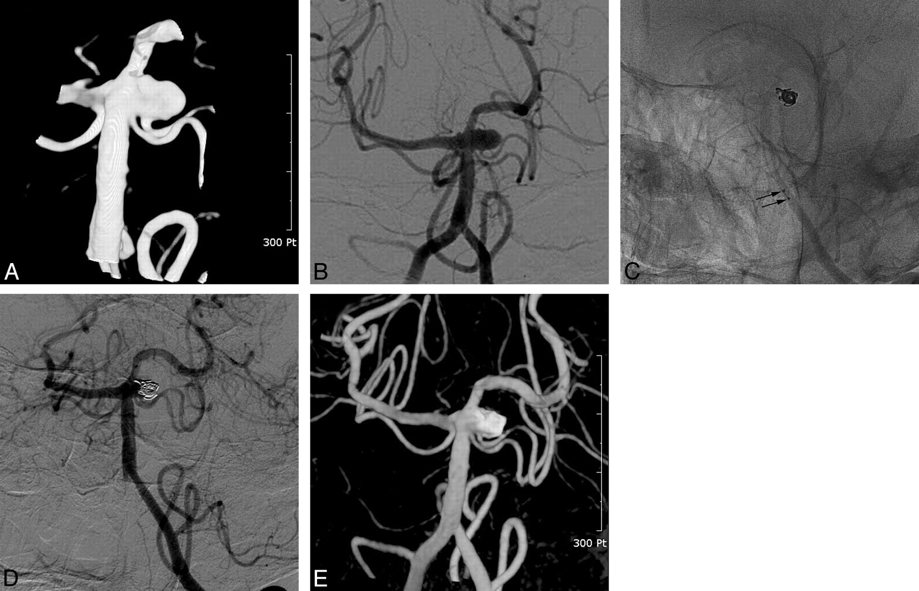
- Fig 1.
A 64-year-old woman with an unruptured aneurysm at the basilar artery−left superior cerebellar artery junction. A and B, 3D reconstruction image (A) and a working-projection image (B) reveal a saccular aneurysm at the basilar artery–duplicated left superior cerebellar artery origin. One of the duplicated left superior cerebellar arteries is incorporated into the sac. C, Coiling of the aneurysm sac is performed by using a 2-microcatheter technique. Note the radiopaque proximal markers of 2 catheters and coils (arrows). D and E, Postembolization control angiogram (D) and 3D reconstruction (E) image reveal near-complete occlusion of the aneurysm sac and a patent superior cerebellar artery incorporated into the sac.
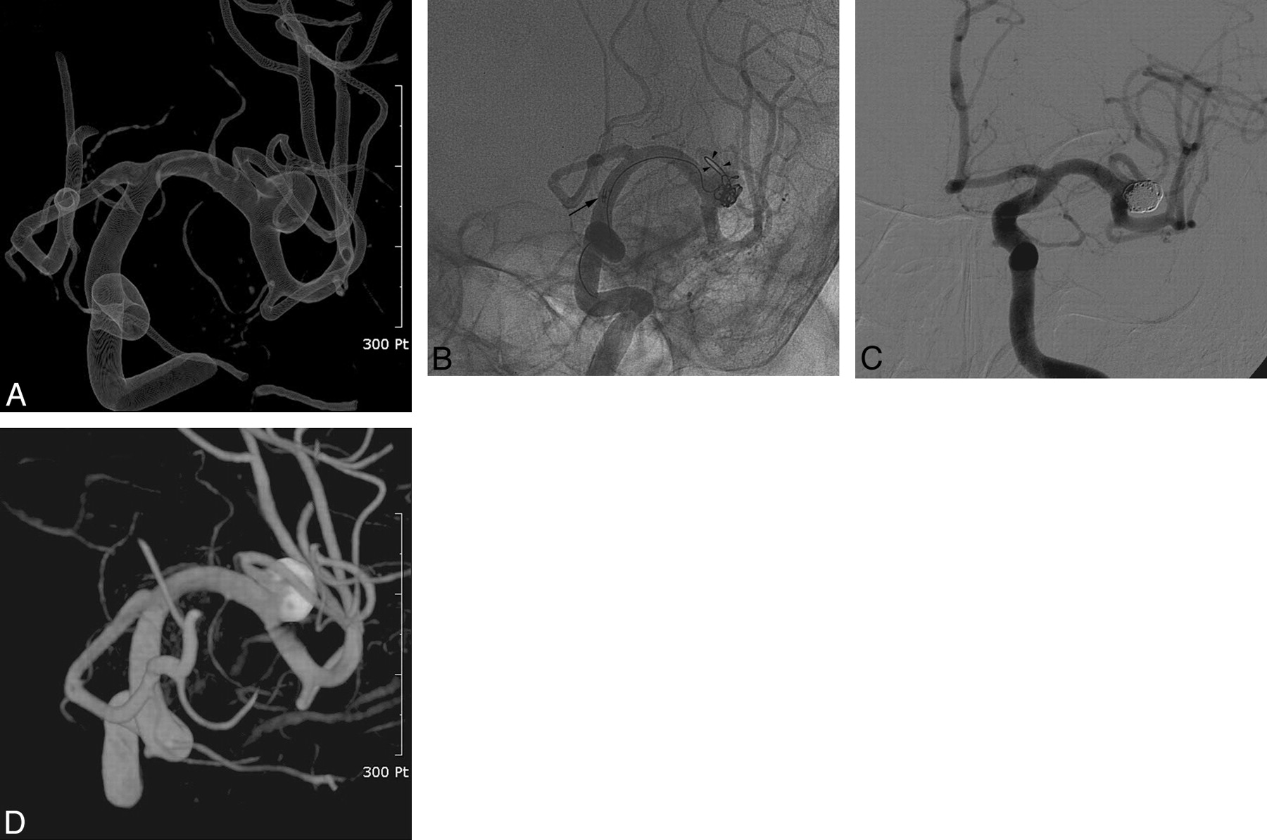
- Fig 2.
A 64-year-old woman with an unruptured aneurysm of the left middle cerebral artery. A, Transparent gradient view of a 3D reconstruction image shows a saccular aneurysm with a superior branch incorporated into the sac. B, Coiling of the aneurysm is performed by using a catheter-supported 2-catheter technique. The incorporated branch is selected by using a microcatheter and a coil (arrowheads). By gently pushing the microcatheter to support the coil mass, we performed coiling by using the other 2 microcatheters. This procedure is repeated during the coiling to preserve the incorporated branch. Note the radiopaque proximal markers of 2 catheters and coils (arrow). C and D, Postembolization control angiogram (C) and a 3D reconstruction (D) image show near-complete occlusion of the aneurysm sac and a patent incorporated branch.
- Fig 3.
A 44-year-old woman with a ruptured aneurysm at the right internal carotid–ophthalmic artery. A and B, Right internal carotid angiogram in a working projection (A) and a 3D reconstruction (B) image reveal a saccular aneurysm with the right ophthalmic artery incorporated into the sac. C, Coiling of the aneurysm by using a balloon-remodeling technique. Coiling is performed while the HyperForm balloon is overinflated and focally herniated (arrow) into the sac to protect the origin of the ophthalmic artery. D, Six-month follow-up angiography reveals a stable state of near-complete occlusion of the aneurysm sac and a patent ophthalmic artery incorporated into it.
- Fig 4.
A 53-year-old woman presenting with subarachnoid hemorrhage. A, Nonenhanced CT scan shows diffuse subarachnoid hemorrhage. B, 3D reconstruction image reveals a small saccular aneurysm with the right ophthalmic artery (arrows) incorporated into the sac. Except for this aneurysm, there is no aneurysm or other vascular malformation responsible for the subarachnoid hemorrhage on follow-up angiography. C, Coiling of the aneurysm by using a stent-assisted technique. D, Six-month follow-up angiogram shows near-complete occlusion of the aneurysm sac and a patent right ophthalmic artery.
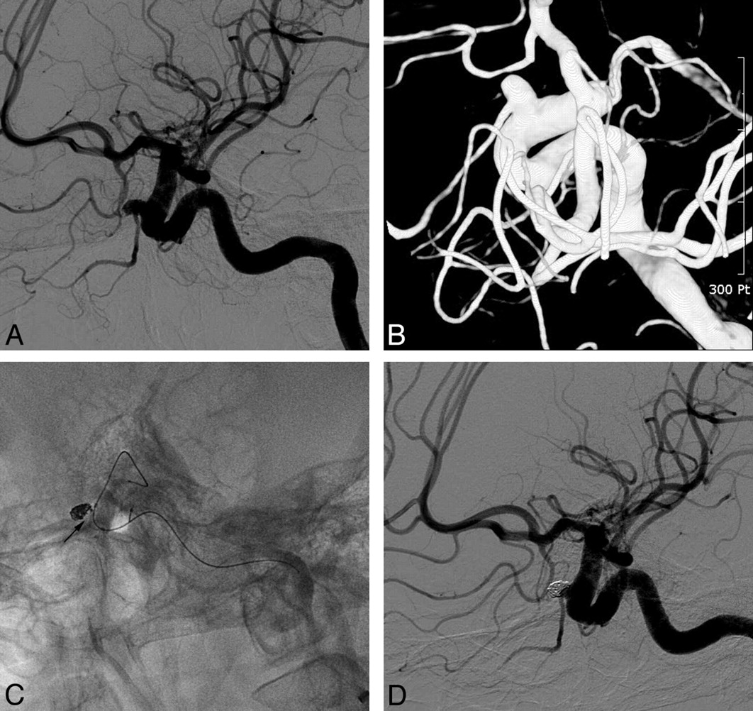
- Fig 5.
A 48-year-old man presenting with subarachnoid hemorrhage. A and B, One-week (not shown) and 2-week (A and B) follow-up angiograms after clipping reveal an increased size of the remnant aneurysm sac due to clip slippage. Note the slipping clip (arrows) and an incorporated branch artery (arrowheads). C, Coil embolization is performed by using a combined balloon- and catheter-assisted technique. Note that a microcatheter and a coil are inserted into the incorporated branch artery (arrowheads). D and E, Postembolization control angiogram subtraction image (D) and a 3D reconstruction (E) image show complete occlusion of the remnant aneurysm sac and preservation of the branch (arrowheads) incorporated into the sac.


Tables
Characteristics of the aneurysms with a branch incorporated into the sac and immediate and follow-up results of coiling
Characteristic No. No. branch-incorporated aneurysms (patients) 79 (69) Presentation Ruptured 26 (25.7%) Unruptured 53 (74.3%) Mean of maximum aneurysm diameter (range) 6.6 mm (2–26) Mean of aneurysm neck (range) 4.0 mm (1.6–8.4) Wide (≥4 mm or dome-to-neck ratio <1) 49 (62.0%) Narrow (<4 mm and dome-to-neck ratio ≥1) 30 (38.0%) Immediate posttreatment control angiography Near complete 71 (89.8%) Neck remnant 4 (5.1%) Incomplete occlusion 4 (5.1%) Treatment-related complications (% of number of patients) Aneurysm rupture 1 (1.4%) Basal ganglia hemorrhage 1 (1.4%) Thromboembolic events during or after treatment 7 (10.1%) Occlusion of the incorporated branch artery 2 (2.9%) Embolic infarct 1 Transient ischemic attack 4 Treatment-related permanent morbidity 4 (5.8%) Treatment-related mortality 0 Follow-up angiography (mean, 15 months; range, 6–50 months) 55 (69.6%) Improved or stable 45 (81.8%) Minor recurrence not requiring retreatment 4 (7.3%) Major recurrence requiring retreatment 6 (10.9%)
In this issue


Jump to section
Related Articles
Cited By...
- Endovascular treatment of unruptured ophthalmic artery aneurysms: clinical usefulness of the balloon occlusion test in predicting vision outcomes after coil embolization
- Combination of Multicatheter Plus Stent or Balloon for Treatment of Complex Aneurysms
- Stent assisted coil embolization of wide-necked bilobed anterior inferior cerebellar artery aneurysm with incorporated artery arising from the dome: a technical note
- Treatment of Intracranial Aneurysms Using the Pipeline Flow-Diverter Embolization Device: A Single-Center Experience with Long-Term Follow-Up Results
- Incidence and Risk Factors of Recurrence After Endovascular Treatment of Intracranial Vertebrobasilar Dissecting Aneurysms