
Article Figures & Data
Figures
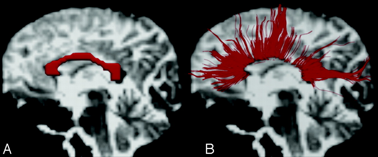
- Fig 1.
Regions of interest (ROIs) of the corpus callosum (CC) and interhemispheric fibers. A, A single ROI is placed in the midline CC in the midsaggital plane for studying callosal fibers on the background of a b = 0 T2-weighted image.29 The ROI is red. B, Left lateral view of a 3D reconstruction of callosal fibers passing through the genu, body, and splenium of the CC in the background of a b = 0 T2-weighted image.
- Fig 2.
ROIs for the projection fibers and long association fibers (LAF) placed on color fractional anisotropy (FA) images. A and B, A 2-ROI approach is used for the projection fibers. One ROI is placed in the posterior limb of the internal capsule (PLIC) and a second ROI is placed in the cerebral peduncle (CP) on the axial plane at the level of the midbrain to generate the projection fibers.29 C and D, A 2-ROI approach is used for LAF (superior longitudinal fasciculus [SLF], inferior fronto-occipital fasciculus [IFOF], and uncinate fasciculus [UF]). One ROI is placed in the coronal plane for the SLF, lateral to the corona radiata,36,42 and a second ROI, on the axial plane at the level of anterior margin of external capsule, where the IFOF and UF pass close to each other.30 All the regions of interest are white.
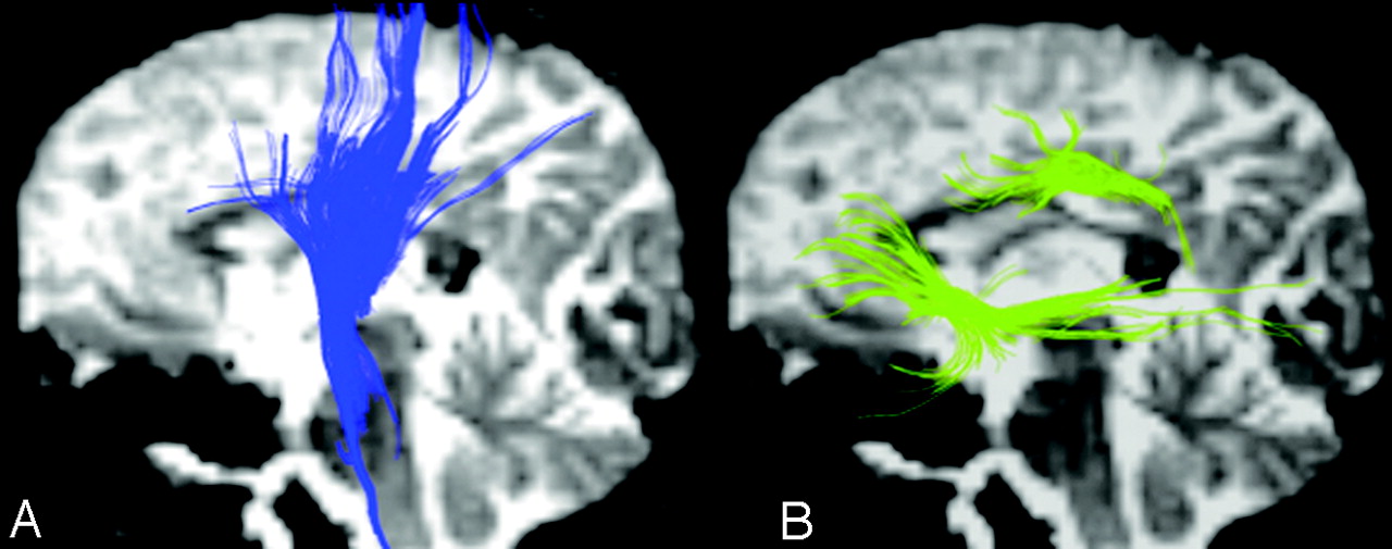
- Fig 3.
Left lateral view of the 3D reconstruction of projection fibers and LAF on the background of a b = 0 T2-weighted image. A, Projection fibers. B, LAF (SLF, IFOF, and UF) pathway.
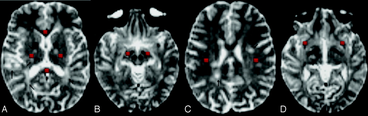
- Fig 4.
Regions of interest in the NAWM are placed on b = 0 T2-weighted images. A, One ROI is placed in the genu of the CC and one, in the splenium of the CC; and bilateral ROIs are placed in the PLIC. B, Bilateral ROIs are placed in the CP at the level of the midbrain. C, Bilateral ROIs for the SLF are placed in the axial plane, one section above the body of the CC lateral to the corona radiata. D, Bilateral ROIs are placed at the level of the anterior margin of the external capsule for the IFOF and UF. All the ROIs are red.
- Fig 5.
Callosal, projection, and association fibers passing through NAWM. A, Callosal fibers from the splenium of the CC passing through NAWM on the background of axial and sagittal planes of b = 0 T2-weighted images. A black arrow points to the T2 lesion on the axial plane, and the arrowhead points to the fibers from the splenium of the CC passing through NAWM. B, Projection fibers pass through NAWM in the background of the axial and sagittal planes of b = 0 T2-weighted images. Black arrows point to the T2 lesions, and the arrowhead points to the projection fibers passing through NAWM. C, The T2 lesion is encircled, and the arrows point to the association fibers passing through NAWM.
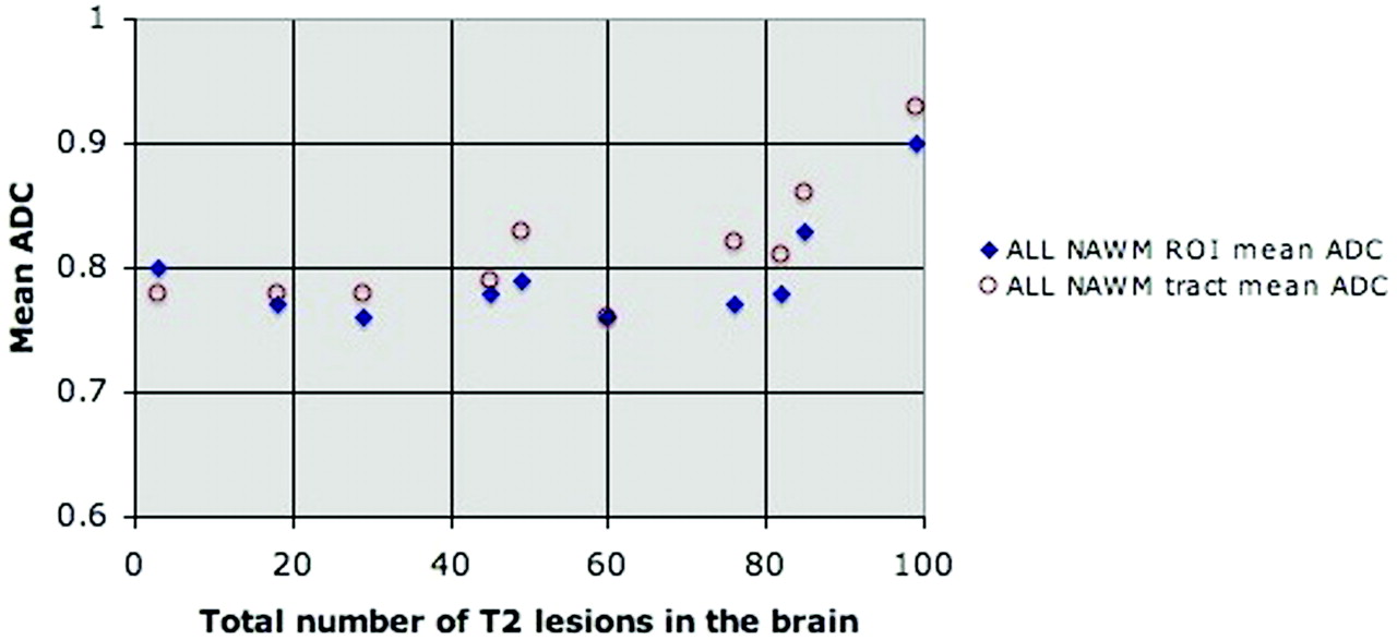
- Fig 6.
Scatterplots of the correlation between all NAWM regions of interest (ROIs) and tract-based mean ADC values of pediatric patients with MS compared with the total number of T2 lesions in the brain. Circles and diamonds represent the mean values.
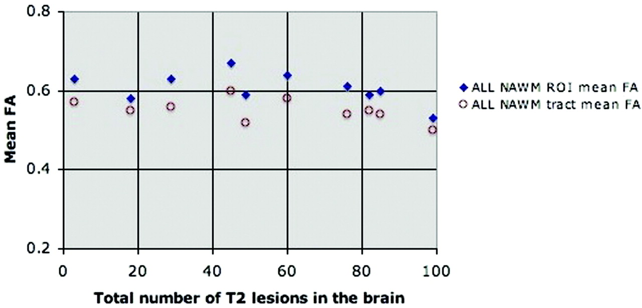
- Fig 7.
Scatterplots of the correlation between all NAWM ROIs and tract-based mean FA values of pediatric patients with MS compared with the total number of T2 lesions in the brain. Circles and diamonds represent the mean values.


Tables
CC PLIC CP LAF ROI mean ADC × 10−3 mm2/s Patients 1.11 ± 0.13 0.73 ± 0.02 0.88 ± 0.10 0.84 ± 0.07 Controls 0.89 ± 0.03 0.70 ± 0.03 0.76 ± 0.05 0.76 ± 0.02 P value 0.0020a 0.021a 0.020a 0.0059a ROI mean FA Patients 0.53 ± 0.06 0.62 ± 0.02 0.65 ± 0.06 0.39 ± 0.04 Controls 0.63 ± 0.06 0.67 ± 0.03 0.69 ± 0.04 0.44 ± 0.02 P value .0039a .0039a .13 .037a Tract mean ADC × 10−3 mm2/s Patients 0.97 ± 0.10 0.76 ± 0.04 0.81 ± 0.06 0.85 ± 0.06 Controls 0.86 ± 0.06 0.71 ± 0.03 0.73 ± 0.02 0.75 ± 0.02 P value .037a .0059a .0020a .0020a Tract mean FA Patients 0.55 ± 0.06 0.56 v 0.03 0.58 ± 0.04 0.42 ± 0.04 Controls 0.62 ± 0.03 0.59 ± 0.02 0.62 ± 0.03 0.48 ± 0.02 P value .0098a .0098a .0039a .0098a -
Note:—ROI indicates region of interest; CC, corpus callosum; PLIC, posterior limb of internal capsule; CP, cerebral peduncle; LAF, long association fibers; ADC, apparent diffusion coefficient; FA, fractional anisotropy.
-
a Statistically significant.
-
Controls Patients P Value Region 1: genu of CC ROI mean ADC 0.77 ± 0.03 0.91 ± 0.14 .002a ROI mean FA 0.80 ± 0.09 0.73 ± 0.10 .28 Tract mean ADC 0.78 ± 0.03 0.87 ± 0.10 .041a Tract mean FA 0.62 ± 0.04 0.57 ± 0.06 .16 Region 2: Splenium of CC ROI mean ADC 0.74 ± 0.04 0.88 ± 0.11 .0039a ROI mean FA 0.86 ± 0.05 0.78 ± 0.05 .0059a Tract mean ADC 0.79 ± 0.05 0.85 ± 0.07 .049a Tract mean FA 0.72 ± 0.03 0.66 ± 0.04 .002a Region 3: PLIC ROI mean ADC 0.69 ± 0.03 0.72 ± 0.02 .0098a ROI mean FA 0.69 ± 0.05 0.66 ± 0.03 .25 Tract mean ADC 0.71 ± 0.03 0.75 ± 0.04 .0098a Tract mean FA 0.61 ± 0.03 0.57 ± 0.03 .0098a Region 4: CP ROI mean ADC 0.71 ± 0.03 0.78 ± 0.05 .037a ROI mean FA 0.73 ± 0.05 0.69 ± 0.04 .19 Tract mean ADC 0.72 ± 0.02 0.78 ± 0.04 .0098a Tract mean FA 0.63 ± 0.03 0.60 ± 0.03 .016a Region 5: Anatomic region for SLF ROI mean ADC 0.71 ± 0.03 0.74 ± 0.04 .014a ROI mean FA 0.60 ± 0.06 0.47 ± 0.07 .0039a Tract mean ADC 0.72 ± 0.03 0.79 ± 0.07 .0039a Tract mean FA 0.54 ± .04 0.45 ± 0.05 .0098a Region 6: Anatomic region for IFOF and UF ROI mean ADC 0.76 ± 0.03 0.82 ± 0.03 .002a ROI mean FA 0.53 ± 0.04 0.45 ± 0.07 .018a Tract mean ADC 0.78 ± 0.03 0.85 ± 0.05 .002a Tract mean FA 0.51 ± 0.03 0.44 ± 0.06 .014a All NAWM ROI mean ADC 0.73 ± 0.02 0.79 ± 0.04 .0020a ROI mean FA 0.68 ± 0.02 0.61 ± 0.04 .0098a Tract mean ADC 0.75 ± 0.03 0.81 ± 0.05 .0098a Tract mean FA 0.61 ± 0.03 0.55 ± 0.03 .002a -
Note:—SLF indicates superior longitudinal fascicle; IFOF, inferior fronto-occipital fascicle; UF, uncinate fascicle; NAWM, normal-appearing white matter; ADC, apparent diffusion co-efficient, ×10−3 mm2/s.
-
a Statistically significant.
-


