
Article Figures & Data
Figures
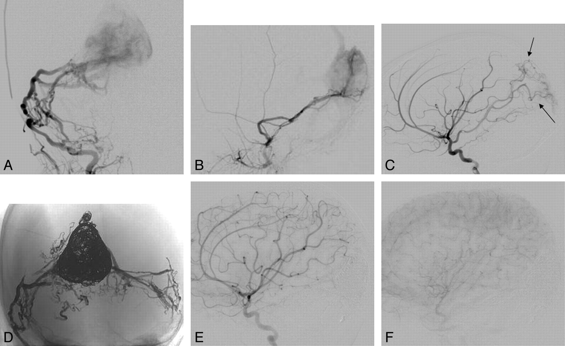
- Fig 1.
A, Frontal view of a left vertebral artery injection. B, Microcatheter injection close to the fistulous point shows instantaneous opacification of the venous collector and absence of nidal vessels, consistent with the mural configuration. C−E, Unsubtracted views after deployment of coils (C) and after Onyx embolization (E). Microcatheter injection at the same point after deployment of detachable coils (D) demonstrates slowing of flow, with a lag before opacification of the venous system. Note how the Onyx cast reproduces the configuration of the vessels leading to the fistula in E. F, Postembolization image demonstrates non-opacification of the venous pouch, verifying closure of the fistula. G and H, No venous embolization was performed, but early regression of the venous varix collecting the shunt flow is seen by comparing a pre-embolization CT (G) with a CT performed 2 weeks later (H, study performed for an unrelated reason).
- Fig 2.
A and B, Arterial supply to this infantile dural arteriovenous fistula involving the torcular in an 11-month-old child was primarily via the middle meningeal arteries bilaterally, which had a fistulous configuration at the torcular junction (frontal view in A, lateral view in B). C, Parasitization of pial vessels, with supply via the posterior cerebral artery (black arrows) and middle cerebral artery branches, is seen as well. D, Unsubtracted image in a frontal view demonstrates the large coil mass in the dysplastic midline torcular and the plexiform Onyx cast recreating the arteriovenous fistula nidus from both middle meningeal and occipital arteries. E and F, Lateral views of a right internal carotid artery injection after embolization (E, early arterial; F, capillary phase) demonstrate nonopacification of the venous collector, with regression of the parasitized pial supply, which was never embolized.
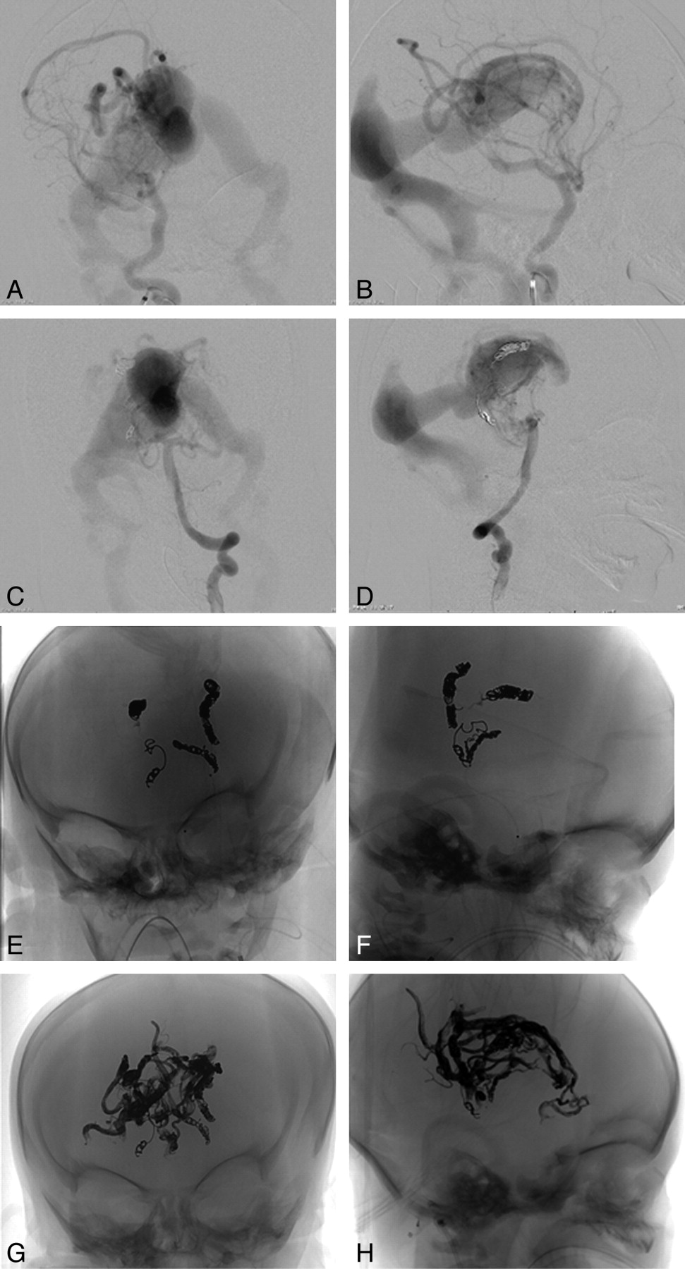
- Fig 3.
A−D, Frontal (A) and lateral (B) views of a right ICA injection and a left vertebral artery injection (C and D) on days 4 and 7 of life demonstrate rapid opacification of the ectatic median vein, making it difficult to appreciate brain parenchymal opacification of these injections. E and F, Frontal (E) and lateral (F) views of the coil mass and small Onyx casts deployed on days 4 and 7 of life. During each of these embolizations, the patient experienced a marked improvement in hemodynamic function, with weaning of the pressors during the procedure. G and H, Frontal (G) and lateral (H) views of the extensive Onyx cast delivered on day 20 of life.
- Fig 4.
A, Frontal unsubtracted view of 2 drops of Onyx lodged in the jugular bulb without any impeding flow and without evidence of jugular stenosis, after Onyx embolization of a Galenic malformation (white arrow demonstrates the detached drops). B, In another vein of Galen malformation case, a thin strand of Onyx was noted to have extended to within the midline varix of the median vein (white arrows), remaining attached to the main cast.


Tables
Patient No. Age Sex Localization Signs/Symptoms at Presentation Signs/Symptoms after Treatment 1 4 d M VoG, c Cardiac failure Improved cardiac function 2 5 mo M VoG, m Prenatal ultrasonic diagnosis Increased intermittent left-sided exotropia 3 7 mo F Dural AVF Incidental finding in work-up for subglottic hemangioma ND 4 9 mo F R frontal AVM Incidental finding in work-up for family history of HHT ND 5 11 mo F Torcular AVF Increasing head circumference ND 6 2 y F VoG, c Gross motor delay ND 7 3 y M VoG, c Macrocephaly, mild dysmetria ND 8 3 y M R frontal AVM Headaches ND 9 6 y M R cerebellar AVM Seizures ND 10 6 y F L para-/intraspinal AVM Headaches Progressive L upper extremity proximal weakness 11 9 y M R parietal AVM Complex partial seizures ND 12 10 y F L frontal AVM Incidental finding on MRI ND 13 12 y F R temporal AVM Altered mental status ND 14 14 y F Splenial AVM Headaches, nausea, vomiting ND 15 18 y M Spinal AVM Klippel-Trenaunay syndrome, leg overgrowth and weakness, cognitive impairment ND -
Note:—mo indicates months; d, days; VoG, vein of Galen malformation; c, choroidal type; m, mural type; ND, no (new) deficits; AVM, arteriovenous malformation; HHT, hereditary hemorrhagic telangiectasia; MRI, MR imaging; AVF, atriovenous fistula; L, left; R, right; y, year.
-
Patient No. Nidus Type Shunt Fistula Retrograde Venous Pressurization 1 Diffuse Mixed N/A No 2 N/A Fistulous Single-hole No 3 N/A Mixed Dural-osseous Reversed flow in deep venous system 4 Compact Plexiform N/A No 5 N/A Mixed Dural Cortical venous drainage via cavernous sinus to SOV, facial, and scalp veins 6 Compact Mixed N/A Reversed flow through deep and superficial veins 7 Diffuse Mixed N/A No 8 Diffuse Fistulous N/A No 9 Compact Plexiform N/A No 10 Diffuse Plexiform N/A No 11 Compact Plexiform N/A No 12 Compact Plexiform N/A No 13 Compact Plexiform N/A No 14 Compact Fistulous N/A No 15 Compact Plexiform N/A No -
Note:—N/A indicates not applicable; SOV, superior ophthalmic vein.
-
Patient No. No. Embolizations Time Interval Between First, Last Treatment Total Amount of Onyx (in mL) Coils/n-BCA Residual/Cure Treatment for Residual Cumulative X-Ray Skin Entrance Dose (mGy) 1 3 16 d 6.7 GDC and Axium coils PE N/A 2665 2 1 N/A 1.3 GDC-18 3D and Nexus (ev3) coils Cure No treatment, asymptomatic 315 3 3 6 mo 1.3 360-GDC soft coils PE Continued embolizations 1225 4 1 N/A 0.8 None NCE Surgery 624 5 6 9 mo 12.4 GDC-18 NCE Continued embolizations 3708 6 5 7 mo 2.8 GDC coils NCE N/A 3992 7 3 4 mo 2.3 Axium coils NCE If feasible, will re-attempt in 2–3 y 1665 8 1 N/A 1.6 None PE Surgery 657 9 1 N/A 1 None PE Surgery 884 10 5 4 mo 19.9 GDC and Axium coils NCE Surgery 2508 11 1 N/A 2.2 None NCE Surgery 1632 12 1 N/A 1.5 None NCE Surgery 501 13 1 N/A 0.4 None Cure None 537 14 1 N/A 2.1 None NCE Surgery 1956 15 2 2 mo 9.9 None NCE Continued embolization 3907 -
Note:—PE indicates partial embolization; NCE, near-complete embolization; n-BCA, n-butyl cyanoacrylate; GDC, Guglielmi detachable coil.
-


