Advertisement

Main menu

  • Home
  • Content
    • Current Issue
    • Accepted Manuscripts
    • Article Preview
    • Past Issue Archive
    • Video Articles
    • AJNR Case Collection
    • Case of the Week Archive
    • Case of the Month Archive
    • Classic Case Archive
  • Special Collections
    • AJNR Awards
    • Low-Field MRI
    • Alzheimer Disease
    • ASNR Foundation Special Collection
    • Photon-Counting CT
    • View All
  • Multimedia
    • AJNR Podcasts
    • AJNR SCANtastic
    • Trainee Corner
    • MRI Safety Corner
    • Imaging Protocols
  • For Authors
    • Submit a Manuscript
    • Submit a Video Article
    • Submit an eLetter to the Editor/Response
    • Manuscript Submission Guidelines
    • Statistical Tips
    • Fast Publishing of Accepted Manuscripts
    • Graphical Abstract Preparation
    • Imaging Protocol Submission
    • Author Policies
  • About Us
    • About AJNR
    • Editorial Board
    • Editorial Board Alumni
  • More
    • Become a Reviewer/Academy of Reviewers
    • Subscribers
    • Permissions
    • Alerts
    • Feedback
    • Advertisers
    • ASNR Home

User menu

  • Alerts
  • Log in

Search

  • Advanced search
American Journal of Neuroradiology

American Journal of Neuroradiology

ASHNR American Society of Functional Neuroradiology ASHNR American Society of Pediatric Neuroradiology ASSR
  • Alerts
  • Log in

Advanced Search

  • Home
  • Content
    • Current Issue
    • Accepted Manuscripts
    • Article Preview
    • Past Issue Archive
    • Video Articles
    • AJNR Case Collection
    • Case of the Week Archive
    • Case of the Month Archive
    • Classic Case Archive
  • Special Collections
    • AJNR Awards
    • Low-Field MRI
    • Alzheimer Disease
    • ASNR Foundation Special Collection
    • Photon-Counting CT
    • View All
  • Multimedia
    • AJNR Podcasts
    • AJNR SCANtastic
    • Trainee Corner
    • MRI Safety Corner
    • Imaging Protocols
  • For Authors
    • Submit a Manuscript
    • Submit a Video Article
    • Submit an eLetter to the Editor/Response
    • Manuscript Submission Guidelines
    • Statistical Tips
    • Fast Publishing of Accepted Manuscripts
    • Graphical Abstract Preparation
    • Imaging Protocol Submission
    • Author Policies
  • About Us
    • About AJNR
    • Editorial Board
    • Editorial Board Alumni
  • More
    • Become a Reviewer/Academy of Reviewers
    • Subscribers
    • Permissions
    • Alerts
    • Feedback
    • Advertisers
    • ASNR Home
  • Follow AJNR on Twitter
  • Visit AJNR on Facebook
  • Follow AJNR on Instagram
  • Join AJNR on LinkedIn
  • RSS Feeds

AJNR Awards, New Junior Editors, and more. Read the latest AJNR updates

Research ArticleHead and Neck

Postsurgical Intracranial Hypotension: Diagnostic and Prognostic Imaging Findings

D.R. Hadizadeh, A. Kovács, H. Tschampa, R. Kristof, J. Schramm and H. Urbach
American Journal of Neuroradiology January 2010, 31 (1) 100-105; DOI: https://doi.org/10.3174/ajnr.A1749
D.R. Hadizadeh
  • Find this author on Google Scholar
  • Find this author on PubMed
  • Search for this author on this site
A. Kovács
  • Find this author on Google Scholar
  • Find this author on PubMed
  • Search for this author on this site
H. Tschampa
  • Find this author on Google Scholar
  • Find this author on PubMed
  • Search for this author on this site
R. Kristof
  • Find this author on Google Scholar
  • Find this author on PubMed
  • Search for this author on this site
J. Schramm
  • Find this author on Google Scholar
  • Find this author on PubMed
  • Search for this author on this site
H. Urbach
  • Find this author on Google Scholar
  • Find this author on PubMed
  • Search for this author on this site
  • Article
  • Figures & Data
  • Info & Metrics
  • Responses
  • References
  • PDF
Loading

Article Figures & Data

Figures

  • Tables
  • Fig 1.
    • Download figure
    • Open in new tab
    • Download powerpoint
    Fig 1.

    Sag ratios defined as maximal anteroposterior midbrain/maximal bipeduncular diameter3 preoperatively (n = 12), on the day after surgery (n = 16), in the first week after surgery (n = 13), and on long-term follow-up examinations (n = 7). Note the temporary significant increase of the sag ratio and complete resolution at long-term follow-up. Error marks indicate SDs of measurements (n = number of patients).

  • Fig 2.
    • Download figure
    • Open in new tab
    • Download powerpoint
    Fig 2.

    Sagittal T1-weighted spin-echo (A) and coronal T2-weighted FSE images (B,C) after depth electrode placement for presurgical work-up. Sagittal (D), axial (E), and coronal (F) T2-weighted FSE images 5 days after left-sided hippocampectomy. Vasogenic edema with increased signal intensity on T2-weighted images and elevated ADC values on diffusion-weighted MR imaging (not shown) is present in the basal ganglia (E; open arrows) and, to a lesser extent, in the brain stem (D; open arrow). CSF loss causes noisy T2-weighted images (D-F) and sagging of the brain stem and cerebellum (D,F). Normally, the iter in the aqueduct is within 2 mm below the incisural line connecting the tuberculum sellae and the entrance of the vein of Galen into the straight sinus (A). With severe CSF loss, the iter and the splenium corporis callosi are displaced downwards (D). Note tonsillar displacement into the foramen magnum (arrows in F compared with B and C).
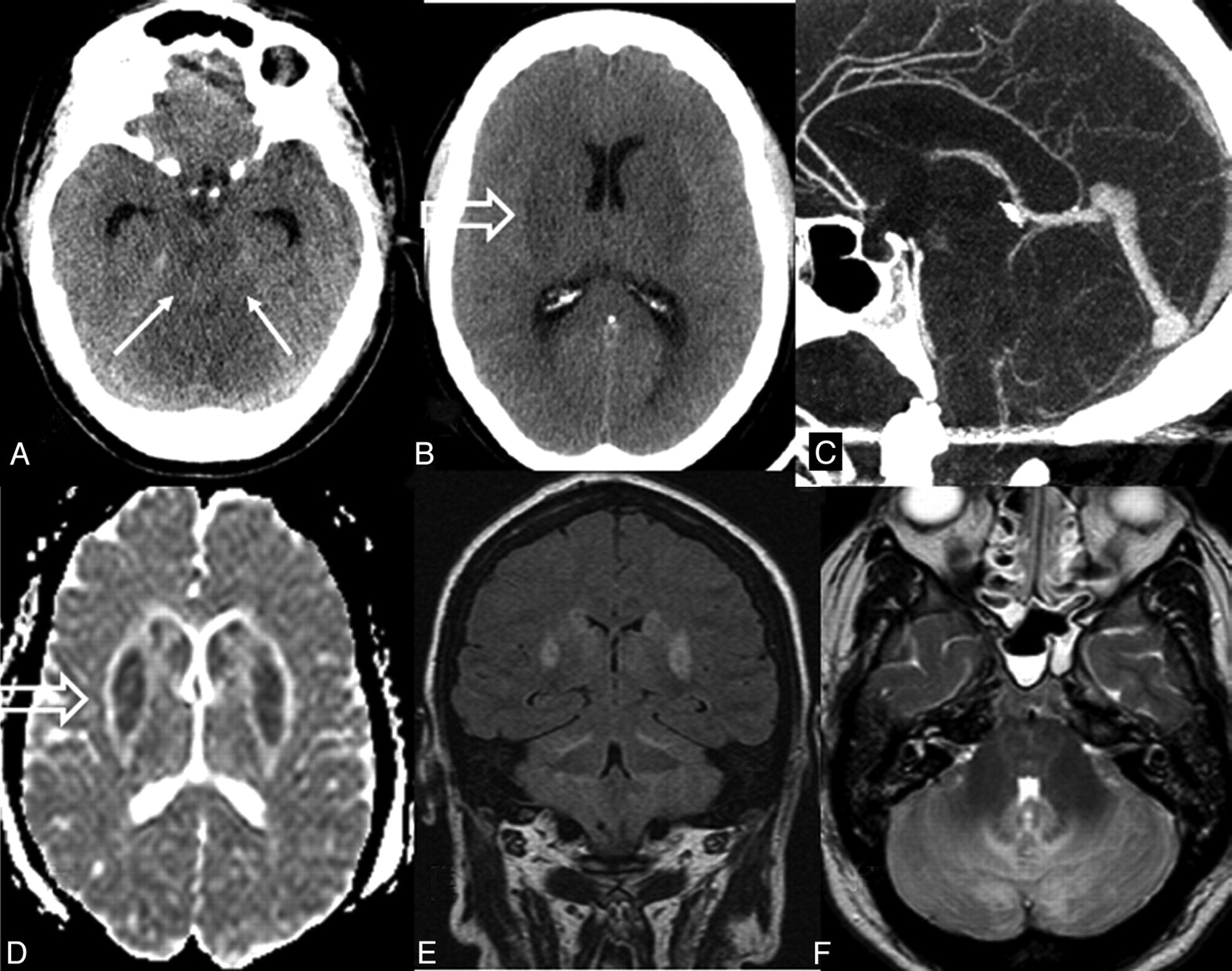
  • Fig 3.
    • Download figure
    • Open in new tab
    • Download powerpoint
    Fig 3.

    Axial FLAIR before right-sided temporal lobe lesionectomy (A,B), 1 day after the lesionectomy (C,D), and 5 years later (E,F). Note that sections in A, B, E, F are angulated along the temporal lobe length axis, whereas sections in C and D are angulated along the c.a.-c.p.-line. However, apart from the resection defect (C; thick arrow), moderate thalamic and basal ganglia hyperintensities (D; open arrow) and compressed ambient cisterns (C; arrows) are clearly visible. From a clinical standpoint, this 36-year-old woman had a complete recovery with resolution of the imaging findings (E,F).

  • Fig 4.
    • Download figure
    • Open in new tab
    • Download powerpoint
    Fig 4.

    Intracranial hypotension after spinal fixation with dural opening. Axial CT images show compressed ambient cisterns (A; arrows) and basal ganglia hypoattenuation (B; open arrow). On midline sagittal reformatted CT angiogram (C), only sparse CSF space is visible in the posterior fossa. Axial ADC map (D) and coronal FLAIR images show mainly cytotoxic edema of the gray matter. Increased signal intensity is also visible in the cerebellum (E,F) and, to a lesser extent, in the pons (F). From a clinical standpoint, this 60-year-old woman who underwent spinal surgery for lumbar spondylodiskitis has recovered fully from symptoms of intracranial hypotension.

Tables

  • Figures
    • View popup
    Table 1:

    Imaging modalities and clinical outcome in 16 patients with intracranial hypotension

    Pt No.SexAge (y)Type of SurgeryImaging ModalitiesClinical OutcomemRS
    1M51Optic gliomaCT, MRIFull recovery0/5
    2M59Oligoastrocytoma WHO grade IICT, MRIFull recovery0/5
    3F362/3 resection of temporal lobeCT, DSA, MRIModerate deficit3/3
    4F37Granulomatous inflammationCT, MRIFull recovery0/5
    5M44Incidental MCA aneurysmCT, DSAFatal6/1
    6M58CavernomaCTFull recovery0/5
    7M67Colloid cystCT, DSAVegetative state5/2
    8M60Recurrent metastasisCTMild deficit2/4
    9M42Hippocampal sclerosisCT, MRI, DSAModerate deficit3/3
    10M39Astrocytoma WHO grade IICT, MRIFatal6/1
    11F44Select amygdaloCT, MRIFatal6/1
    12M59AVM, Spetzler & Martin grade ICT, MRI, DSAMild deficit2/4
    13F34Oligodendroglioma WHO grade IIICT, MRIModerate deficit3/3
    14F70Spondylodiskitis T6/7CT, MRIFull recovery0/5
    15F71Spondylodiskitis L4/5, spinal fixationCT, MRIFatal6/1
    16M88Bilat. subdural hematomas: hollow screwsCT, MRIMild deficit2/4
    • Note:—mRS indicates modified Rankin scale/Glasgow outcome score; DSA, digital subtraction angiography; MCA, middle cerebral artery; select amygdalo, selective amygdalohippocampectomy; AVM, arteriovenous malformation; M, male; F, female; WHO, World Health Organization; bilat, bilateral; MRI, MR imaging.

    • View popup
    Table 2:

    Imaging findings in 16 patients with intracranial hypotension

    Pt No.tcesdfbscerangsagSag Ratiodilbgenl
    1n.d.yn.d.n.d.n.d.n0.83n.d.+n.d.
    2n.d.yn.d.n.d.n.d.y0.90n+n.d.
    3n.d.yny88y0.92n++n.d.
    4n.d.ynnn.d.n1.00n.d.+y
    5n.d.nn.d.n.d.n.d.y1.15n.d.+n.d.
    6n.d.nn.d.n.d.n.d.y0.92n.d.+n.d.
    7n.d.nn.d.n.d.70n1.09n.d.++n.d.
    8n.d.yn.d.n.d.n.d.y1.00n.d.+n.d.
    9n.d.nn.d.n.d.58y0.83n.d.++n.d.
    10n.d.nnyn.d.n0.87n.d.++n.d.
    11n.d.nyy44y1.11n+++y
    12n.d.nyyn.d.y0.88n++n.d.
    13n.d.nn.d.n.d.n.d.y0.92n.d.+n.d.
    14nyyy102y1.00n+n
    15nyyy130n0.96n+++n
    16yyyy105y1.00y++n.d.
    • Note:—tce indicates dural thickening and enhancement; sdf, subdural fluid collections; bs, brain stem hyperintensity; cer, cerebellar hyperintensities; ang, angle of vein of Galen to straight sinus; sag, brain sagging; sag ratio, maximal anterior-posterior midbrain diameter divided by maximal bipeduncular diameter; dil, dilation of intracranial dural sinuses and/or spinal epidural plexuses; bg, hyperintense thalamic/basal ganglia signal; enl, enlargement of pituitary gland; y, yes; n, no; n.d., not determinable.

    • View popup
    Table 3:

    Correlation of imaging findings with clinical outcome parameters in 16 patients with intracranial hypotension

    mRSP ValueGOSP Valuen
    sdf0.360.171−0.381.14616
    bs0.258.537−0.206.6248
    cer0.756.030b−0.637.089c8
    ang−0.049.9160.049.9167
    sag−0.026.9230.120.65916
    Sag ratio0.442.087c−0.478.061c16
    dil0.059.9000.043.9277
    bg0.632.009a−0.646.007a16
    enl−0.408.5920.302.6984
    • a Statistically highly significant (P < .01).

    • b Statistically significant (P < .05).

    • c Strong tendency but not statistically significant.

PreviousNext
Back to top

In this issue

American Journal of Neuroradiology: 31 (1)
American Journal of Neuroradiology
Vol. 31, Issue 1
1 Jan 2010
  • Table of Contents
  • Index by author
Advertisement
Print
Download PDF
Email Article

Thank you for your interest in spreading the word on American Journal of Neuroradiology.

NOTE: We only request your email address so that the person you are recommending the page to knows that you wanted them to see it, and that it is not junk mail. We do not capture any email address.

Enter multiple addresses on separate lines or separate them with commas.
Postsurgical Intracranial Hypotension: Diagnostic and Prognostic Imaging Findings
(Your Name) has sent you a message from American Journal of Neuroradiology
(Your Name) thought you would like to see the American Journal of Neuroradiology web site.
CAPTCHA
This question is for testing whether or not you are a human visitor and to prevent automated spam submissions.
Cite this article
D.R. Hadizadeh, A. Kovács, H. Tschampa, R. Kristof, J. Schramm, H. Urbach
Postsurgical Intracranial Hypotension: Diagnostic and Prognostic Imaging Findings
American Journal of Neuroradiology Jan 2010, 31 (1) 100-105; DOI: 10.3174/ajnr.A1749

Citation Manager Formats

  • BibTeX
  • Bookends
  • EasyBib
  • EndNote (tagged)
  • EndNote 8 (xml)
  • Medlars
  • Mendeley
  • Papers
  • RefWorks Tagged
  • Ref Manager
  • RIS
  • Zotero
0 Responses
Respond to this article
Share
Bookmark this article
Postsurgical Intracranial Hypotension: Diagnostic and Prognostic Imaging Findings
D.R. Hadizadeh, A. Kovács, H. Tschampa, R. Kristof, J. Schramm, H. Urbach
American Journal of Neuroradiology Jan 2010, 31 (1) 100-105; DOI: 10.3174/ajnr.A1749
del.icio.us logo Twitter logo Facebook logo Mendeley logo
  • Tweet Widget
  • Facebook Like
  • Google Plus One
Purchase

Jump to section

  • Article
    • Abstract
    • Materials and Methods
    • Results
    • Discussion
    • Conclusions
    • Acknowledgment
    • References
  • Figures & Data
  • Info & Metrics
  • Responses
  • References
  • PDF

Related Articles

  • No related articles found.
  • PubMed
  • Google Scholar

Cited By...

  • Coma from wall suction-induced CSF leak complicating spinal surgery
  • Crossref (36)
  • Google Scholar

This article has been cited by the following articles in journals that are participating in Crossref Cited-by Linking.

  • Spontaneous Intracranial Hypotension: Dilemmas in Diagnosis
    Maryam Rahman, Sharatchandra S Bidari, Ron G Quisling, William A Friedman
    Neurosurgery 2011 69 1
  • Intracranial hypotension
    Horst Urbach
    Current Opinion in Neurology 2014 27 4
  • Intracranial Hypotension and Intracranial Hypertension
    Esther L. Yuh, William P. Dillon
    Neuroimaging Clinics of North America 2010 20 4
  • Spontaneous intracranial hypotension: key features for a frequently misdiagnosed disorder
    Carlos Perez-Vega, Pilar Robles-Lomelin, Isabel Robles-Lomelin, Victor Garcia Navarro
    Neurological Sciences 2020 41 9
  • Postoperative intracranial hypotension-associated venous congestion: Case report and literature review
    Alexander I. Evins, Davide Boeris, Justin C. Burrell, Alessandro Ducati
    Clinical Neurology and Neurosurgery 2013 115 10
  • Early awareness of cerebrospinal fluid hypovolemia after craniotomy for microsurgical aneurysmal clipping
    Ichiro Kawahara, Keisuke Tsutsumi, Yuki Matsunaga, Hideaki Takahata, Tomonori Ono, Keisuke Toda, Hiroshi Baba
    Acta Neurochirurgica 2013 155 8
  • Prompt Recognition and Management of Postoperative Intracranial Hypotension-Associated Venous Congestion: A Case Report
    Kendall A. Snyder, Michelle J. Clarke, Julie R. Gilbertson, Sara E. Hocker
    Neurocritical Care 2016 24 3
  • Pseudohypoxic Brain Swelling: Report of 2 Cases and Introduction of the Lentiform Rim Sign as Potential MRI Marker
    Houman Sotoudeh, Philip R. Chapman, Ehsan Sotoudeh, Aparna Singhal, Gagandeep Choudhary, Omid Shafaat
    World Neurosurgery 2020 133
  • Spinal dementia: Don’t miss it, it’s treatable
    Horst Urbach, A El Rahal, K Wolf, C Zander, T Demerath, F Volz, J Beck, N Lützen
    Neuroradiology 2024 66 10
  • The frequency and severity of intracranial hypotension post-intraoperative lumbar drainage using a Tuohy needle and the traditional needle
    M. Maher Hulou, Muhammad M. Abd-El-Barr, William B. Gormley, Amir A. Zamani, Ian F. Dunn, Ossama Al-Mefty
    British Journal of Neurosurgery 2016 30 4

More in this TOC Section

  • Correlation of Apparent Diffusion Coefficient at 3T with Prognostic Parameters of Retinoblastoma
  • Parathyroid Lesions: Characterization with Dual-Phase Arterial and Venous Enhanced CT of the Neck
  • MR Diagnosis of Facial Neuritis: Diagnostic Performance of Contrast-Enhanced 3D-FLAIR Technique Compared with Contrast-Enhanced 3D-T1-Fast-Field Echo with Fat Suppression
Show more HEAD AND NECK

Similar Articles

Advertisement

Indexed Content

  • Current Issue
  • Accepted Manuscripts
  • Article Preview
  • Past Issues
  • Editorials
  • Editor's Choice
  • Fellows' Journal Club
  • Letters to the Editor
  • Video Articles

Cases

  • Case Collection
  • Archive - Case of the Week
  • Archive - Case of the Month
  • Archive - Classic Case

More from AJNR

  • Trainee Corner
  • Imaging Protocols
  • MRI Safety Corner

Multimedia

  • AJNR Podcasts
  • AJNR Scantastics

Resources

  • Turnaround Time
  • Submit a Manuscript
  • Submit a Video Article
  • Submit an eLetter to the Editor/Response
  • Manuscript Submission Guidelines
  • Statistical Tips
  • Fast Publishing of Accepted Manuscripts
  • Graphical Abstract Preparation
  • Imaging Protocol Submission
  • Evidence-Based Medicine Level Guide
  • Publishing Checklists
  • Author Policies
  • Become a Reviewer/Academy of Reviewers
  • News and Updates

About Us

  • About AJNR
  • Editorial Board
  • Editorial Board Alumni
  • Alerts
  • Permissions
  • Not an AJNR Subscriber? Join Now
  • Advertise with Us
  • Librarian Resources
  • Feedback
  • Terms and Conditions
  • AJNR Editorial Board Alumni

American Society of Neuroradiology

  • Not an ASNR Member? Join Now

© 2025 by the American Society of Neuroradiology All rights, including for text and data mining, AI training, and similar technologies, are reserved.
Print ISSN: 0195-6108 Online ISSN: 1936-959X

Powered by HighWire