
Article Figures & Data
Figures
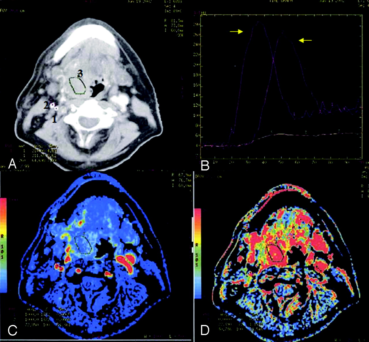
- Fig 1.
PCT imaging with ICA reference of a patient with right tongue base squamous cell carcinoma. A, Axial contrast-enhanced CT scan shows a mass involving the right tongue base. Regions of interest have been placed in the ICA (1), the internal jugular vein (2), and the tumor (3). B, Time-enhancement curve for the patient shown in A. The first peak is arterial enhancement; the second is venous (arrows). The flat curve at the bottom represents tissue region-of-interest (3) enhancement. C, Fractional tissue BF map for the patient in A. There is increased fractional tissue BF in the region of the mass lesion compared with surrounding tissue. D, PS map for the same patient. The tumor site displays increased capillary permeability compared with surrounding tissue.
- Fig 2.
PCT imaging with ECA reference of the same patient depicted in Fig 1. A, Regions of interest have been placed in the ECA (1), the internal jugular vein (2), and tumor (3). B, Time-enhancement curve for the patient shown in A. The first peak is ECA enhancement; the second is venous enhancement (arrows). C, The fractional tissue BF map for the patient in A generated with ECA reference is similar to that generated by using the ICA as the reference function. D, PS map generated with ECA reference demonstrates increased capillary permeability similar to that generated by using the ICA as reference.
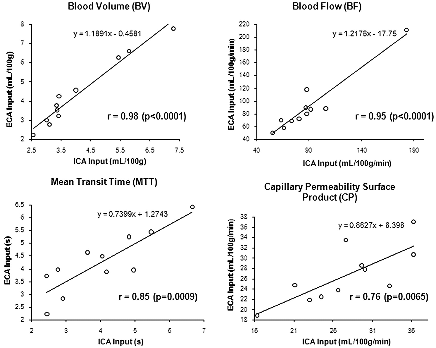
- Fig 3.
Scatterplots of perfusion parameter measurements derived with an ECA arterial reference compared with the ICA. r values are the Pearson product moment correlation coefficients. All correlations are significant (P < .05).


Tables
Patient No. Age (yr) Sex Tumor Location Stage 1 59 M Left tongue base T4N2cM0 3 50 F Oral tongue T3N0M0 4 49 F Epiglottis T2N0M0 5 40 M Right floor of mouth T4N2bM1 8 66 M Epiglottis T3N0M0 9 41 M Left buccal area T3N0M0 10 56 M Vallecula T2N2aM0 11 56 M Right tonsil T4N2bM0 12 59 M Right tonsil T4N2bM0 13 72 F Left tongue base T4N0M0 14 46 M Left tonsil T1N2aM0 Values ICA ECA Difference between Means P Value BF (mL/100 g/min) 89.25 ± 34.42 90.93 ± 43.94 −1.67 ± 15.17 .7221 BV (mL/100 g) 4.07 ± 1.46 4.38 ± 1.78 −0.31 ± 0.46 .0488 MTT (s) 4.03 ± 1.36 4.26 ± 1.19 −0.23 ± 0.72 .3190 Capillary PS (mL/100 g/min) 27.68 ± 6.25 26.74 ± 5.45 0.94 ± 4.12 .4676 -
Note:—ICA indicates internal carotid artery; ECA, external carotid artery; BF, blood flow; BV, blood volume; MTT, mean transit time; PS, permeability surface-area product.
-
* Data are mean ± SD.
-


