
Article Figures & Data
Figures
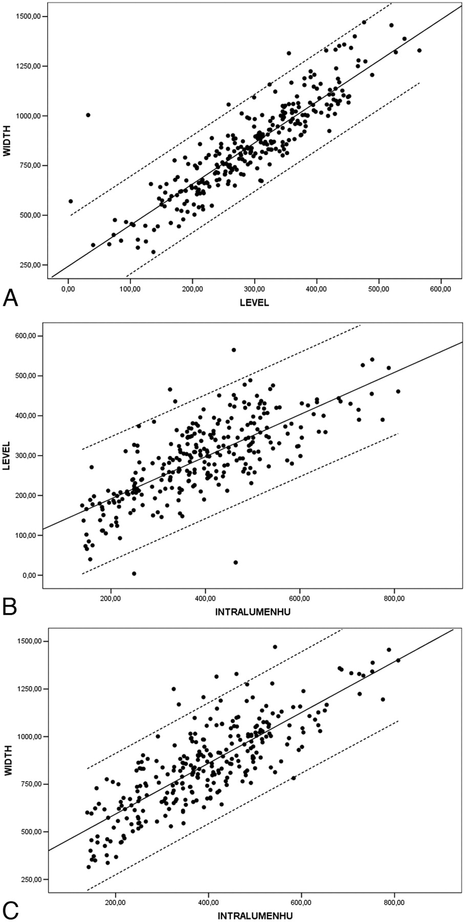
- Fig 1.
A, Scatterplot between width and level (NASCET II-V) with linear regression through the origin, with a 99% mean predictive interval and 99% individual predictive interval. Width = 2.81 × Level. B, Scatterplot between intraluminal Hounsfield unit values (IntraluminHU) and level (NASCET II-V) with linear regression through the origin with a 99% mean predictive interval and a 99% individual predictive interval. Level = 0.72 × Intraluminal HU. C, Scatterplot between intraluminal Hounsfield unit value and width (NASCET II-V) with linear regression through the origin with a 99% mean predictive interval and 99% individual predictive interval. Width = 2.07 × Intraluminal HU Value.
- Fig 2.
Bland-Altman plots of intraobserver variability in the window width (A), window level (B), and intraluminal Hounsfield unit measurements (C). Obs indicates observer. Intralumin-HU indicates intraluminal Hounsfield unit.
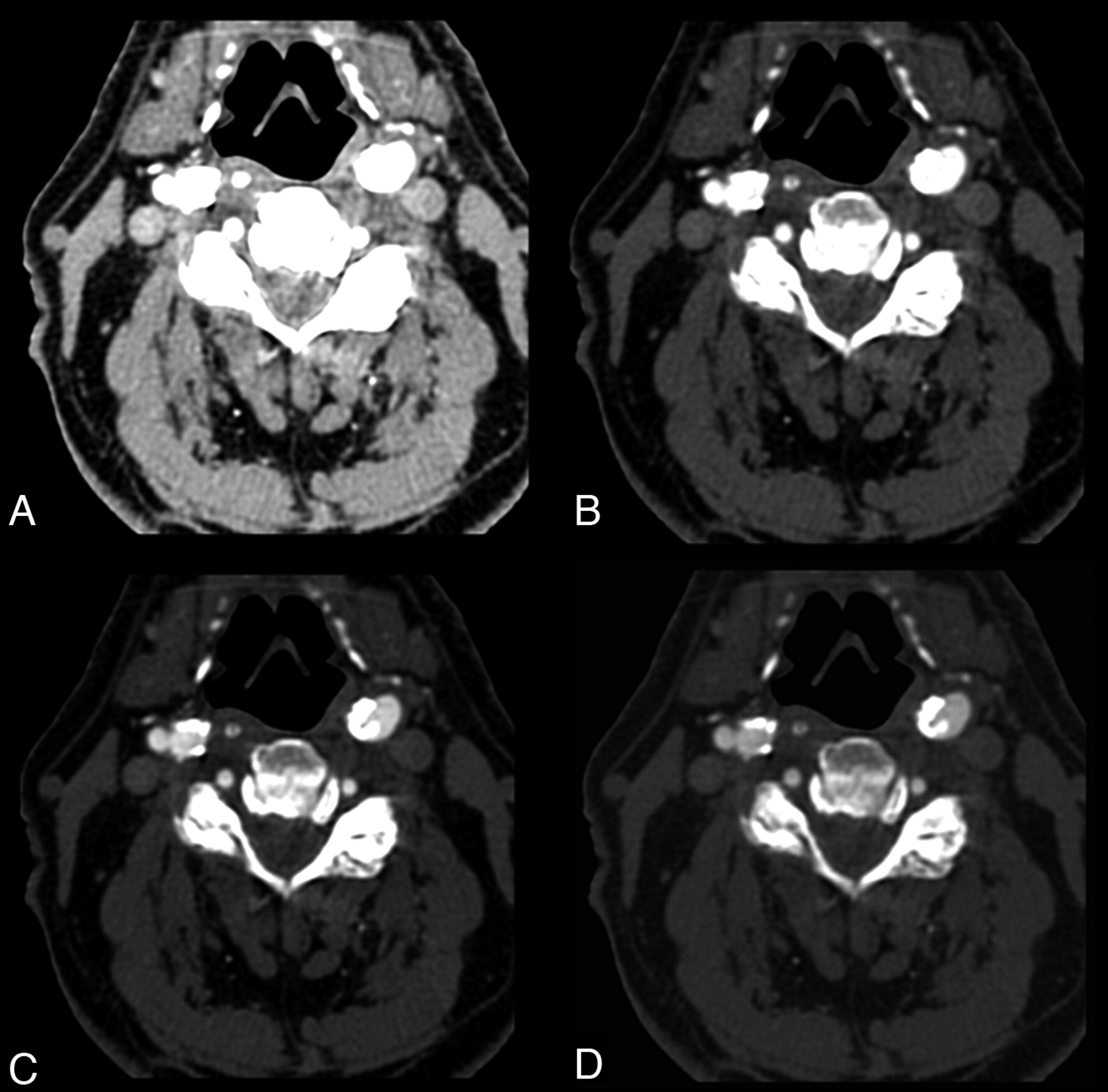
- Fig 3.
A 68-year-old man. CT axial images applying different window parameters. A and B, Intraluminal Hounsfield unit value is 547 (right bulbus). With a 275 width and 30 level (A) and 600/200 (B), it is not possible to differentiate lumen from calcification. C, With 800/300, it is possible to differentiate opacified lumen and calcification. D, By using equations described in this article, we obtained window parameters of 1132/394; a clear differentiation between lumen and parietal calcifications is visible.


Tables
- Table 1:
κ statistics for the comparison of stenosis grading between observer 1 and observer 2
NASCET Class Observer 2 Total II III IV V Observer 1 II 12 0 0 0 12 III 1 28 1 1 31 IV 0 2 28 3 33 V 0 0 3 62 65 Total 13 30 32 66 141 Note:—NASCET indicates North American Symptomatic Carotid Endarterectomy Trial.
*Measurements were made by selecting a plane perpendicular to the lumen center line.
- Table 2:
Pearson correlation calculation between window level, window width, and intraluminal Hounsfield unit values in NASCET classes*
Pearson Correlation Variables NASCET II-V (r) NASCET II (r) NASCET III (r) NASCET IV (r) NASCET V (r) Window level−window width 0.891 0.928 0.890 0.905 0.881 IntraluminHU value−window level 0.743 0.753 0.718 0.689 0.763 IntraluminHU value−window width 0.808 0.724 0.738 0.786 0.836 Note:—IntraluminHU indicates intraluminal Hounsfield unit.
* Correlation is significant at the 0.01 level (2-tailed).
In this issue


Jump to section
Related Articles
Cited By...
- Assessment of Attenuation in Pericarotid Fat among Patients with Carotid Plaque and Spontaneous Carotid Dissection
- Computed tomography analysis of vulnerable carotid atherosclerotic plaque and relationship to clinical characteristics
- Impact Analysis of Different CT Configurations of Carotid Artery Plaque Calcifications on Cerebrovascular Events
- Perivascular Fat Density and Contrast Plaque Enhancement: Does a Correlation Exist?
- Carotid Intraplaque-Hemorrhage Volume and Its Association with Cerebrovascular Events
- Carotid Plaque CTA Analysis in Symptomatic Subjects with Bilateral Intraplaque Hemorrhage: A Preliminary Analysis
- Carotid Artery Wall Imaging: Perspective and Guidelines from the ASNR Vessel Wall Imaging Study Group and Expert Consensus Recommendations of the American Society of Neuroradiology
- CT Attenuation Analysis of Carotid Intraplaque Hemorrhage
- Semiautomated and Automated Algorithms for Analysis of the Carotid Artery Wall on Computed Tomography and Sonography: A Correlation Study
- Carotid Artery Plaque Characterization Using CT Multienergy Imaging
- Carotid Artery Wall Thickness Measured Using CT: Inter- and Intraobserver Agreement Analysis
- Association between Carotid Artery Plaque Type and Cerebral Microbleeds
- Carotid Artery Wall Thickness and Leukoaraiosis: Preliminary Results Using Multidetector Row CT Angiography