Advertisement

Main menu

  • Home
  • Content
    • Current Issue
    • Accepted Manuscripts
    • Article Preview
    • Past Issue Archive
    • Video Articles
    • AJNR Case Collection
    • Case of the Week Archive
    • Case of the Month Archive
    • Classic Case Archive
  • Special Collections
    • AJNR Awards
    • Low-Field MRI
    • Alzheimer Disease
    • ASNR Foundation Special Collection
    • Photon-Counting CT
    • View All
  • Multimedia
    • AJNR Podcasts
    • AJNR SCANtastic
    • Trainee Corner
    • MRI Safety Corner
    • Imaging Protocols
  • For Authors
    • Submit a Manuscript
    • Submit a Video Article
    • Submit an eLetter to the Editor/Response
    • Manuscript Submission Guidelines
    • Statistical Tips
    • Fast Publishing of Accepted Manuscripts
    • Graphical Abstract Preparation
    • Imaging Protocol Submission
    • Author Policies
  • About Us
    • About AJNR
    • Editorial Board
    • Editorial Board Alumni
  • More
    • Become a Reviewer/Academy of Reviewers
    • Subscribers
    • Permissions
    • Alerts
    • Feedback
    • Advertisers
    • ASNR Home

User menu

  • Alerts
  • Log in

Search

  • Advanced search
American Journal of Neuroradiology

American Journal of Neuroradiology

ASHNR American Society of Functional Neuroradiology ASHNR American Society of Pediatric Neuroradiology ASSR
  • Alerts
  • Log in

Advanced Search

  • Home
  • Content
    • Current Issue
    • Accepted Manuscripts
    • Article Preview
    • Past Issue Archive
    • Video Articles
    • AJNR Case Collection
    • Case of the Week Archive
    • Case of the Month Archive
    • Classic Case Archive
  • Special Collections
    • AJNR Awards
    • Low-Field MRI
    • Alzheimer Disease
    • ASNR Foundation Special Collection
    • Photon-Counting CT
    • View All
  • Multimedia
    • AJNR Podcasts
    • AJNR SCANtastic
    • Trainee Corner
    • MRI Safety Corner
    • Imaging Protocols
  • For Authors
    • Submit a Manuscript
    • Submit a Video Article
    • Submit an eLetter to the Editor/Response
    • Manuscript Submission Guidelines
    • Statistical Tips
    • Fast Publishing of Accepted Manuscripts
    • Graphical Abstract Preparation
    • Imaging Protocol Submission
    • Author Policies
  • About Us
    • About AJNR
    • Editorial Board
    • Editorial Board Alumni
  • More
    • Become a Reviewer/Academy of Reviewers
    • Subscribers
    • Permissions
    • Alerts
    • Feedback
    • Advertisers
    • ASNR Home
  • Follow AJNR on Twitter
  • Visit AJNR on Facebook
  • Follow AJNR on Instagram
  • Join AJNR on LinkedIn
  • RSS Feeds

AJNR Awards, New Junior Editors, and more. Read the latest AJNR updates

OtherBRAIN

MR Imaging Identification of Oligodendroglial Hyperplasia

B.E. Hamilton and G.M. Nesbit
American Journal of Neuroradiology August 2009, 30 (7) 1412-1413; DOI: https://doi.org/10.3174/ajnr.A1522
B.E. Hamilton
  • Find this author on Google Scholar
  • Find this author on PubMed
  • Search for this author on this site
G.M. Nesbit
  • Find this author on Google Scholar
  • Find this author on PubMed
  • Search for this author on this site
  • Article
  • Figures & Data
  • Info & Metrics
  • Responses
  • References
  • PDF
Loading

Abstract

SUMMARY: We report a case of oligodendroglial hyperplasia detected by using high-resolution high-field MR imaging. This disorder is considered part of the spectrum of cortical migrational abnormalities and is found with increased incidence in patients with epilepsy. Surgery offers the best chance for cure in patients with medically refractory partial complex epilepsy. Accurate localization and detection of the full lesion extent by using a high-resolution imaging technique such as 3T MR imaging is important to surgical success. Detection of subtle dysplastic lesions such as oligodendroglial hyperplasia may be clinically relevant.

We report a case of oligodendroglial hyperplasia detected by using high-resolution high-field MR imaging. This disorder is considered in the spectrum of cortical migrational abnormalities and is found with increased incidence in patients with epilepsy. Because it may be amenable to surgical resection and possible cure, its detection by using 3T MR imaging is clinically important.

Case Report

A 21-year-old man presented with a 10-year history of partial complex epilepsy that had been refractory to multiple antiepileptic medications. At the time of surgical consultation, these included oxcarbazepine (Trileptal), levetiracetam (Keppra), and topiramate (Topamax). His seizures occurred with a frequency of approximately 2–3 times per week.

Semiologic and electroencephalographic (EEG) findings both supported a right frontal lobe epileptogenic focus. An interictal single-photon emission CT study suggested a right frontal lobe focus that correlated with a right frontal lobe focus also suspected on video EEG monitoring. Findings of a 1.5T MR imaging study at our institution revealed no abnormality. Subsequent 3T findings revealed an abnormality corresponding to the right frontal lobe electrophysiologic focus (Figs 1 and 2). The decision was made with the family for the patient to undergo surgical resection, given the good location and limited size of the anatomic abnormality. A subdural electrode grid was placed, and surgical resection of the right frontal lesion was performed without complication.

Fig 1.
  • Download figure
  • Open in new tab
  • Download powerpoint
Fig 1.

Coronal 3D T1 echo-spoiled gradient-echo image demonstrates an area of cortical thickening and indistinctness involving the right frontal lobe (thin arrow). Compare with the normal left frontal cortex (wide arrow).

Fig 2.
  • Download figure
  • Open in new tab
  • Download powerpoint
Fig 2.

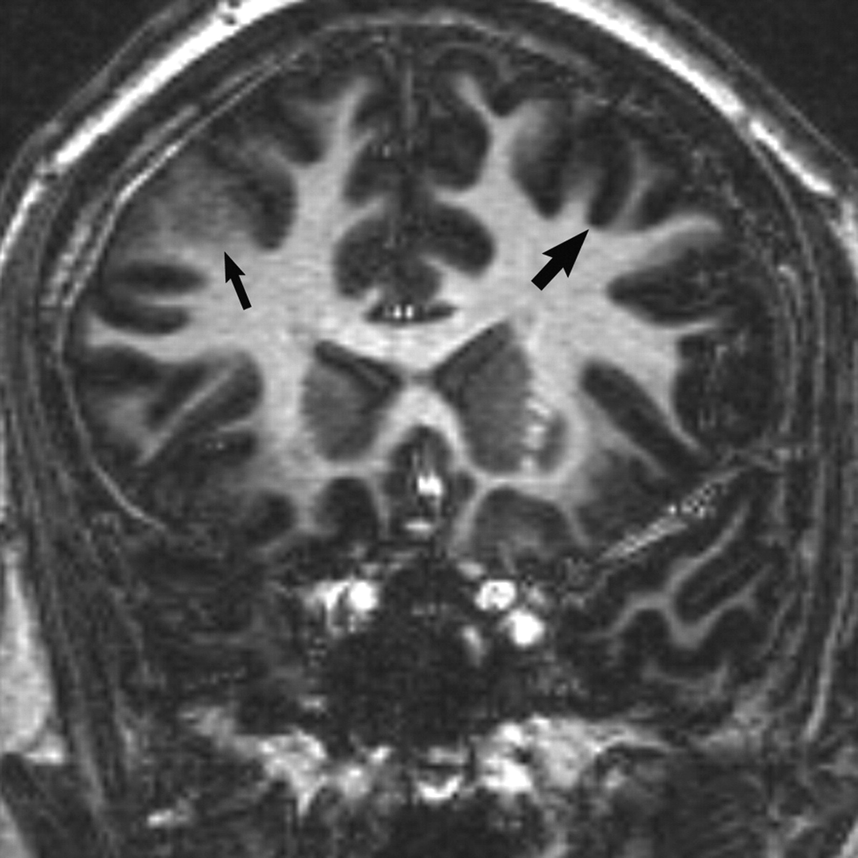
Axial T2-weighted MR image reveals subtle cortical thickening and blurring of the gray-white matter junction (open arrow) in the right frontal lobe. Subtle gyral expansion is questioned. Note areas of normal thin-appearing cortex in the ipsilateral and contralateral hemisphere for comparison (closed arrows).

Histology revealed oligodendroglial hyperplasia, which is characterized by an increased number of oligodendrocytes in the juxtacortical white matter. Nuclei were uniform with no nucleoli. Immunoperoxidase stain for Ki-67 showed no staining, yielding a labeling index of essentially zero. Dr Peter Burger at Johns Hopkins Reference Laboratories, Baltimore, Md, also provided a consultative review, which concurred with the original diagnosis. The results of fluorescent in situ hybridization showed no evidence for 1p and 19q chromosomal deletions. No evidence for neoplasia was identified.

The patient experienced a decrease in seizure frequency immediately following surgery, though his seizures were not completely eliminated. Postoperative imaging suggested a small area of residual abnormality that may have accounted for incomplete resolution of seizures, though at the time of this review, no repeat resection had been performed.

Discussion

Oligodendroglial hyperplasia is a benign disorder, which is found with increased frequency in patients with epilepsy. It has previously been considered an occult lesion on imaging.1 Although benign, it may be the underlying cause for seizures in some patients. The ability to identify its presence with imaging allows the possibility of surgical cure.

Surgical resection offers the best hope for durable seizure control in patients with partial complex-type epilepsy, with the potential to decrease or even eliminate antiepileptic medication use. Surgical resection is the preferred treatment technique in patients refractory to medication.2 Many patients also experience negative drug-related side effects, and long-term medical therapy is expensive, so surgery is an attractive option.3 However, surgery relies on accurate lesion localization, so good-quality imaging is important for preoperative planning to allow complete resection.4–6

Histologically, oligodendroglial hyperplasia is characterized by an increased number of normal-appearing oligodendroglial cells in the cortex and juxtacortical white matter. It is considered at the mildest end of the spectrum of neuronal migrational disorders or microdysgenesis. Cortical migrational abnormalities, including forms of microdysgenesis, may be responsible for epilepsy. As with other disorders of cortical migration, oligodendroglial hyperplasia can also be found in patients without a history of seizures, so correlation with the patient's clinical presentation is important.1

High-resolution high-field MR imaging best evaluates epilepsy. Our practice requires good-quality imaging with whole-brain coverage, because we usually do not know the physiologic localization before scanning. We include the following unenhanced sequences on our 3T scanners by using an 8-channel parallel imaging−enabled head coil for epilepsy evaluation:

  1. 3D coronal T1-weighted turbo field-echo (TR, 8.5 ms/TE, 3.9 ms); acquisition matrix, 240 × 168 (reconstructed isovoxel size, 1 mm); FOV, 200–230 mm; section thickness, 1 mm (0 gap); section (frequency) reduction factor, 2; time, 6 minutes 16 seconds.

  2. Axial turbo spin-echo T2-weighted sequence (TR, 3000 ms/TE, 80 ms; acquisition matrix, 416 × 271 (reconstructed voxel size, 0.43 in-plane); FOV, 200–230 mm; flip angle, 90°; section thickness, 4.0 mm (0.4-mm gap); phase (P) reduction factor, 1.7; time, 1 minute 57 seconds.

  3. Coronal 2D short τ inversion recovery (TR, 8250 ms/TE, 20 ms; TI, 250 ms; acquisition matrix, 420 × 279 (reconstructed voxel size, 0.45 mm in-plane); FOV, 180–220 mm; section thickness, 2.0 mm (0.2-mm gap); P reduction factor, 3; phase oversampling, 2; time, 5 minutes 30 seconds.

  4. Coronal fluid-attenuated inversion recovery (FLAIR) (TR, 11,000 ms/TE, 120 ms; TI, 2800 ms); acquisition matrix, 352 × 209 (reconstructed voxel size, 0.45 mm in-plane); FOV, 220–240 mm; section thickness, 4.0 mm (0.4-mm gap); P reduction factor, 1.7; time, 3 minutes 51 seconds.

  5. Sagittal T1 FLAIR (TR, 2000 ms/TE, 10 ms; TI, 800 ms); FOV, 200–230 mm; section thickness, 4 mm (0.4-mm gap); acquisition matrix, 272 × 232 (reconstructed voxel size, 0.43 mm in-plane); P reduction factor, 1.7; time, 4 minutes 50 seconds.

In summary, the current case demonstrated blurring and thickening of cortex in the affected area at 3T imaging (though not visible on 1.5T MR imaging), which likely reflected the increased number of oligodendrocytes within juxtacortical white matter. Higher field MR imaging may be important for identifying forms of microdysgenesis such as oligodendroglial hyperplasia, which may be responsible for epilepsy.

References

  1. ↵
    Burger PC, Scheithauer BW, Vogel FS. The brain: surgery for seizures. In: Surgical Pathology of the Nervous System and Its Coverings. 4th ed. New York: Churchill Livingstone;2002
  2. ↵
    Phal PM, Usmanov A, Nesbit GM, et al. Qualitative comparison of 3-T and 1.5-T MRI in the evaluation of epilepsy. AJR Am J Roentgenol 2008;191:890–95
    CrossRefPubMed
  3. ↵
    Cascino GD. Improving quality of life with epilepsy surgery: the seizure outcome is the key to success. Neurology 2007;68:1967–68
    FREE Full Text
  4. ↵
    Langfitt JT, Holloway RG, McDermott MP, et al. Health care costs decline after successful epilepsy surgery. Neurology 2007;68:1290–98
    Abstract/FREE Full Text
  5. Wyllie E, Lachhwani DK, Gupta A, et al. Successful surgery for epilepsy due to early brain lesions despite generalized EEG findings. Neurology 2007;69:389–97
    Abstract/FREE Full Text
  6. ↵
    Urbach H, Binder D, von Lehe M, et al. Correlation of MRI and histopathology in epileptogenic parietal and occipital lobe lesions. Seizure 2007;16:608–14. Epub 2007 Jun 11
    CrossRefPubMed
  • Received December 15, 2008.
  • Accepted after revision December 22, 2008.
  • American Society of Neuroradiology
View Abstract
PreviousNext
Back to top

In this issue

American Journal of Neuroradiology: 30 (7)
American Journal of Neuroradiology
Vol. 30, Issue 7
August 2009
  • Table of Contents
  • Index by author
Advertisement
Print
Download PDF
Email Article

Thank you for your interest in spreading the word on American Journal of Neuroradiology.

NOTE: We only request your email address so that the person you are recommending the page to knows that you wanted them to see it, and that it is not junk mail. We do not capture any email address.

Enter multiple addresses on separate lines or separate them with commas.
MR Imaging Identification of Oligodendroglial Hyperplasia
(Your Name) has sent you a message from American Journal of Neuroradiology
(Your Name) thought you would like to see the American Journal of Neuroradiology web site.
CAPTCHA
This question is for testing whether or not you are a human visitor and to prevent automated spam submissions.
Cite this article
B.E. Hamilton, G.M. Nesbit
MR Imaging Identification of Oligodendroglial Hyperplasia
American Journal of Neuroradiology Aug 2009, 30 (7) 1412-1413; DOI: 10.3174/ajnr.A1522

Citation Manager Formats

  • BibTeX
  • Bookends
  • EasyBib
  • EndNote (tagged)
  • EndNote 8 (xml)
  • Medlars
  • Mendeley
  • Papers
  • RefWorks Tagged
  • Ref Manager
  • RIS
  • Zotero
0 Responses
Respond to this article
Share
Bookmark this article
MR Imaging Identification of Oligodendroglial Hyperplasia
B.E. Hamilton, G.M. Nesbit
American Journal of Neuroradiology Aug 2009, 30 (7) 1412-1413; DOI: 10.3174/ajnr.A1522
del.icio.us logo Twitter logo Facebook logo Mendeley logo
  • Tweet Widget
  • Facebook Like
  • Google Plus One
Purchase

Jump to section

  • Article
    • Abstract
    • Case Report
    • Discussion
    • References
  • Figures & Data
  • Info & Metrics
  • Responses
  • References
  • PDF

Related Articles

  • No related articles found.
  • PubMed
  • Google Scholar

Cited By...

  • MRI, Magnetoencephalography, and Surgical Outcome of Oligodendrocytosis versus Focal Cortical Dysplasia Type I
  • Crossref (17)
  • Google Scholar

This article has been cited by the following articles in journals that are participating in Crossref Cited-by Linking.

  • Mild Malformation of Cortical Development with Oligodendroglial Hyperplasia in Frontal Lobe Epilepsy: A New Clinico‐Pathological Entity
    Johannes Schurr, Roland Coras, Karl Rössler, Tom Pieper, Manfred Kudernatsch, Hans Holthausen, Peter Winkler, Friedrich Woermann, Christian G. Bien, Tilman Polster, Reinhard Schulz, Thilo Kalbhenn, Horst Urbach, Albert Becker, Thomas Grunwald, Hans‐Juergen Huppertz, Antonio Gil‐Nagel, Rafael Toledano, Martha Feucht, Angelika Mühlebner, Thomas Czech, Ingmar Blümcke
    Brain Pathology 2017 27 1
  • Development and Dysgenesis of the Cerebral Cortex: Malformations of Cortical Development
    Charles Raybaud, Elysa Widjaja
    Neuroimaging Clinics of North America 2011 21 3
  • Age-related MR characteristics in mild malformation of cortical development with oligodendroglial hyperplasia and epilepsy (MOGHE)
    Till Hartlieb, Peter Winkler, Roland Coras, Tom Pieper, H. Holthausen, Ingmar Blümcke, Martin Staudt, Manfred Kudernatsch
    Epilepsy & Behavior 2019 91
  • Magnetic resonance imaging findings and clinical characteristics in mild malformation of cortical development with oligodendroglial hyperplasia and epilepsy in a predominantly adult cohort
    Vanessa C. Mendes Coelho, Marcia Morita‐Sherman, Clarissa L. Yasuda, Marina M. K. Alvim, Barbara Juarez Amorim, Helder Tedeschi, Enrico Ghizoni, Fabio Rogerio, Fernando Cendes
    Epilepsia 2021 62 6
  • Clinical characteristics and postoperative seizure outcome in patients with mild malformation of cortical development and oligodendroglial hyperplasia
    Ahmed Gaballa, Friedrich G. Woermann, Thomas Cloppenborg, Thilo Kalbhenn, Ingmar Blümcke, Christian G. Bien, Susanne Fauser
    Epilepsia 2021 62 12
  • High-Field Magnetic Resonance Imaging for Epilepsy
    Horst Urbach
    Neuroimaging Clinics of North America 2012 22 2
  • Mild malformation of cortical development with oligodendroglial hyperplasia and epilepsy (MOGHE): a widespread disease with an apparently focal epilepsy
    Raghavendra Seetharam, Javeria Nooraine, Radhika Mhatre, Jaychandran Ramachandran, Rajesh B. Iyer, Anita Mahadevan
    Epileptic Disorders 2021 23 2
  • MRI, Magnetoencephalography, and Surgical Outcome of Oligodendrocytosis versus Focal Cortical Dysplasia Type I
    D. Mata-Mbemba, Y. Iimura, L.-N. Hazrati, A. Ochi, H. Otsubo, O.C. Snead, J. Rutka, E. Widjaja
    American Journal of Neuroradiology 2018 39 12
  • Clinical characteristics and surgical outcomes in children with mild malformation of cortical development and oligodendroglial hyperplasia in epilepsy
    Xianyu Liu, Ying Zhu, Qingzhu Liu, Shuang Zhang, Pengxia Wu, Yu Sun, Jie Zhang, Ruofan Wang, Taoyun Ji, Shuang Wang, Xiaoyan Liu, Yuwu Jiang, Lixin Cai, Ye Wu
    Epilepsia Open 2023 8 3
  • Epileptic Patient with Mild Malformation of Cortical Development with Oligodendroglial Hyperplasia and Epilepsy (MOGHE): A Case Report and Review of the Literature
    A. Verentzioti, I. Blumcke, A. Alexoudi, P. Patrikelis, A. Siatouni, S. Korfias, D. Sakas, S. Gatzonis
    Case Reports in Neurological Medicine 2019 2019

More in this TOC Section

  • Predictors of Reperfusion in Patients with Acute Ischemic Stroke
  • Enhanced Axonal Metabolism during Early Natalizumab Treatment in Relapsing-Remitting Multiple Sclerosis
  • SWI or T2*: Which MRI Sequence to Use in the Detection of Cerebral Microbleeds? The Karolinska Imaging Dementia Study
Show more BRAIN

Similar Articles

Advertisement

Indexed Content

  • Current Issue
  • Accepted Manuscripts
  • Article Preview
  • Past Issues
  • Editorials
  • Editor's Choice
  • Fellows' Journal Club
  • Letters to the Editor
  • Video Articles

Cases

  • Case Collection
  • Archive - Case of the Week
  • Archive - Case of the Month
  • Archive - Classic Case

More from AJNR

  • Trainee Corner
  • Imaging Protocols
  • MRI Safety Corner
  • Book Reviews

Multimedia

  • AJNR Podcasts
  • AJNR Scantastics

Resources

  • Turnaround Time
  • Submit a Manuscript
  • Submit a Video Article
  • Submit an eLetter to the Editor/Response
  • Manuscript Submission Guidelines
  • Statistical Tips
  • Fast Publishing of Accepted Manuscripts
  • Graphical Abstract Preparation
  • Imaging Protocol Submission
  • Evidence-Based Medicine Level Guide
  • Publishing Checklists
  • Author Policies
  • Become a Reviewer/Academy of Reviewers
  • News and Updates

About Us

  • About AJNR
  • Editorial Board
  • Editorial Board Alumni
  • Alerts
  • Permissions
  • Not an AJNR Subscriber? Join Now
  • Advertise with Us
  • Librarian Resources
  • Feedback
  • Terms and Conditions
  • AJNR Editorial Board Alumni

American Society of Neuroradiology

  • Not an ASNR Member? Join Now

© 2025 by the American Society of Neuroradiology All rights, including for text and data mining, AI training, and similar technologies, are reserved.
Print ISSN: 0195-6108 Online ISSN: 1936-959X

Powered by HighWire