Advertisement

Main menu

  • Home
  • Content
    • Current Issue
    • Accepted Manuscripts
    • Article Preview
    • Past Issue Archive
    • Video Articles
    • AJNR Case Collection
    • Case of the Week Archive
    • Case of the Month Archive
    • Classic Case Archive
  • Special Collections
    • AJNR Awards
    • Low-Field MRI
    • Alzheimer Disease
    • ASNR Foundation Special Collection
    • Photon-Counting CT
    • View All
  • Multimedia
    • AJNR Podcasts
    • AJNR SCANtastic
    • Trainee Corner
    • MRI Safety Corner
    • Imaging Protocols
  • For Authors
    • Submit a Manuscript
    • Submit a Video Article
    • Submit an eLetter to the Editor/Response
    • Manuscript Submission Guidelines
    • Statistical Tips
    • Fast Publishing of Accepted Manuscripts
    • Graphical Abstract Preparation
    • Imaging Protocol Submission
    • Author Policies
  • About Us
    • About AJNR
    • Editorial Board
    • Editorial Board Alumni
  • More
    • Become a Reviewer/Academy of Reviewers
    • Subscribers
    • Permissions
    • Alerts
    • Feedback
    • Advertisers
    • ASNR Home

User menu

  • Alerts
  • Log in

Search

  • Advanced search
American Journal of Neuroradiology

American Journal of Neuroradiology

ASHNR American Society of Functional Neuroradiology ASHNR American Society of Pediatric Neuroradiology ASSR
  • Alerts
  • Log in

Advanced Search

  • Home
  • Content
    • Current Issue
    • Accepted Manuscripts
    • Article Preview
    • Past Issue Archive
    • Video Articles
    • AJNR Case Collection
    • Case of the Week Archive
    • Case of the Month Archive
    • Classic Case Archive
  • Special Collections
    • AJNR Awards
    • Low-Field MRI
    • Alzheimer Disease
    • ASNR Foundation Special Collection
    • Photon-Counting CT
    • View All
  • Multimedia
    • AJNR Podcasts
    • AJNR SCANtastic
    • Trainee Corner
    • MRI Safety Corner
    • Imaging Protocols
  • For Authors
    • Submit a Manuscript
    • Submit a Video Article
    • Submit an eLetter to the Editor/Response
    • Manuscript Submission Guidelines
    • Statistical Tips
    • Fast Publishing of Accepted Manuscripts
    • Graphical Abstract Preparation
    • Imaging Protocol Submission
    • Author Policies
  • About Us
    • About AJNR
    • Editorial Board
    • Editorial Board Alumni
  • More
    • Become a Reviewer/Academy of Reviewers
    • Subscribers
    • Permissions
    • Alerts
    • Feedback
    • Advertisers
    • ASNR Home
  • Follow AJNR on Twitter
  • Visit AJNR on Facebook
  • Follow AJNR on Instagram
  • Join AJNR on LinkedIn
  • RSS Feeds

AJNR Awards, New Junior Editors, and more. Read the latest AJNR updates

OtherReview articles
Open Access

Functional Imaging of Central Nervous System Involvement in Complex Regional Pain Syndrome

P. Schwenkreis, C. Maier and M. Tegenthoff
American Journal of Neuroradiology August 2009, 30 (7) 1279-1284; DOI: https://doi.org/10.3174/ajnr.A1630
P. Schwenkreis
  • Find this author on Google Scholar
  • Find this author on PubMed
  • Search for this author on this site
C. Maier
  • Find this author on Google Scholar
  • Find this author on PubMed
  • Search for this author on this site
M. Tegenthoff
  • Find this author on Google Scholar
  • Find this author on PubMed
  • Search for this author on this site
  • Article
  • Figures & Data
  • Info & Metrics
  • Responses
  • References
  • PDF
Loading

Article Figures & Data

Figures

  • Fig 1.
    • Download figure
    • Open in new tab
    • Download powerpoint
    Fig 1.

    Correlation analysis. BOLD contrast negatively correlates to mean sustained pain levels (A) and 2-point-discrimination thresholds (B). Pain-related BOLD contrast is found in Brodmann area (BA) 1 of the S1 (A), whereas perception-related BOLD contrast is found in BA 2 of S1 (B). Both analyses reveal BOLD contrast localized in the same region of S2. NRS indicates numeric rating scale. Reprinted with permission of Elsevier from Pleger et al.32
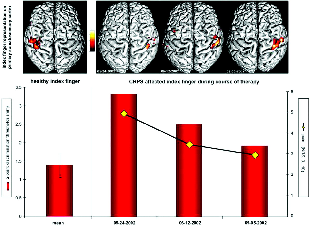
  • Fig 2.
    • Download figure
    • Open in new tab
    • Download powerpoint
    Fig 2.

    Changes in S1 representation, pain intensity, and discrimination ability during the course of therapy in a single patient. BOLD contrast received from cortical maps on S1 contralateral to the healthy (left image) and to the CRPS-affected side (right, images of 3 consecutive measurements). Contrast maps are shown from above, projected on the individual rendered T1-weighted MR imaging dataset. The diagram below shows 2-point-discrimination thresholds of the healthy index finger (left column) and of the CRPS-affected index finger (right, columns indicate values of 3 consecutive measurements) as well as the intensity of CRPS pain (yellow rhombus, 3 consecutive evaluations). NRS indicates numeric rating scale. Reprinted with permission of Wiley-Liss from Pleger et al.34

  • Fig 3.
    • Download figure
    • Open in new tab
    • Download powerpoint
    Fig 3.

    A, Contrast map of the general linear model (GLM) calculated by subtracting the tapping condition of controls (right hand) from the tapping condition in patients (CRPS-affected side [ie, right hand]). Brain sections are referred to by the superior-inferior level in millimeters relative to a line through the anterior and posterior commissure. Z-score indicates level of significance. B, Contrast map of the GLM calculated by subtracting the tapping condition on the unaffected side (ie, left hand) from the affected side (ie, right hand) in patients with CRPS. Brain sections are referred to by the superior-inferior level in millimeters relative to a line through the anterior and posterior commissure. Z-score indicates level of significance. C, Implementation of the individual degrees of motor impairment (z-values of tapping frequencies [ie, values of the individual maximum tapping frequencies normalized to the mean maximum tapping frequencies of controls]) as regressors in the GLM model for the condition “tapping on the CRPS-affected side.” With this approach, brain areas correlating with the degree of motor impairment are isolated. Key areas of activations are indicated according to the following abbreviations: SFC indicates superior frontal cortex; MFC, middle frontal cortex; IFC, inferior frontal cortex; AIP, anterior intraparietal area, MIP, medial intraparietal area; mtc, middle temporal cortex; loc, lateral occipital complex. Reprinted with permission of Oxford University Press from Maihöfner et al.48

PreviousNext
Back to top

In this issue

American Journal of Neuroradiology: 30 (7)
American Journal of Neuroradiology
Vol. 30, Issue 7
August 2009
  • Table of Contents
  • Index by author
Advertisement
Print
Download PDF
Email Article

Thank you for your interest in spreading the word on American Journal of Neuroradiology.

NOTE: We only request your email address so that the person you are recommending the page to knows that you wanted them to see it, and that it is not junk mail. We do not capture any email address.

Enter multiple addresses on separate lines or separate them with commas.
Functional Imaging of Central Nervous System Involvement in Complex Regional Pain Syndrome
(Your Name) has sent you a message from American Journal of Neuroradiology
(Your Name) thought you would like to see the American Journal of Neuroradiology web site.
CAPTCHA
This question is for testing whether or not you are a human visitor and to prevent automated spam submissions.
Cite this article
P. Schwenkreis, C. Maier, M. Tegenthoff
Functional Imaging of Central Nervous System Involvement in Complex Regional Pain Syndrome
American Journal of Neuroradiology Aug 2009, 30 (7) 1279-1284; DOI: 10.3174/ajnr.A1630

Citation Manager Formats

  • BibTeX
  • Bookends
  • EasyBib
  • EndNote (tagged)
  • EndNote 8 (xml)
  • Medlars
  • Mendeley
  • Papers
  • RefWorks Tagged
  • Ref Manager
  • RIS
  • Zotero
0 Responses
Respond to this article
Share
Bookmark this article
Functional Imaging of Central Nervous System Involvement in Complex Regional Pain Syndrome
P. Schwenkreis, C. Maier, M. Tegenthoff
American Journal of Neuroradiology Aug 2009, 30 (7) 1279-1284; DOI: 10.3174/ajnr.A1630
del.icio.us logo Twitter logo Facebook logo Mendeley logo
  • Tweet Widget
  • Facebook Like
  • Google Plus One
Purchase

Jump to section

  • Article
    • Abstract
    • Somatosensory System
    • Motor System
    • Conclusions
    • References
  • Figures & Data
  • Info & Metrics
  • Responses
  • References
  • PDF

Related Articles

  • No related articles found.
  • PubMed
  • Google Scholar

Cited By...

  • Comparison of subjective motor imagery abilities in CRPS compared to chronic limb pain and healthy controls: a cross sectional study
  • Comparison of subjective motor imagery abilities in CRPS compared to chronic limb pain and health controls: a cross sectional study
  • Comparison of subjective motor imagery abilities in CRPS compared to chronic limb pain and health controls: a cross sectional study
  • Atypical influence of biomechanical knowledge in Complex Regional Pain Syndrome-Towards a different perspective on body representation
  • CRPS: Central aspects related to locus of pain, pathophysiology, and mood
  • Crossref (67)
  • Google Scholar

This article has been cited by the following articles in journals that are participating in Crossref Cited-by Linking.

  • Pain and the brain: Specificity and plasticity of the brain in clinical chronic pain
    Vania A. Apkarian, Javeria A. Hashmi, Marwan N. Baliki
    Pain 2011 152 3
  • Primary Somatosensory Cortex Function in Complex Regional Pain Syndrome: A Systematic Review and Meta-Analysis
    Flavia Di Pietro, James H. McAuley, Luke Parkitny, Martin Lotze, Benedict M. Wand, G. Lorimer Moseley, Tasha R. Stanton
    The Journal of Pain 2013 14 10
  • Underlying Pathology and Associated Factors of Hemiplegic Shoulder Pain
    Leonid Kalichman, Motti Ratmansky
    American Journal of Physical Medicine & Rehabilitation 2011 90 9
  • Rapid treatment-induced brain changes in pediatric CRPS
    Nathalie Erpelding, Laura Simons, Alyssa Lebel, Paul Serrano, Melissa Pielech, Sanjay Prabhu, Lino Becerra, David Borsook
    Brain Structure and Function 2016 221 2
  • Brain Neuroplastic Changes Accompany Anxiety and Memory Deficits in a Model of Complex Regional Pain Syndrome
    Maral Tajerian, David Leu, Yani Zou, Peyman Sahbaie, Wenwu Li, Hamda Khan, Vivian Hsu, Wade Kingery, Ting Ting Huang, Lino Becerra, J. David Clark
    Anesthesiology 2014 121 4
  • Nociception Affects Motor Output
    Jo Nijs, Liesbeth Daenen, Patrick Cras, Filip Struyf, Nathalie Roussel, Rob A.B. Oostendorp
    The Clinical Journal of Pain 2012 28 2
  • Primary Motor Cortex Function in Complex Regional Pain Syndrome: A Systematic Review and Meta-Analysis
    Flavia Di Pietro, James H. McAuley, Luke Parkitny, Martin Lotze, Benedict M. Wand, G. Lorimer Moseley, Tasha R. Stanton
    The Journal of Pain 2013 14 11
  • Neuroimaging Chronic Pain: What Have We Learned and Where Are We Going?
    Katherine T Martucci, Pamela Ng, Sean Mackey
    Future Neurology 2014 9 6
  • Cognitive correlates of “neglect-like syndrome” in patients with complex regional pain syndrome
    Leonie Kolb, Christoph Lang, Frank Seifert, Christian Maihöfner
    Pain 2012 153 5
  • Effects of Mirror Therapy in Stroke Patients With Complex Regional Pain Syndrome Type 1: A Randomized Controlled Study
    Secil Pervane Vural, Guldal Funda Nakipoglu Yuzer, Didem Sezgin Ozcan, Sibel Demir Ozbudak, Nese Ozgirgin
    Archives of Physical Medicine and Rehabilitation 2016 97 4

More in this TOC Section

  • An Atlas of Neonatal Neurovascular Imaging Anatomy as Depicted with Microvascular Imaging: The Intracranial Arteries
  • An Atlas of Neonatal Neurovascular Imaging Anatomy as Depicted with Microvascular Imaging: The Intracranial Veins
  • Clinical Translation of Hyperpolarized 13C Metabolic Probes for Glioma Imaging
Show more Review articles

Similar Articles

Advertisement

Indexed Content

  • Current Issue
  • Accepted Manuscripts
  • Article Preview
  • Past Issues
  • Editorials
  • Editor's Choice
  • Fellows' Journal Club
  • Letters to the Editor
  • Video Articles

Cases

  • Case Collection
  • Archive - Case of the Week
  • Archive - Case of the Month
  • Archive - Classic Case

More from AJNR

  • Trainee Corner
  • Imaging Protocols
  • MRI Safety Corner
  • Book Reviews

Multimedia

  • AJNR Podcasts
  • AJNR Scantastics

Resources

  • Turnaround Time
  • Submit a Manuscript
  • Submit a Video Article
  • Submit an eLetter to the Editor/Response
  • Manuscript Submission Guidelines
  • Statistical Tips
  • Fast Publishing of Accepted Manuscripts
  • Graphical Abstract Preparation
  • Imaging Protocol Submission
  • Evidence-Based Medicine Level Guide
  • Publishing Checklists
  • Author Policies
  • Become a Reviewer/Academy of Reviewers
  • News and Updates

About Us

  • About AJNR
  • Editorial Board
  • Editorial Board Alumni
  • Alerts
  • Permissions
  • Not an AJNR Subscriber? Join Now
  • Advertise with Us
  • Librarian Resources
  • Feedback
  • Terms and Conditions
  • AJNR Editorial Board Alumni

American Society of Neuroradiology

  • Not an ASNR Member? Join Now

© 2025 by the American Society of Neuroradiology All rights, including for text and data mining, AI training, and similar technologies, are reserved.
Print ISSN: 0195-6108 Online ISSN: 1936-959X

Powered by HighWire