Advertisement

Main menu

  • Home
  • Content
    • Current Issue
    • Accepted Manuscripts
    • Article Preview
    • Past Issue Archive
    • Video Articles
    • AJNR Case Collection
    • Case of the Week Archive
    • Case of the Month Archive
    • Classic Case Archive
  • Special Collections
    • AJNR Awards
    • Low-Field MRI
    • Alzheimer Disease
    • ASNR Foundation Special Collection
    • Photon-Counting CT
    • View All
  • Multimedia
    • AJNR Podcasts
    • AJNR SCANtastic
    • Trainee Corner
    • MRI Safety Corner
    • Imaging Protocols
  • For Authors
    • Submit a Manuscript
    • Submit a Video Article
    • Submit an eLetter to the Editor/Response
    • Manuscript Submission Guidelines
    • Statistical Tips
    • Fast Publishing of Accepted Manuscripts
    • Graphical Abstract Preparation
    • Imaging Protocol Submission
    • Author Policies
  • About Us
    • About AJNR
    • Editorial Board
    • Editorial Board Alumni
  • More
    • Become a Reviewer/Academy of Reviewers
    • Subscribers
    • Permissions
    • Alerts
    • Feedback
    • Advertisers
    • ASNR Home

User menu

  • Alerts
  • Log in

Search

  • Advanced search
American Journal of Neuroradiology

American Journal of Neuroradiology

ASHNR American Society of Functional Neuroradiology ASHNR American Society of Pediatric Neuroradiology ASSR
  • Alerts
  • Log in

Advanced Search

  • Home
  • Content
    • Current Issue
    • Accepted Manuscripts
    • Article Preview
    • Past Issue Archive
    • Video Articles
    • AJNR Case Collection
    • Case of the Week Archive
    • Case of the Month Archive
    • Classic Case Archive
  • Special Collections
    • AJNR Awards
    • Low-Field MRI
    • Alzheimer Disease
    • ASNR Foundation Special Collection
    • Photon-Counting CT
    • View All
  • Multimedia
    • AJNR Podcasts
    • AJNR SCANtastic
    • Trainee Corner
    • MRI Safety Corner
    • Imaging Protocols
  • For Authors
    • Submit a Manuscript
    • Submit a Video Article
    • Submit an eLetter to the Editor/Response
    • Manuscript Submission Guidelines
    • Statistical Tips
    • Fast Publishing of Accepted Manuscripts
    • Graphical Abstract Preparation
    • Imaging Protocol Submission
    • Author Policies
  • About Us
    • About AJNR
    • Editorial Board
    • Editorial Board Alumni
  • More
    • Become a Reviewer/Academy of Reviewers
    • Subscribers
    • Permissions
    • Alerts
    • Feedback
    • Advertisers
    • ASNR Home
  • Follow AJNR on Twitter
  • Visit AJNR on Facebook
  • Follow AJNR on Instagram
  • Join AJNR on LinkedIn
  • RSS Feeds

AJNR Awards, New Junior Editors, and more. Read the latest AJNR updates

OtherBRAIN

Retained Surgical Sponges after Craniotomies: Imaging Appearances and Complications

A.K. Kim, E.B. Lee, L.J. Bagley and L.A. Loevner
American Journal of Neuroradiology June 2009, 30 (6) 1270-1272; DOI: https://doi.org/10.3174/ajnr.A1469
A.K. Kim
  • Find this author on Google Scholar
  • Find this author on PubMed
  • Search for this author on this site
E.B. Lee
  • Find this author on Google Scholar
  • Find this author on PubMed
  • Search for this author on this site
L.J. Bagley
  • Find this author on Google Scholar
  • Find this author on PubMed
  • Search for this author on this site
L.A. Loevner
  • Find this author on Google Scholar
  • Find this author on PubMed
  • Search for this author on this site
  • Article
  • Figures & Data
  • Info & Metrics
  • Responses
  • References
  • PDF
Loading

Abstract

SUMMARY: Imaging within 24 to 48 hours after most neurosurgical procedures is a routine practice. Nonresorbable surgical sponges have radiopaque filaments readily visible on CT scans and plain film radiographs. However, the proton-poor barium sulfate responsible for this radio-opacity is generally not detectable on MR imaging in the immediate post-operative period. Findings on MR imaging become more evident with elapsing time and when a foreign-body reaction to the sponge manifests as a mass lesion, which can mimic residual or recurrent intracranial tumor or abscess. Although preventive measures by our surgical colleagues to ensure accurate and correct sponge counts before and after wound closure is paramount, even the most fastidious efforts may rarely result in an inadvertently retained surgical sponge. The role of the radiologist is to recognize the imaging findings of this entity and its potential complications so that appropriate and prompt management can be initiated.

Retained surgical materials and their potential adverse outcomes are infrequent but important complications that have been well described after abdominal and pelvic surgery.1–2 In contrast, a limited number of studies have been reported regarding retained surgical sponges after craniotomies. It is important that the radiologist be aware of this entity; its imaging appearance; and its potential complications, which include the formation of granulomas and abscesses. These complications may be mistaken for residual or recurrent neoplasms. We discuss the imaging appearance of retained surgical sponges after neurosurgical procedures in 3 patients and review the potential complications should these go unidentified.

Case Reports

Case 1

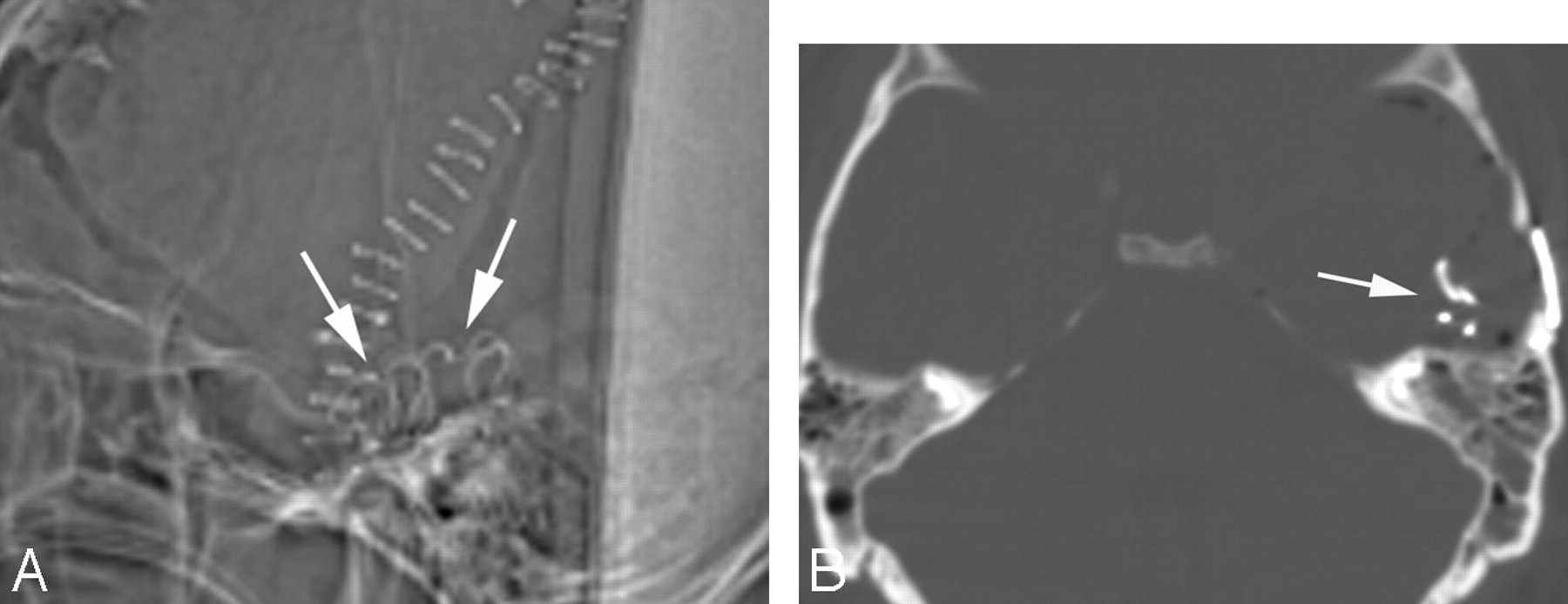
A 44-year-old man with no significant medical history was admitted from the emergency department with a 3-week history of fever; headache; and progressive cognitive decline, including memory loss and confusion. Initial brain MR imaging showed increased T2-weighted fluid-attenuated inversion recovery signal intensity in the left hippocampus with regions of leptomeningeal enhancement. Differential considerations included herpes encephalitis, limbic encephalitis, or infiltrating glioma. The patient was empirically treated with acyclovir for possible herpes encephalitis, with no improvement in his clinical condition. Analysis of CSF and hematologic evaluation were negative for malignant or infectious sources. The unclear cause of the patient's symptoms prompted a brain biopsy, results of which revealed acute hemorrhagic leukoencephalopathy. Routine postoperative head CT scan performed within 12 hours of surgery showed a serpentine hyperattenuated object in the surgical bed suspicious for a retained surgical sponge (Fig 1). Surgical exploration confirmed a retained cottonoid sponge that was removed.

Fig 1.
  • Download figure
  • Open in new tab
  • Download powerpoint
Fig 1.

A, Sagittal topogram from unenhanced CT scan shows a hyperattenuated curvilinear object overlying the surgical field, representing a retained surgical sponge. B, Axial CT scan in bone window shows the hyperattenuated serpiginous barium-impregnated filaments of the retained cottonoid sponge in the left middle cranial fossa.

Case 2

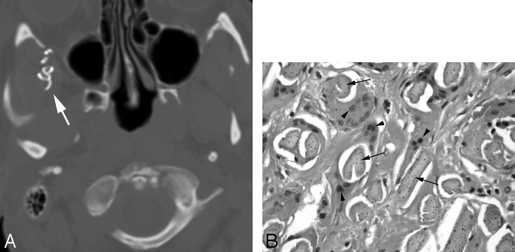
A 52-year-old woman underwent a right pterional craniotomy for resection of a right frontal atypical meningioma. Routine postoperative CT scan performed within 12 hours of surgery revealed multiple curvilinear hyperattenuated structures in the right infratemporal fossa, thought to represent surgical clips (Fig 2). On routine postoperative MR imaging performed within 12 hours of surgery, these curvilinear structures in the right infratemporal fossa surgical bed were not identified because they were obscured by susceptibility artifacts caused by blood products and air. Routine follow-up brain MR imaging study performed at an outside institution 4 months after the initial surgery showed a nodular enhancing “mass” in the right infratemporal fossa, which was interpreted as concerning for recurrent meningioma. The patient underwent a second craniotomy. On intraoperative inspection, the mass was found encased within the right temporalis muscle and, on gross inspection, had an attenuated fibrous capsule with apparent tufts of cottonoid material embedded within it. Gross pathologic evaluation demonstrated several portions of blue plastic foreign material (surgical sponge) present within a rubbery, tan-pink, soft tissue mass. On microscopic inspection, foreign material with suture admixed in a giant-cell granulomatous response was identified. No neoplasm was present.

Fig 2.
  • Download figure
  • Open in new tab
  • Download powerpoint
Fig 2.

A, Axial CT scan in bone window at the level of the infratemporal fossa shows a hyperattenuated serpiginous structure (arrow) in the right infratemporal fossa compatible with a retained surgical sponge. B, Microphotograph at high power demonstrates multinucleated foreign-body giant cells (arrowheads) engulfing foreign-body material (arrows; hematoxylin-eosin, original magnification ×40).

Case 3

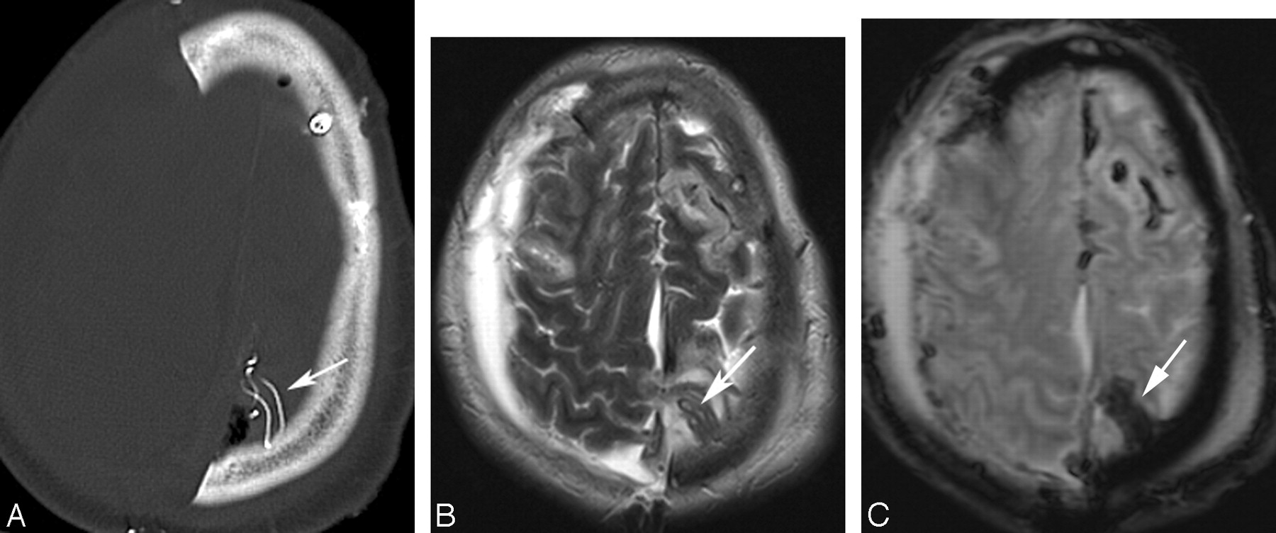
A 65-year-old man was admitted from the trauma bay after sustaining a major head traumatic injury from an assault. Initial CT examination of the head showed multiple calvarial fractures, extensive intracranial hemorrhage, and edema with mass effect. Increasing parenchymal edema and mass effect on serial CT studies in combination with declining responsiveness prompted decompressive right hemispheric craniectomy 5 days after admission. During the operation, there was considerable bleeding from the superior sagittal sinus that was controlled with multiple cottonoid sponges. Before wound closure, all but 1 of the cottonoid sponges could be safely removed. Routine postoperative head CT scan performed within 12 hours of the surgery revealed a serpentine hyperattenuated retained surgical sponge adjacent to the superior sagittal sinus at the vertex (Fig 3A). MR imaging examination showed the surgical sponge as a curvilinear T2-weighted hypointense structure (Fig 3B) that demonstrated susceptibility artifacts on the gradient-echo sequence. The sponge could not be removed, and the patient was eventually discharged to a rehabilitation center.

Fig 3.
  • Download figure
  • Open in new tab
  • Download powerpoint
Fig 3.

A, Axial unenhanced CT study shows the characteristic hyperattenuated serpentine object (arrow) representing the barium-impregnated filaments of a retained cottonoid sponge. B, Axial T2-weighted image shows the retained surgical sponge (arrow) as a hypointense serpentine foreign body that shows susceptibility effects (arrow) on the gradient-echo sequence (C).

Discussion

Gossypiboma is a term used to describe a foreign-body reaction surrounding material made of cotton matrix. The term is derived from the Latin word gossypium, meaning “cotton” and the Swahili term boma, meaning “place of concealment” and historically has been used, along with the term textiloma, to describe the foreign-body granulomatous reactions that occur in response to inadvertently retained surgical sponges. Other less commonly used terms are gauzoma (from surgical gauze) and muslinoma (from muslin, a woven cotton fabric).

Two types of foreign-body reactions generally occur in response to a retained surgical sponge. There can be an aseptic fibrous reaction that involves adhesion formation, encapsulation, and granulomatous pseudotumoral formation as in the patient in case 2. Alternatively, there can be an exudative reaction resulting in abscess formation. Although exudative responses typically manifest in the early postoperative stage, granulomatous responses can remain clinically silent for years.3

Neurosurgeons and other surgical subspecialists routinely use hemostatic resorbable and nonresorbable agents. Bioabsorbable agents may be left in the surgical bed to maintain hemostasis after wound closure. Three commonly used resorbable hemostatic agents are Gelfoam gelatin foam (Pfizer, New York, NY), Surgicel oxidized cellulose (Ethicon Endo-Surgery, Cincinnati, Ohio), and Avitene microfibrillar collagen (Davol, Cranston, RI).4 Nonresorbable agents are removed before wound closure. An exception to the nonresorbable agents is muslin, which is a cotton fabric occasionally used to reinforce aneurysmal walls during surgical repair of intracranial aneurysms. Although there are case reports describing an excessive inflammatory response to resorbable hemostatic agents5 and muslin,6 this is more commonly observed with the nonresorbable materials. Nonresorbable hemostatic agents commonly used in neurosurgical procedures are synthetic rayon fibers fashioned into strips or pledgets (cottonoids and kites) containing filaments impregnated with radiopaque barium sulfate that allow visualization on plain radiographs or on CT scans.4

Imaging within 24 to 48 hours after most neurosurgical procedures is routine practice. Whereas resorbable hemostatic agents may be difficult to appreciate on CT, especially in the early postoperative phase, nonresorbable surgical sponges have radiopaque filaments readily visible on CT scans and on plain radiographs. These filaments appear as curvilinear hyperattenuated structures. However, the proton-poor barium sulfate responsible for this radio-opacity is generally not detectable on MR imaging. The MR appearance of retained foreign-body material will depend on the time elapsed since surgery, the type of foreign-body reaction that occurs, and on the substance of the foreign material itself. Imaging findings on MR imaging become more evident when the foreign-body reaction manifests as a mass lesion, which can mimic residual or recurrent intracranial tumor or abscess.4,7

Cotton and gauze pads used in the surgical field should always have a tag that allows them to be easily located and removed, and all materials that are placed within the wound temporarily must be counted with meticulous care.8 Although preventive measures by our surgical colleagues to ensure accurate and correct sponge counts before and after wound closure is paramount, even the most fastidious efforts may result in an inadvertently retained surgical sponge. The role of the radiologist is to recognize the imaging findings of this entity and its potential complications so that appropriate and prompt management can be initiated.

Footnotes

  • Previously presented as a poster at: Annual Meeting of the American Society of Neuroradiology, June 2–5, 2008, New Orleans, La.

References

  1. ↵
    O'Connor AR, Coakley FD, Meng MV, et al. Imaging of retained surgical sponges in the abdomen and pelvis. AJR Am J Roentgenol 2003;180:481–89
    PubMed
  2. ↵
    Gawande AA, Studdert DM, Orav EJ, et al. Risk factors for retained instruments and sponges after surgery. N Engl J Med 2003;348:229–35
    CrossRefPubMed
  3. ↵
    Sakayama K, Fujibuchi T, Sugawara Y, et al. A 40-year-old gossypiboma (foreign body granuloma) mimicking a malignant femoral surface tumor. Skel Radiol 2005;34:221–24
    CrossRefPubMed
  4. ↵
    Ribalta T, McCutcheon IE, Neto AG, et al. Textiloma (gossypiboma) mimicking recurrent intracranial tumor. Arch Pathol Lab Med 2004;128:749–58
    PubMed
  5. ↵
    Kothbauer KF, Jallo GI, Siffert J, et al. Foreign body reaction to hemostatic materials mimicking recurrent brain tumor. J Neurosurg 2001;95:503–06
    PubMed
  6. ↵
    Brochert A, Reynolds T, Baker R. MRI in a case of muslin-induced granuloma. Neuroradiology 2003;45:82–84
    PubMed
  7. ↵
    Nishio Y, Hayashi N, Hamada H, et al. A case of delayed brain abscess due to a retained intracranial wooden foreign body: a case report and review of the last 20 years. Acta Neurochir 2004;146:847–50
    PubMed
  8. ↵
    Gibbs VC, Coakley FD, Reines HD. Preventable errors in the operating room: retained foreign bodies after surgery –part I. Curr Probl Surg 2007;44:281–337
    CrossRefPubMed
  • Received November 17, 2008.
  • Accepted after revision November 19, 2008.
  • Copyright © American Society of Neuroradiology
View Abstract
PreviousNext
Back to top

In this issue

American Journal of Neuroradiology: 30 (6)
American Journal of Neuroradiology
Vol. 30, Issue 6
June 2009
  • Table of Contents
  • Index by author
Advertisement
Print
Download PDF
Email Article

Thank you for your interest in spreading the word on American Journal of Neuroradiology.

NOTE: We only request your email address so that the person you are recommending the page to knows that you wanted them to see it, and that it is not junk mail. We do not capture any email address.

Enter multiple addresses on separate lines or separate them with commas.
Retained Surgical Sponges after Craniotomies: Imaging Appearances and Complications
(Your Name) has sent you a message from American Journal of Neuroradiology
(Your Name) thought you would like to see the American Journal of Neuroradiology web site.
CAPTCHA
This question is for testing whether or not you are a human visitor and to prevent automated spam submissions.
Cite this article
A.K. Kim, E.B. Lee, L.J. Bagley, L.A. Loevner
Retained Surgical Sponges after Craniotomies: Imaging Appearances and Complications
American Journal of Neuroradiology Jun 2009, 30 (6) 1270-1272; DOI: 10.3174/ajnr.A1469

Citation Manager Formats

  • BibTeX
  • Bookends
  • EasyBib
  • EndNote (tagged)
  • EndNote 8 (xml)
  • Medlars
  • Mendeley
  • Papers
  • RefWorks Tagged
  • Ref Manager
  • RIS
  • Zotero
0 Responses
Respond to this article
Share
Bookmark this article
Retained Surgical Sponges after Craniotomies: Imaging Appearances and Complications
A.K. Kim, E.B. Lee, L.J. Bagley, L.A. Loevner
American Journal of Neuroradiology Jun 2009, 30 (6) 1270-1272; DOI: 10.3174/ajnr.A1469
del.icio.us logo Twitter logo Facebook logo Mendeley logo
  • Tweet Widget
  • Facebook Like
  • Google Plus One
Purchase

Jump to section

  • Article
    • Abstract
    • Case Reports
    • Discussion
    • Footnotes
    • References
  • Figures & Data
  • Info & Metrics
  • Responses
  • References
  • PDF

Related Articles

  • No related articles found.
  • PubMed
  • Google Scholar

Cited By...

  • Imaging Foreign Bodies
  • Imaging Features of a Gelatin-Thrombin Matrix Hemostatic Agent in the Intracranial Surgical Bed: A Unique Space-Occupying Pseudomass
  • Crossref (17)
  • Google Scholar

This article has been cited by the following articles in journals that are participating in Crossref Cited-by Linking.

  • Imaging Features of a Gelatin-Thrombin Matrix Hemostatic Agent in the Intracranial Surgical Bed: A Unique Space-Occupying Pseudomass
    K. O. Learned, S. Mohan, I. Z. Hyder, L. J. Bagley, S. Wang, J. Y. Lee
    American Journal of Neuroradiology 2014 35 4
  • Automatic detection of foreign body objects in neurosurgery using a deep learning approach on intraoperative ultrasound images: From animal models to first in-human testing
    Haley G. Abramson, Eli J. Curry, Griffin Mess, Rasika Thombre, Kelley M. Kempski-Leadingham, Shivang Mistry, Subhiksha Somanathan, Laura Roy, Nancy Abu-Bonsrah, George Coles, Joshua C. Doloff, Henry Brem, Nicholas Theodore, Judy Huang, Amir Manbachi
    Frontiers in Surgery 2022 9
  • Intracranial gossypiboma 9 years after intracranial pressure bolt insertion: illustrative case
    Ryan T. S. Loh, Tomasz Matys, Kieren S. J. Allinson, Thomas Santarius
    Journal of Neurosurgery: Case Lessons 2022 3 8
  • Intruding implements: a pictorial review of retained surgical foreign objects in neuroradiology
    Alessandra D’Amico, Teresa Perillo, Lorenzo Ugga, Renato Cuocolo, Arturo Brunetti
    Insights into Imaging 2019 10 1
  • MRI findings for diagnosis of postoperative foreign body granulomas versus recurrent tumours in patients of brain tumour surgery
    L.-Y. Lin, S.-C. Lin, H.-J. Lee, S.-T. Chen, H.-M. Wu, Y.-W. Chen, H.-H. Chen, C.-B. Luo, W.-Y. Guo, F.-C. Chang
    Clinical Radiology 2021 76 4
  • Retained foreign bodies and associated risk factors and outcomes in pediatric surgical patients
    Angelina L. Schwartz, Maziar M. Nourian, Brian T. Bucher
    Journal of Pediatric Surgery 2019 54 4
  • Imaging appearance of surgical sponges at 1.5T MRI: An in vitro study
    Lisa M. Ho, Elmar M. Merkle, Paul C. Kuo, Erik K. Paulson
    European Journal of Radiology 2011 80 2
  • Radiographic Detectability of Retained Neuropatties in a Cadaver Model
    Wangjian Thomas Luo, Robert Almack, John B. Mawson, David Douglas Cochrane
    World Neurosurgery 2015 84 2
  • A Textiloma on the Pterion : A Rarely Occurred Craniotomy Complication
    Ealmaan Kim
    Journal of Korean Neurosurgical Society 2013 53 4
  • Complications in Neurosurgery
    Anton V. Kalinovskiy, Jamil A. Rzaev
    2023 130

More in this TOC Section

  • Enhanced Axonal Metabolism during Early Natalizumab Treatment in Relapsing-Remitting Multiple Sclerosis
  • SWI or T2*: Which MRI Sequence to Use in the Detection of Cerebral Microbleeds? The Karolinska Imaging Dementia Study
  • Progression of Microstructural Damage in Spinocerebellar Ataxia Type 2: A Longitudinal DTI Study
Show more BRAIN

Similar Articles

Advertisement

Indexed Content

  • Current Issue
  • Accepted Manuscripts
  • Article Preview
  • Past Issues
  • Editorials
  • Editor's Choice
  • Fellows' Journal Club
  • Letters to the Editor
  • Video Articles

Cases

  • Case Collection
  • Archive - Case of the Week
  • Archive - Case of the Month
  • Archive - Classic Case

More from AJNR

  • Trainee Corner
  • Imaging Protocols
  • MRI Safety Corner
  • Book Reviews

Multimedia

  • AJNR Podcasts
  • AJNR Scantastics

Resources

  • Turnaround Time
  • Submit a Manuscript
  • Submit a Video Article
  • Submit an eLetter to the Editor/Response
  • Manuscript Submission Guidelines
  • Statistical Tips
  • Fast Publishing of Accepted Manuscripts
  • Graphical Abstract Preparation
  • Imaging Protocol Submission
  • Evidence-Based Medicine Level Guide
  • Publishing Checklists
  • Author Policies
  • Become a Reviewer/Academy of Reviewers
  • News and Updates

About Us

  • About AJNR
  • Editorial Board
  • Editorial Board Alumni
  • Alerts
  • Permissions
  • Not an AJNR Subscriber? Join Now
  • Advertise with Us
  • Librarian Resources
  • Feedback
  • Terms and Conditions
  • AJNR Editorial Board Alumni

American Society of Neuroradiology

  • Not an ASNR Member? Join Now

© 2025 by the American Society of Neuroradiology All rights, including for text and data mining, AI training, and similar technologies, are reserved.
Print ISSN: 0195-6108 Online ISSN: 1936-959X

Powered by HighWire