
Abstract
SUMMARY: Vestibular schwannomas are common, and gamma knife radiosurgery is a treatment option of symptomatic tumors. Hydrocephalus may be a complication of gamma knife treatment of vestibular schwannoma, though the cause-and-effect relationship can be debated because tumors can cause hydrocephalus without treatment. We present an MR imaging study of an unusual case of communicating hydrocephalus after gamma knife radiosurgery of a vestibular schwannoma in which the timeline of events strongly suggests that gamma knife played a contributory role in the development of hydrocephalus. We discuss risk factors for the development of hydrocephalus after radiation therapy and the role of MR CSF cine-flow study in the evaluation of treatment options for hydrocephalus in this setting.
Hydrocephalus is an uncommon and controversial complication of gamma knife radiosurgery for vestibular schwannomas. We report a complete MR imaging study including pretreatment and posttreatment imaging and MR CSF cine-flow study of such a case. The findings are consistent with a communicating hydrocephalus secondary to tumor necrosis, likely accelerated or worsened by gamma knife treatment.
Case Report
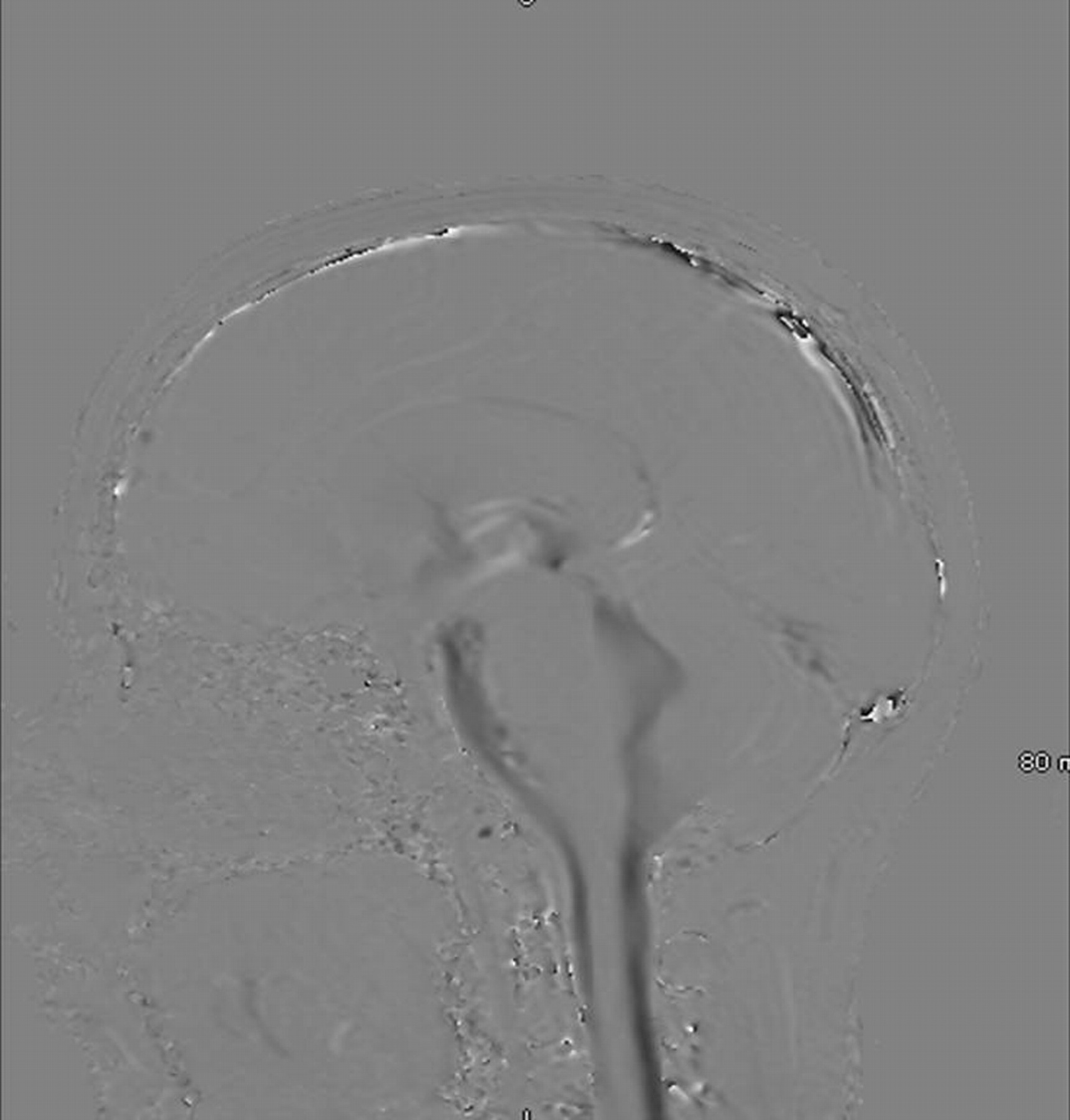
The patient was a 22-year-old previously healthy woman with a long history of left-sided tinnitus and decreased hearing. An MR imaging study was performed and demonstrated a 3-cm enhancing mass that was closely associated with the left vestibular nerve, with extension into the left porous acousticus. The appearance was consistent with vestibular schwannoma. After reviewing her options, the patient and her parents elected gamma knife radiosurgery instead of surgical removal. Nine months after the gamma knife procedure, the patient presented with headache, 2+ papilledema bilaterally, and a partial third nerve palsy. Another MR imaging scan was obtained, which demonstrated severe ventriculomegaly, with enlargement of the lateral, third, and fourth ventricles. No intraventricular point of obstruction of CSF flow was identified. The 3-cm vestibular schwannoma was again demonstrated, now with decreased central enhancement consistent with central necrosis (Figs 1–⇓3). A lumbar puncture procedure was performed, and CSF demonstrated a protein level of 127 mg/dL (nmL 15–60 mg/dL) and trace red blood cells (128/mm3 and 66/mm3, tubes 1 and 2). White blood cells were slightly elevated at 19/mm3 (nmL range 1-5) and glucose levels were 56 mg/dL. Gram smear and culture results were negative, and there was no clinical evidence of infection. MR imaging cine-flow study was performed and confirmed free flow of CSF, without evidence of intraventricular obstruction (Fig 4). Endoscopic third ventriculostomy was performed without effect. A ventriculoperitoneal shunt was placed with relief of symptoms.

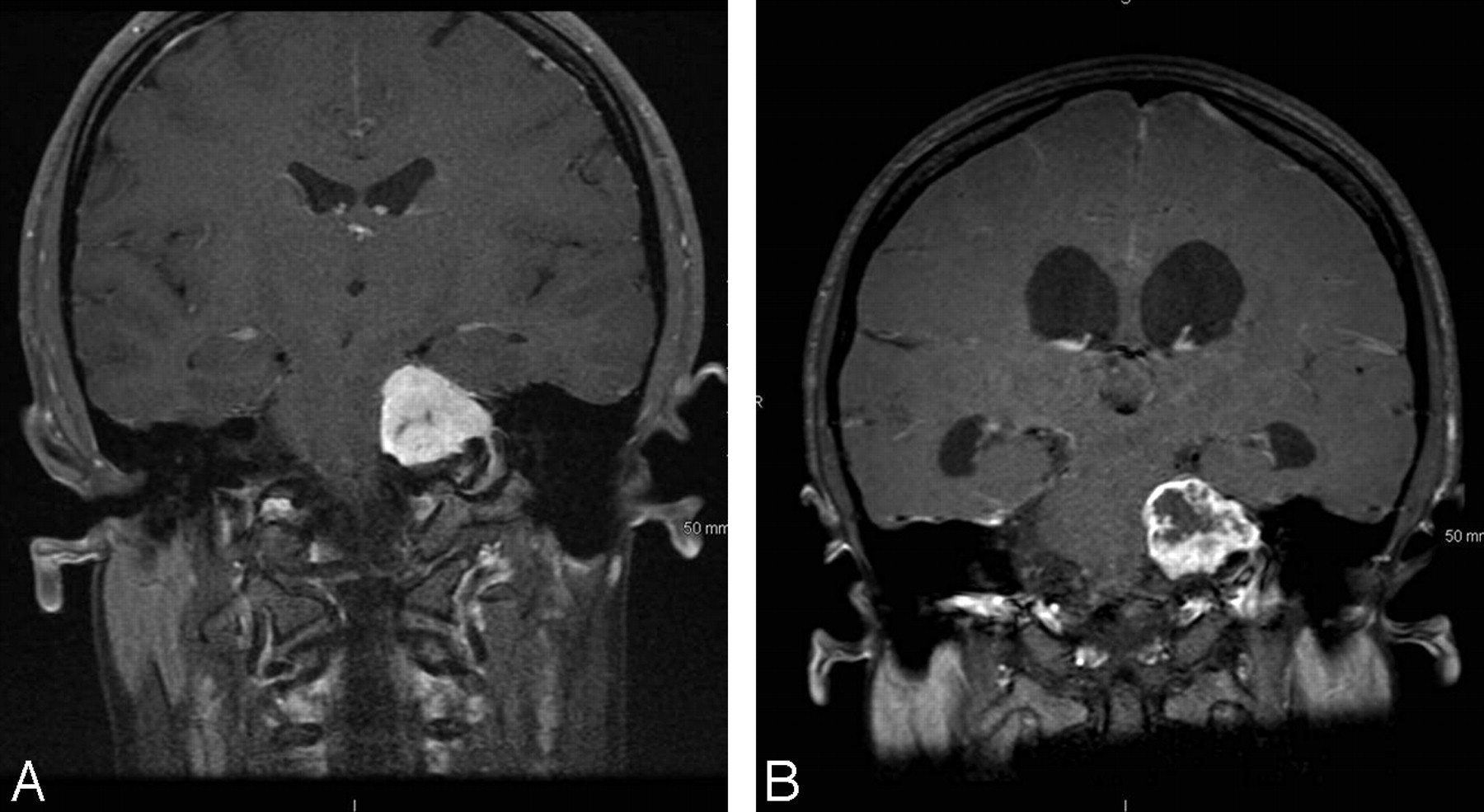
Axial T1-weighted MR imaging postgadolinium (A) 2 weeks before gamma knife treatment and (B) 9 months after gamma knife treatment demonstrates a 3-cm acoustic schwannoma. Central necrosis is seen in the posttreatment tumor.

Axial T1-weighted MR imaging postgadolinium (A) 2 weeks before gamma knife treatment and (B) 9 months after gamma knife treatment.

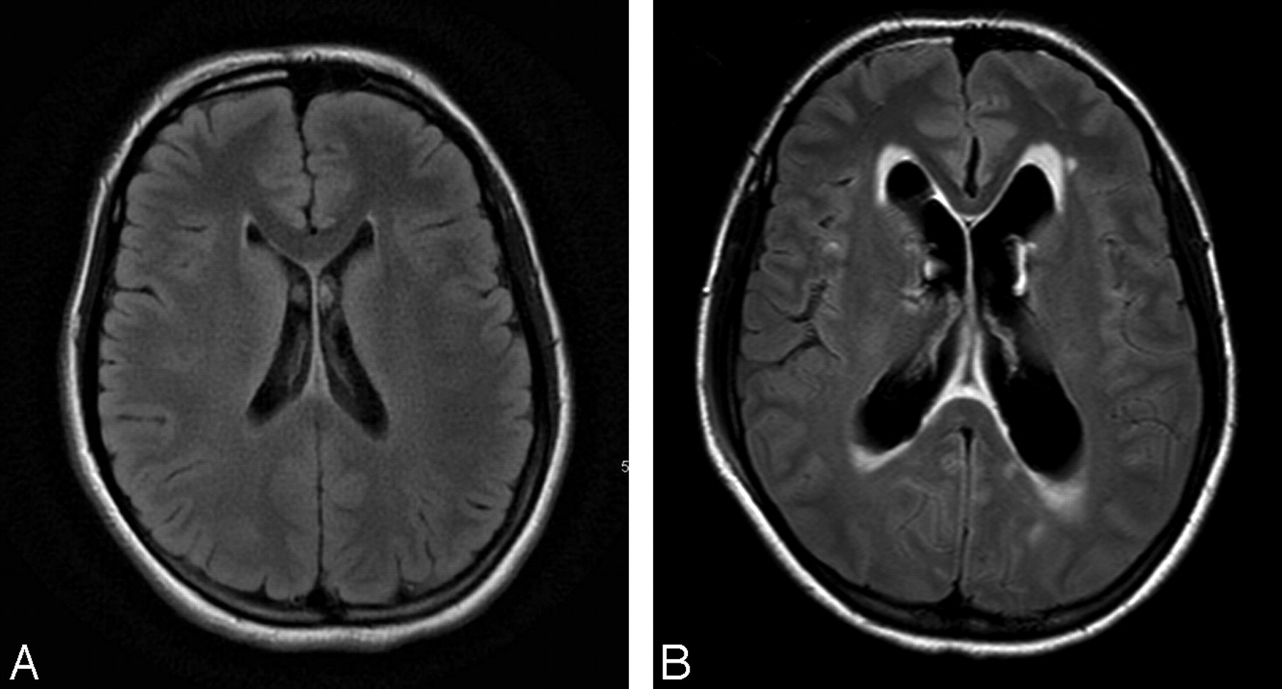
Axial fluid-attenuated inversion recovery MR images (A) 2 weeks before gamma knife treatment and (B) 9 months after gamma knife treatment. Ventriculomegaly is demonstrated and was noted to extend to the third and fourth ventricles. Note transependymal flow of CSF in Fig 3B.

Sagittal MR CSF cine-flow study (gradient-echo, TE, 7.9 ms; TR, 21 ms). This study was performed when the patient returned with ventriculomegaly 9 months after gamma knife treatment. CSF cine flow demonstrated free flow of CSF without evidence of intraventricular obstruction.
Discussion
Hydrocephalus is a known complication of an acoustic schwannoma, occurring in approximately 14% of cases and in 4% to 6% of cases after gamma knife treatment.1,2 Although it has been reported that gamma knife radiosurgery may contribute to the development of hydrocephalus, a causal relationship has not been established and remains controversial.1–7 The time course of events in this case suggests a contributory role of gamma knife treatment in the development or exacerbation of hydrocephalus. Indeed, in the few reported cases in which a time line is documented, the time course of events is similar, with hydrocephalus developing 4 to 18 months after radiosurgery.1–3
Of note, the patient had a markedly elevated CSF protein level. It has been proposed that communicating hydrocephalus accompanying an acoustic schwannoma is caused by tumor necrosis, with subsequent elevation of CSF protein concentration. Elevated CSF protein levels are thought to then obstruct CSF resorption at the level of the arachnoid granulations.1 These events are reported to occur in acoustic schwannomas without radiosurgical treatment, though radiosurgery may exacerbate these events in some patients.
Hydrocephalus after a gamma knife procedure is an infrequent event and likely reflects subtle differences in the tumor rather than differences in radiosurgical technique. It is generally accepted that vestibular schwannomas demonstrate variable growth rates.8 Our case is unusual with regard to both tumor size and patient age. The incidence of hydrocephalus, including nonobstructive hydrocephalus, has been shown to be greater with larger tumors, and 3 cm is considered the maximal size for gamma knife radiosurgery by some criteria.9,10 Acoustic schwannomas typically present in the sixth decade of life,1,10 with only a handful of patients presenting younger than 30 years. Such cases are frequently seen in the setting of neurofibromatosis type 2,1,10 though no such history was present in the case of our patient. Although histologic examination is not available in such cases, tumor necrosis and hydrocephalus may arise from faster-growing tumors, possibly correlating with patient age. Although not all studies agree, 2 studies suggest that faster growth rates are seen in younger, female patients.11,12 Faster growth rates and larger tumors are likely at increased risk for posttreatment tumor necrosis and the development of hydrocephalus.
Acoustic schwannomas can cause either communicating or noncommunicating obstructive hydrocephalus, by different mechanisms.13 Third ventriculostomy is an option for treatment of a noncommunicating hydrocephalus13 but would not be expected to offer palliation for communicating hydrocephalus, as in the case of our patient. MR CSF cine-flow study may be helpful in making this determination.
Conclusions
We presented an MR imaging study of communicating hydrocephalus after gamma knife treatment of acoustic schwannoma. Gamma knife treatment may exacerbate the development of hydrocephalus in some cases of acoustic schwannoma. MR CSF cine-flow study may help to determine the type of hydrocephalus and assist in directing management.
References
- Received August 1, 2008.
- Accepted after revision September 19, 2008.
- Copyright © American Society of Neuroradiology


