Advertisement

Main menu

  • Home
  • Content
    • Current Issue
    • Accepted Manuscripts
    • Article Preview
    • Past Issue Archive
    • Video Articles
    • AJNR Case Collection
    • Case of the Week Archive
    • Case of the Month Archive
    • Classic Case Archive
  • Special Collections
    • Advancing NeuroMRI with High-Relaxivity Contrast Agents
    • AJNR Awards
    • Low-Field MRI
    • Alzheimer Disease
    • ASNR Foundation Special Collection
    • Photon-Counting CT
    • View All
  • Multimedia
    • AJNR Podcasts
    • AJNR SCANtastic
    • Trainee Corner
    • MRI Safety Corner
    • Imaging Protocols
  • For Authors
    • Submit a Manuscript
    • Submit a Video Article
    • Submit an eLetter to the Editor/Response
    • Manuscript Submission Guidelines
    • Statistical Tips
    • Fast Publishing of Accepted Manuscripts
    • Graphical Abstract Preparation
    • Imaging Protocol Submission
    • Author Policies
  • About Us
    • About AJNR
    • Editorial Board
    • Editorial Board Alumni
  • More
    • Become a Reviewer/Academy of Reviewers
    • Subscribers
    • Permissions
    • Alerts
    • Feedback
    • Advertisers
    • ASNR Home

User menu

  • Alerts
  • Log in

Search

  • Advanced search
American Journal of Neuroradiology

American Journal of Neuroradiology

ASHNR American Society of Functional Neuroradiology ASHNR American Society of Pediatric Neuroradiology ASSR
  • Alerts
  • Log in

Advanced Search

  • Home
  • Content
    • Current Issue
    • Accepted Manuscripts
    • Article Preview
    • Past Issue Archive
    • Video Articles
    • AJNR Case Collection
    • Case of the Week Archive
    • Case of the Month Archive
    • Classic Case Archive
  • Special Collections
    • Advancing NeuroMRI with High-Relaxivity Contrast Agents
    • AJNR Awards
    • Low-Field MRI
    • Alzheimer Disease
    • ASNR Foundation Special Collection
    • Photon-Counting CT
    • View All
  • Multimedia
    • AJNR Podcasts
    • AJNR SCANtastic
    • Trainee Corner
    • MRI Safety Corner
    • Imaging Protocols
  • For Authors
    • Submit a Manuscript
    • Submit a Video Article
    • Submit an eLetter to the Editor/Response
    • Manuscript Submission Guidelines
    • Statistical Tips
    • Fast Publishing of Accepted Manuscripts
    • Graphical Abstract Preparation
    • Imaging Protocol Submission
    • Author Policies
  • About Us
    • About AJNR
    • Editorial Board
    • Editorial Board Alumni
  • More
    • Become a Reviewer/Academy of Reviewers
    • Subscribers
    • Permissions
    • Alerts
    • Feedback
    • Advertisers
    • ASNR Home
  • Follow AJNR on Twitter
  • Visit AJNR on Facebook
  • Follow AJNR on Instagram
  • Join AJNR on LinkedIn
  • RSS Feeds

AJNR Awards, New Junior Editors, and more. Read the latest AJNR updates


Improved Turnaround Times | Median time to first decision: 12 days

LetterLetter

Heat Stroke: Increased Signal Intensity in the Bilateral Cerebellar Dentate Nuclei and Splenium on Diffusion-Weighted MR Imaging

J.S. Lee, J.C. Choi, S.-Y. Kang, J.-H. Kang and J.-K. Park
American Journal of Neuroradiology April 2009, 30 (4) e58; DOI: https://doi.org/10.3174/ajnr.A1432
J.S. Lee
  • Find this author on Google Scholar
  • Find this author on PubMed
  • Search for this author on this site
J.C. Choi
  • Find this author on Google Scholar
  • Find this author on PubMed
  • Search for this author on this site
S.-Y. Kang
  • Find this author on Google Scholar
  • Find this author on PubMed
  • Search for this author on this site
J.-H. Kang
  • Find this author on Google Scholar
  • Find this author on PubMed
  • Search for this author on this site
J.-K. Park
  • Find this author on Google Scholar
  • Find this author on PubMed
  • Search for this author on this site
  • Article
  • Figures & Data
  • Info & Metrics
  • Responses
  • References
  • PDF
Loading

Heat stroke is a critical illness characterized by hyperthermia (elevated core body temperature >40°C) with accompanying life-threatening complications. Previous MR imaging findings of heat stroke included diffuse atrophy of the cerebellum1; hyperintense lesions in the cerebellum,1–3 external capsule and adjacent lateral putamen,2 medial thalamus,2 and hippocampus3; and patchy cortical lesions in the frontal and parietal lobe.3 However, to our knowledge, the findings of diffusion-weighted imaging (DWI) and apparent diffusion coefficient (ADC) mapping in patients in the early stage of heat stroke have not been reported. We describe a patient with heat stroke, with increased signal intensity in the cerebellar dentate nuclei and splenium on DWI.

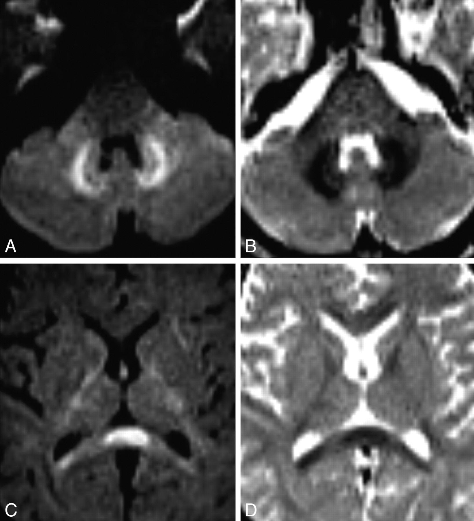
A 41-year-old man was admitted to the hospital in a comatose state, with high fever and respiratory failure. He had been working in a greenhouse for 3 hours on a hot and humid day just before the admission. DWI and ADC mapping on day 4 revealed restricted water diffusion in the bilateral dentate nuclei (312 × 10−6 mm2/s, Fig 1A, -B) and splenium of the corpus callosum (338 × 10−6 mm2/s, Fig 1C, -D). T2-weighted imaging (T2WI) showed slightly increased signal intensity only in the splenium of the corpus callosum. Follow-up DWI and T2WI, performed on the 21st day, revealed no signal intensity abnormality. However, the last follow-up MR imaging, performed on the 90th day, revealed cerebellar atrophy.

The present case showed symmetric and bilateral lesions in the splenium and cerebellar dentate nuclei. The involvement of dentate nucleus and splenium in our patient was very similar to the case of metronidazole-induced encephalopathy.4 Our patient had restricted water diffusion in the cerebellar dentate nuclei and splenium, which suggests that cytotoxic edema occurs early in developing heat stroke. The mechanism of cytotoxic edema in heat stroke is controversial. It may be related to hyperthermia-induced vasoconstriction and resultant cerebral ischemia or direct cytotoxicity of heat or both. In our patient, the last follow-up MR imaging, performed on the 90th day, revealed tissue loss in the cerebellar structures. These findings indicate that cytotoxic edema leads to delayed irreversible neuronal damage in the cerebellum. In conclusion, our case demonstrated that heat stroke causes irreversible neuronal damage, especially in cerebellum.

  Fig 1.
  • Download figure
  • Open in new tab
  • Download powerpoint
Fig 1.

Initial DWI and ADC map reveal restricted water diffusion in the bilateral dentate nuclei (A and B) and splenium of the corpus callosum (C and D).

References

  1. ↵
    Albukrek D, Bakon M, Moran DS, et al. Heat-stroke-induced cerebellar atrophy: clinical course, CT and MRI findings. Neuroradiology 1997;39:195–97
    CrossRefPubMed
  2. ↵
    McLaughlin CT, Kane AG, Auber AE. MR imaging of heat stroke: external capsule and thalamic T1 shortening and cerebellar injury. AJNR Am J Neuroradiol 2003;24:1372–75
    Abstract/FREE Full Text
  3. ↵
    Sudhakar PJ, Al-Hashimi H. Bilateral hippocampal hyperintensities: a new finding in MR imaging of heat stroke. Pediatr Radiol 2007;37:1289–91
    CrossRefPubMed
  4. ↵
    Kim E, Na DG, Kim EY, et al. MR imaging of metronidazole-induced encephalopathy: lesion distribution and diffusion-weighted imaging findings. AJNR Am J Neuroradiol 2007;28:1652–58. Epub 2007 Sep 20
    Abstract/FREE Full Text
  • Copyright © American Society of Neuroradiology
PreviousNext
Back to top

In this issue

American Journal of Neuroradiology: 30 (4)
American Journal of Neuroradiology
Vol. 30, Issue 4
April 2009
  • Table of Contents
  • Index by author
Advertisement
Print
Download PDF
Email Article

Thank you for your interest in spreading the word on American Journal of Neuroradiology.

NOTE: We only request your email address so that the person you are recommending the page to knows that you wanted them to see it, and that it is not junk mail. We do not capture any email address.

Enter multiple addresses on separate lines or separate them with commas.
Heat Stroke: Increased Signal Intensity in the Bilateral Cerebellar Dentate Nuclei and Splenium on Diffusion-Weighted MR Imaging
(Your Name) has sent you a message from American Journal of Neuroradiology
(Your Name) thought you would like to see the American Journal of Neuroradiology web site.
CAPTCHA
This question is for testing whether or not you are a human visitor and to prevent automated spam submissions.
Cite this article
J.S. Lee, J.C. Choi, S.-Y. Kang, J.-H. Kang, J.-K. Park
Heat Stroke: Increased Signal Intensity in the Bilateral Cerebellar Dentate Nuclei and Splenium on Diffusion-Weighted MR Imaging
American Journal of Neuroradiology Apr 2009, 30 (4) e58; DOI: 10.3174/ajnr.A1432

Citation Manager Formats

  • BibTeX
  • Bookends
  • EasyBib
  • EndNote (tagged)
  • EndNote 8 (xml)
  • Medlars
  • Mendeley
  • Papers
  • RefWorks Tagged
  • Ref Manager
  • RIS
  • Zotero
0 Responses
Respond to this article
Share
Bookmark this article
Heat Stroke: Increased Signal Intensity in the Bilateral Cerebellar Dentate Nuclei and Splenium on Diffusion-Weighted MR Imaging
J.S. Lee, J.C. Choi, S.-Y. Kang, J.-H. Kang, J.-K. Park
American Journal of Neuroradiology Apr 2009, 30 (4) e58; DOI: 10.3174/ajnr.A1432
del.icio.us logo Twitter logo Facebook logo Mendeley logo
  • Tweet Widget
  • Facebook Like
  • Google Plus One
Purchase

Jump to section

  • Article
    • References
  • Figures & Data
  • Info & Metrics
  • Responses
  • References
  • PDF

Related Articles

  • No related articles found.
  • PubMed
  • Google Scholar

Cited By...

  • Thermal Cycling-Hyperthermia Attenuates Rotenone-Induced Cell Injury in SH-SY5Y Cells through Heat-Activated Mechanisms
  • Thermal cycling-hyperthermia ameliorates cognitive impairment of intracerebroventricular A{beta}25-35-induced Alzheimers disease in C57BL/6 mice
  • Crossref
  • Google Scholar

This article has not yet been cited by articles in journals that are participating in Crossref Cited-by Linking.

More in this TOC Section

  • Letter to the Editor regarding “Automated Volumetric Software in Dementia: Help or Hindrance to the Neuroradiologist?”
  • Reply:
  • Brain AVM’s Nidus: What if We Hadn’t Understood Anything?
Show more LETTERS

Similar Articles

Advertisement

Indexed Content

  • Current Issue
  • Accepted Manuscripts
  • Article Preview
  • Past Issues
  • Editorials
  • Editor's Choice
  • Fellows' Journal Club
  • Letters to the Editor
  • Video Articles

Cases

  • Case Collection
  • Archive - Case of the Week
  • Archive - Case of the Month
  • Archive - Classic Case

More from AJNR

  • Trainee Corner
  • Imaging Protocols
  • MRI Safety Corner
  • Book Reviews

Multimedia

  • AJNR Podcasts
  • AJNR Scantastics

Resources

  • Turnaround Time
  • Submit a Manuscript
  • Submit a Video Article
  • Submit an eLetter to the Editor/Response
  • Manuscript Submission Guidelines
  • Statistical Tips
  • Fast Publishing of Accepted Manuscripts
  • Graphical Abstract Preparation
  • Imaging Protocol Submission
  • Evidence-Based Medicine Level Guide
  • Publishing Checklists
  • Author Policies
  • Become a Reviewer/Academy of Reviewers
  • News and Updates

About Us

  • About AJNR
  • Editorial Board
  • Editorial Board Alumni
  • Alerts
  • Permissions
  • Not an AJNR Subscriber? Join Now
  • Advertise with Us
  • Librarian Resources
  • Feedback
  • Terms and Conditions
  • AJNR Editorial Board Alumni

American Society of Neuroradiology

  • Not an ASNR Member? Join Now

© 2025 by the American Society of Neuroradiology All rights, including for text and data mining, AI training, and similar technologies, are reserved.
Print ISSN: 0195-6108 Online ISSN: 1936-959X

Powered by HighWire