
Article Figures & Data
Figures
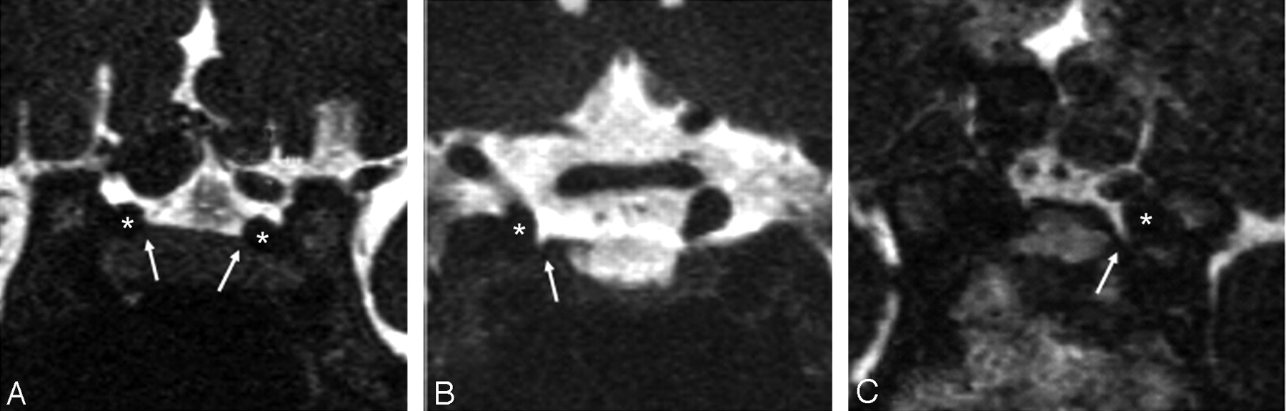
- Fig 1.
Configuration of the carotid cave (arrow) by 3D-CISS coronal images. The depth of the carotid cave is classified as having no dent (A), a shallow dent (B), and a deep dent (C). The asterisk indicates the ICA.
- Fig 2.
Visualization of the ICA border in the cavernous sinus. On the 3D-CISS image (A), the right ICA (arrowhead) is rated as grade I (no visible boundary), and the left ICA (white arrows) is rated as grade II (vaguely visible boundary). On the fusion image with 3D-CISS and MRA (B), both ICAs are rated as grade III (obvious boundary with clear delineation). The optic nerve (blue arrow) and ophthalmic artery (yellow arrow) were easy to differentiate with fusion image.
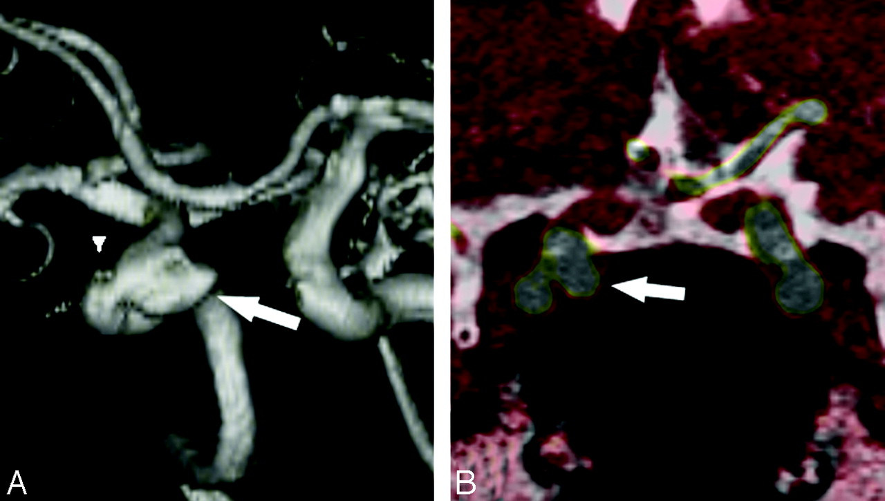
- Fig 3.
Right ICA aneurysm projection medially. The MRA VR image (A) shows that the aneurysm (arrow) is located just distal to the ophthalmic artery (arrowhead), indicating the aneurysm to be intradural. However, on fusion images (B), the aneurysm (arrow) dose not contact with the CSF of the suprasellar cistern and was diagnosed as an extradural aneurysm.
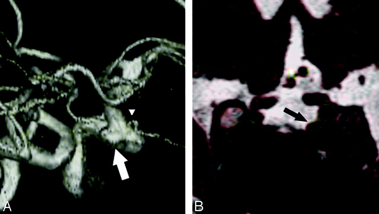
- Fig 4.
Left ICA aneurysm projection medially. A, MRA VR image. B, Fusion images. The aneurysm is located at lateral aspect of the ICA, the same level of the origin of the ophthalmic artery. The aneurysm (arrow) on the fusion image is faced to the CSF space and is diagnosed as an intradural paraclinoid (carotid cave) aneurysm.
- Fig 5.
Large right ICA aneurysm. A, Fusion coronal image. B, Fusion sagittal image. C, VR images. The upper dome of the aneurysm contacts to the CSF space, but the lower dome of the aneurysm is seen in the cavernous sinus. The white line is the assumed line of the DDR. This aneurysm (arrow) is classified as a transdural type.
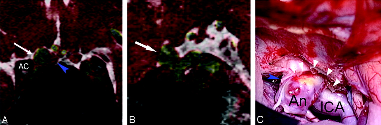
- Fig 6.
Anterior wall aneurysm of the right ICA. The MR images ([A] fusion coronal image and [B] fusion sagittal image) show that the aneurysm (arrow) is located just distal to the DDR (intradural) and is attached to the anterior clinoid process (AC) laterally. The operative view after removing the anterior clinoid process and sectioning of the DDR shows the aneurysm to be distal to the DDR. An indicates aneurysm; ICA, distal internal carotid artery; white arrowheads, tag ends of the DDR; blue arrowhead, optic nerve.


Tables
Differentiation of aneurysms from extradural to intradural by MR fusion images and relationship to the origin of the ophthalmic artery
MR Fusion Findings Ophthalmic artery Intradural Transdural Extradural Total Distal 16 2 1 19 Same level 4 1 3 8 Proximal 3 8 11 Not visible 1 1 Total 21 6 12 39
In this issue


Jump to section
Related Articles
Cited By...
- No citing articles found.