Advertisement

Main menu

  • Home
  • Content
    • Current Issue
    • Accepted Manuscripts
    • Article Preview
    • Past Issue Archive
    • Video Articles
    • AJNR Case Collection
    • Case of the Week Archive
    • Case of the Month Archive
    • Classic Case Archive
  • Special Collections
    • AJNR Awards
    • Low-Field MRI
    • Alzheimer Disease
    • ASNR Foundation Special Collection
    • Photon-Counting CT
    • View All
  • Multimedia
    • AJNR Podcasts
    • AJNR SCANtastic
    • Trainee Corner
    • MRI Safety Corner
    • Imaging Protocols
  • For Authors
    • Submit a Manuscript
    • Submit a Video Article
    • Submit an eLetter to the Editor/Response
    • Manuscript Submission Guidelines
    • Statistical Tips
    • Fast Publishing of Accepted Manuscripts
    • Graphical Abstract Preparation
    • Imaging Protocol Submission
    • Author Policies
  • About Us
    • About AJNR
    • Editorial Board
    • Editorial Board Alumni
  • More
    • Become a Reviewer/Academy of Reviewers
    • Subscribers
    • Permissions
    • Alerts
    • Feedback
    • Advertisers
    • ASNR Home

User menu

  • Alerts
  • Log in

Search

  • Advanced search
American Journal of Neuroradiology

American Journal of Neuroradiology

ASHNR American Society of Functional Neuroradiology ASHNR American Society of Pediatric Neuroradiology ASSR
  • Alerts
  • Log in

Advanced Search

  • Home
  • Content
    • Current Issue
    • Accepted Manuscripts
    • Article Preview
    • Past Issue Archive
    • Video Articles
    • AJNR Case Collection
    • Case of the Week Archive
    • Case of the Month Archive
    • Classic Case Archive
  • Special Collections
    • AJNR Awards
    • Low-Field MRI
    • Alzheimer Disease
    • ASNR Foundation Special Collection
    • Photon-Counting CT
    • View All
  • Multimedia
    • AJNR Podcasts
    • AJNR SCANtastic
    • Trainee Corner
    • MRI Safety Corner
    • Imaging Protocols
  • For Authors
    • Submit a Manuscript
    • Submit a Video Article
    • Submit an eLetter to the Editor/Response
    • Manuscript Submission Guidelines
    • Statistical Tips
    • Fast Publishing of Accepted Manuscripts
    • Graphical Abstract Preparation
    • Imaging Protocol Submission
    • Author Policies
  • About Us
    • About AJNR
    • Editorial Board
    • Editorial Board Alumni
  • More
    • Become a Reviewer/Academy of Reviewers
    • Subscribers
    • Permissions
    • Alerts
    • Feedback
    • Advertisers
    • ASNR Home
  • Follow AJNR on Twitter
  • Visit AJNR on Facebook
  • Follow AJNR on Instagram
  • Join AJNR on LinkedIn
  • RSS Feeds

AJNR Awards, New Junior Editors, and more. Read the latest AJNR updates

OtherPediatric Neuroimaging
Open Access

Pediatric MR Imaging with Automated Spine Survey Iterative Scan Technique (ASSIST)

K.L. Weiss, D. Sun and J.L. Weiss
American Journal of Neuroradiology April 2009, 30 (4) 821-824; DOI: https://doi.org/10.3174/ajnr.A1430
K.L. Weiss
  • Find this author on Google Scholar
  • Find this author on PubMed
  • Search for this author on this site
D. Sun
  • Find this author on Google Scholar
  • Find this author on PubMed
  • Search for this author on this site
J.L. Weiss
  • Find this author on Google Scholar
  • Find this author on PubMed
  • Search for this author on this site
  • Article
  • Figures & Data
  • Info & Metrics
  • Responses
  • References
  • PDF
Loading

Article Figures & Data

Figures

  • Fig 1.
    • Download figure
    • Open in new tab
    • Download powerpoint
    Fig 1.

    Midline sagittal ASSIST (from left to right). A, A 7-year-old boy with ADHD demonstrating mild motion artifacts. B, A 9-year-old boy with Asperger syndrome. Diaphragmatic contour (arrow) is well defined, suggesting good breath-hold. C, A 10-year-old girl with developmental delay and short stature. Diaphragmatic contour (arrow) is well defined, suggesting good breath-hold. D, A 12-year-old girl with prominent scoliosis. Examination resulted in autolabeling failure. E, A 16-year-old adolescent boy with mild Scheuermann disease at T6-T8 (arrows) and L4 superior endplate deformity (arrow). F, A 17-year-old adolescent boy with lumbarized S1 (L6) vertebrae (closed arrow) and enlarged pituitary gland (open arrow). G, An 18-year-old woman with mild kyphoscoliosis.

  • Fig 2.
    • Download figure
    • Open in new tab
    • Download powerpoint
    Fig 2.

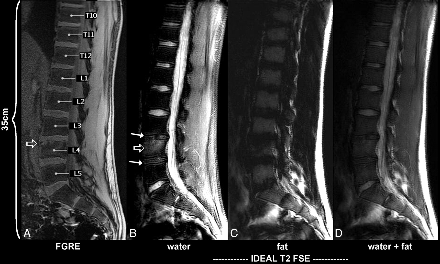
    Midline sagittal ASSIST lower station in a 16-year-old adolescent boy (same subject as in 1E). From left to right, autolabeled out-of-plane FGE (TR, 57 ms; TE, 1.4 ms; flip angle, 30°; bandwidth, ± 62.5 kHz; matrix, 512 × 352; breath-hold, 21 sec), IDEAL T2 FSE (TR, 2050 ms; TE, 61.7 ms; ETL, 9; bandwidth, ± 25 kHz; matrix, 512 × 288; 3.33 minutes).

  • Fig 3.
    • Download figure
    • Open in new tab
    • Download powerpoint
    Fig 3.

    Left of midline sagittal ASSIST upper station (10 mm) in a 9-year-old boy (same subject as in 1B). Note vertically oriented neurocentral synchondrosis visualized on this image from T2 (upper arrow) to T11 (lower arrow).

PreviousNext
Back to top

In this issue

American Journal of Neuroradiology: 30 (4)
American Journal of Neuroradiology
Vol. 30, Issue 4
April 2009
  • Table of Contents
  • Index by author
Advertisement
Print
Download PDF
Email Article

Thank you for your interest in spreading the word on American Journal of Neuroradiology.

NOTE: We only request your email address so that the person you are recommending the page to knows that you wanted them to see it, and that it is not junk mail. We do not capture any email address.

Enter multiple addresses on separate lines or separate them with commas.
Pediatric MR Imaging with Automated Spine Survey Iterative Scan Technique (ASSIST)
(Your Name) has sent you a message from American Journal of Neuroradiology
(Your Name) thought you would like to see the American Journal of Neuroradiology web site.
CAPTCHA
This question is for testing whether or not you are a human visitor and to prevent automated spam submissions.
Cite this article
K.L. Weiss, D. Sun, J.L. Weiss
Pediatric MR Imaging with Automated Spine Survey Iterative Scan Technique (ASSIST)
American Journal of Neuroradiology Apr 2009, 30 (4) 821-824; DOI: 10.3174/ajnr.A1430

Citation Manager Formats

  • BibTeX
  • Bookends
  • EasyBib
  • EndNote (tagged)
  • EndNote 8 (xml)
  • Medlars
  • Mendeley
  • Papers
  • RefWorks Tagged
  • Ref Manager
  • RIS
  • Zotero
0 Responses
Respond to this article
Share
Bookmark this article
Pediatric MR Imaging with Automated Spine Survey Iterative Scan Technique (ASSIST)
K.L. Weiss, D. Sun, J.L. Weiss
American Journal of Neuroradiology Apr 2009, 30 (4) 821-824; DOI: 10.3174/ajnr.A1430
del.icio.us logo Twitter logo Facebook logo Mendeley logo
  • Tweet Widget
  • Facebook Like
  • Google Plus One
Purchase

Jump to section

  • Article
    • Abstract
    • Materials and Methods
    • Results
    • Discussion
    • Conclusions
    • Acknowledgments
    • Footnotes
    • References
  • Figures & Data
  • Info & Metrics
  • Responses
  • References
  • PDF

Related Articles

  • No related articles found.
  • PubMed
  • Google Scholar

Cited By...

  • Subminute Fat-Water-Separated Dual-Echo Automated Spine Survey Iterative Scan Technique
  • Crossref (2)
  • Google Scholar

This article has been cited by the following articles in journals that are participating in Crossref Cited-by Linking.

  • Rapid MRI Detection of Vertebral Numeric Variation
    Jason J. Akbar, Kenneth L. Weiss, Muhammad A. Saafir, Jane L. Weiss
    American Journal of Roentgenology 2010 195 2
  • Subminute Fat-Water-Separated Dual-Echo Automated Spine Survey Iterative Scan Technique
    K.L. Weiss, C.R. Richards, D. Sun, J.L. Weiss
    American Journal of Neuroradiology 2009 30 10

More in this TOC Section

  • CHARGE fetal MRI clival cleft
  • fetal brain development of 10 weeks gestation
  • Neuroimaging Delineation and Progression of SLSMD
Show more Pediatric Neuroimaging

Similar Articles

Advertisement

Indexed Content

  • Current Issue
  • Accepted Manuscripts
  • Article Preview
  • Past Issues
  • Editorials
  • Editor's Choice
  • Fellows' Journal Club
  • Letters to the Editor
  • Video Articles

Cases

  • Case Collection
  • Archive - Case of the Week
  • Archive - Case of the Month
  • Archive - Classic Case

More from AJNR

  • Trainee Corner
  • Imaging Protocols
  • MRI Safety Corner
  • Book Reviews

Multimedia

  • AJNR Podcasts
  • AJNR Scantastics

Resources

  • Turnaround Time
  • Submit a Manuscript
  • Submit a Video Article
  • Submit an eLetter to the Editor/Response
  • Manuscript Submission Guidelines
  • Statistical Tips
  • Fast Publishing of Accepted Manuscripts
  • Graphical Abstract Preparation
  • Imaging Protocol Submission
  • Evidence-Based Medicine Level Guide
  • Publishing Checklists
  • Author Policies
  • Become a Reviewer/Academy of Reviewers
  • News and Updates

About Us

  • About AJNR
  • Editorial Board
  • Editorial Board Alumni
  • Alerts
  • Permissions
  • Not an AJNR Subscriber? Join Now
  • Advertise with Us
  • Librarian Resources
  • Feedback
  • Terms and Conditions
  • AJNR Editorial Board Alumni

American Society of Neuroradiology

  • Not an ASNR Member? Join Now

© 2025 by the American Society of Neuroradiology All rights, including for text and data mining, AI training, and similar technologies, are reserved.
Print ISSN: 0195-6108 Online ISSN: 1936-959X

Powered by HighWire