Advertisement

Main menu

  • Home
  • Content
    • Current Issue
    • Accepted Manuscripts
    • Article Preview
    • Past Issue Archive
    • Video Articles
    • AJNR Case Collection
    • Case of the Week Archive
    • Case of the Month Archive
    • Classic Case Archive
  • Special Collections
    • Advancing NeuroMRI with High-Relaxivity Contrast Agents
    • AJNR Awards
    • Low-Field MRI
    • Alzheimer Disease
    • ASNR Foundation Special Collection
    • Photon-Counting CT
    • View All
  • Multimedia
    • AJNR Podcasts
    • AJNR SCANtastic
    • Trainee Corner
    • MRI Safety Corner
    • Imaging Protocols
  • For Authors
    • Submit a Manuscript
    • Submit a Video Article
    • Submit an eLetter to the Editor/Response
    • Manuscript Submission Guidelines
    • Statistical Tips
    • Fast Publishing of Accepted Manuscripts
    • Graphical Abstract Preparation
    • Imaging Protocol Submission
    • Author Policies
  • About Us
    • About AJNR
    • Editorial Board
    • Editorial Board Alumni
  • More
    • Become a Reviewer/Academy of Reviewers
    • Subscribers
    • Permissions
    • Alerts
    • Feedback
    • Advertisers
    • ASNR Home

User menu

  • Alerts
  • Log in

Search

  • Advanced search
American Journal of Neuroradiology

American Journal of Neuroradiology

ASHNR American Society of Functional Neuroradiology ASHNR American Society of Pediatric Neuroradiology ASSR
  • Alerts
  • Log in

Advanced Search

  • Home
  • Content
    • Current Issue
    • Accepted Manuscripts
    • Article Preview
    • Past Issue Archive
    • Video Articles
    • AJNR Case Collection
    • Case of the Week Archive
    • Case of the Month Archive
    • Classic Case Archive
  • Special Collections
    • Advancing NeuroMRI with High-Relaxivity Contrast Agents
    • AJNR Awards
    • Low-Field MRI
    • Alzheimer Disease
    • ASNR Foundation Special Collection
    • Photon-Counting CT
    • View All
  • Multimedia
    • AJNR Podcasts
    • AJNR SCANtastic
    • Trainee Corner
    • MRI Safety Corner
    • Imaging Protocols
  • For Authors
    • Submit a Manuscript
    • Submit a Video Article
    • Submit an eLetter to the Editor/Response
    • Manuscript Submission Guidelines
    • Statistical Tips
    • Fast Publishing of Accepted Manuscripts
    • Graphical Abstract Preparation
    • Imaging Protocol Submission
    • Author Policies
  • About Us
    • About AJNR
    • Editorial Board
    • Editorial Board Alumni
  • More
    • Become a Reviewer/Academy of Reviewers
    • Subscribers
    • Permissions
    • Alerts
    • Feedback
    • Advertisers
    • ASNR Home
  • Follow AJNR on Twitter
  • Visit AJNR on Facebook
  • Follow AJNR on Instagram
  • Join AJNR on LinkedIn
  • RSS Feeds

AJNR Awards, New Junior Editors, and more. Read the latest AJNR updates


Improved Turnaround Times | Median time to first decision: 12 days

OtherHead and Neck Imaging

Transdural Spread of Glioblastoma through the Foramen Ovale with Presentation as a Masticator Space Mass

R. Kwak and D. Shatzkes
American Journal of Neuroradiology April 2009, 30 (4) 808-810; DOI: https://doi.org/10.3174/ajnr.A1349
R. Kwak
  • Find this author on Google Scholar
  • Find this author on PubMed
  • Search for this author on this site
D. Shatzkes
  • Find this author on Google Scholar
  • Find this author on PubMed
  • Search for this author on this site
  • Article
  • Figures & Data
  • Info & Metrics
  • Responses
  • References
  • PDF
Loading

Article Figures & Data

Figures

  • Tables
  • Fig 1.
    • Download figure
    • Open in new tab
    • Download powerpoint
    Fig 1.

    A 53-year-old man with history of glioblastoma presented with chronic otomastoiditis and mandibular nerve denervation. A, Coronal T2-weighted MR image demonstrates a transcranial mass extending through the left foramen ovale, with the largest component in the masticator space (white arrowheads). Subacute denervation of the left medial pterygoid and masseter muscles is evidenced by their increased signal intensity and decreased volume (white arrows). Chronic postoperative and treatment-related changes are noted in the left middle cranial fossa and left temporal lobe (black arrow). B, Axial contrast-enhanced fat-suppressed T1-weighted MR image demonstrates the extracranial portion of the mass interposed between the left medial and lateral pterygoid muscles (white arrowheads). The left temporalis and masseter muscles demonstrate asymmetrically decreased volume (white arrows). Note the mass effect on the left eustachian tube orifice and abnormal signal within the left mastoid air cells, likely on an obstructive basis (black arrow).
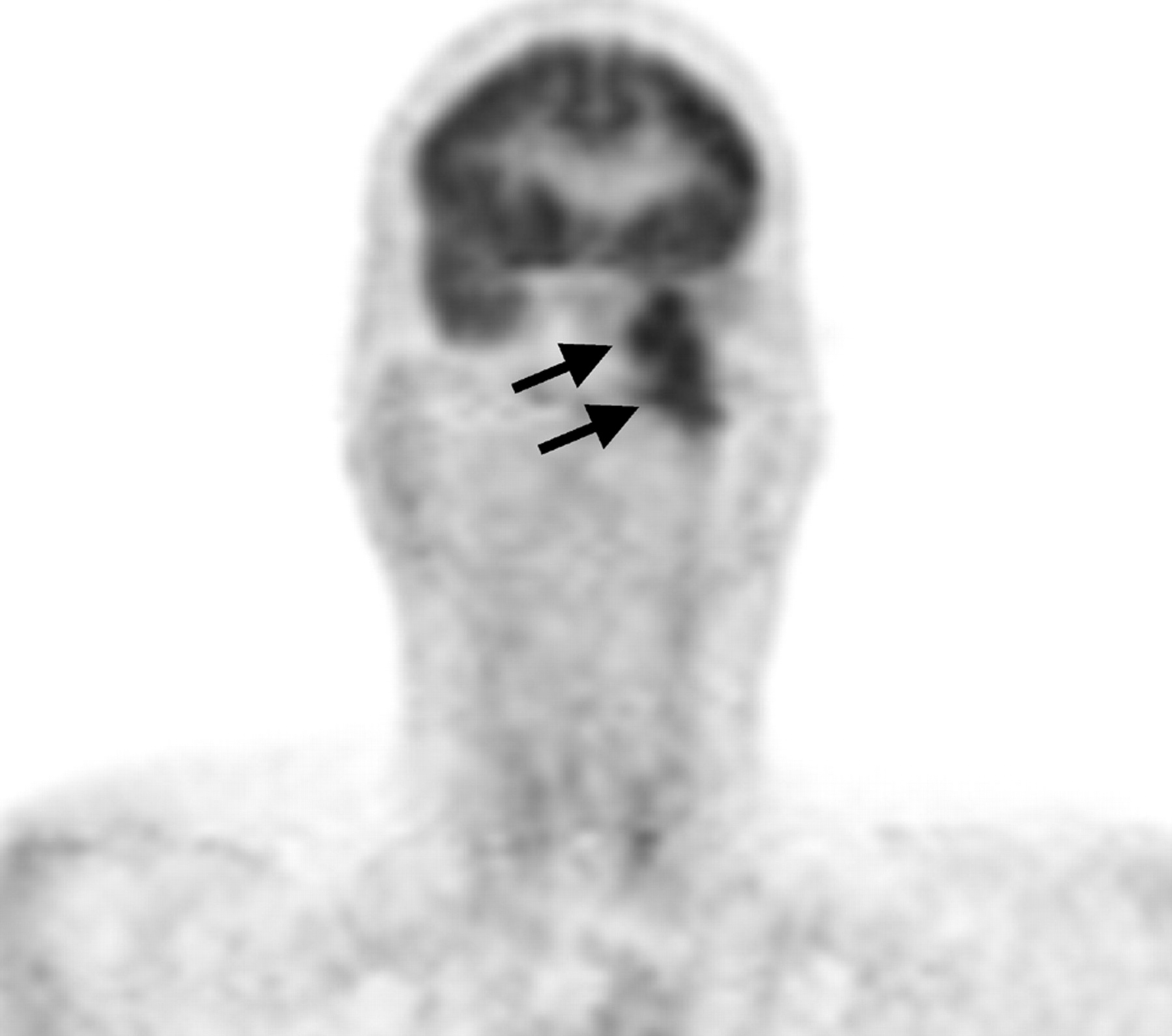
  • Fig 2.
    • Download figure
    • Open in new tab
    • Download powerpoint
    Fig 2.

    PET images in the coronal plane clearly show transcranial tumor extending from the floor of the left middle cranial fossa into the soft tissues of the left deep face (black arrows).

Tables

  • Figures
  • Spontaneous extradural spread of glioblastoma through the skull base

    InvestigatorAge (yr), SexSite of PrimarySite of Extradural SpreadPrior Treatment
    Sanerkin, 1962665, ML temporal lobeL middle cranial fossaNone
    Nager, 1967741, MLarge R temporalR temporal bone, presenting as a mass in the R external auditory canalNo prior craniotomy or radiotherapy
    Hoyt et al, 19728–FrontotemporalAnterior fossa
    Liwnicz and Rubinstein, 1979326, MLarge L temporal lobeL middle cranial fossaRadiation and surgery approx 8 months prior
    Aoyama et al, 1980930 FR frontalAnterior cranial fossa into nasal cavityNone
    Shuangshoti et al, 19871030, MR frontotemporalR sphenoid wing, posterior wall of frontal sinusNone
    Shuangshoti et al, 19871025, FR frontotemporalSphenoid wing, ethmoid, posterior orbitNone
    Bigner et al, 1989117, FL medial temporalL sphenoid wing and sella turcica into nasopharynxLeft ventriculopleural shunt
    Lampl et al, 19901232, MR frontoparietalEthmoid sinus, frontal sinusResection 8 months prior with chemoradiation
    Pompili et al, 19931340, MBifrontalL orbit, ethmoid, maxillary sinus, and nasal fossaPartial resection and radiation 21 months prior
    Horiuchi et al, 19961441, FR temporal lobeR anterior and middle cranial fossae into orbit, nasal cavity, and oral cavity2 prior resections
    Rainov et al, 19961566, FL temporal lobeAnterior and middle cranial fossaeNone
    • Note:—R, L indicate right and left, respectively; approx, approximately.

PreviousNext
Back to top

In this issue

American Journal of Neuroradiology: 30 (4)
American Journal of Neuroradiology
Vol. 30, Issue 4
April 2009
  • Table of Contents
  • Index by author
Advertisement
Print
Download PDF
Email Article

Thank you for your interest in spreading the word on American Journal of Neuroradiology.

NOTE: We only request your email address so that the person you are recommending the page to knows that you wanted them to see it, and that it is not junk mail. We do not capture any email address.

Enter multiple addresses on separate lines or separate them with commas.
Transdural Spread of Glioblastoma through the Foramen Ovale with Presentation as a Masticator Space Mass
(Your Name) has sent you a message from American Journal of Neuroradiology
(Your Name) thought you would like to see the American Journal of Neuroradiology web site.
CAPTCHA
This question is for testing whether or not you are a human visitor and to prevent automated spam submissions.
Cite this article
R. Kwak, D. Shatzkes
Transdural Spread of Glioblastoma through the Foramen Ovale with Presentation as a Masticator Space Mass
American Journal of Neuroradiology Apr 2009, 30 (4) 808-810; DOI: 10.3174/ajnr.A1349

Citation Manager Formats

  • BibTeX
  • Bookends
  • EasyBib
  • EndNote (tagged)
  • EndNote 8 (xml)
  • Medlars
  • Mendeley
  • Papers
  • RefWorks Tagged
  • Ref Manager
  • RIS
  • Zotero
0 Responses
Respond to this article
Share
Bookmark this article
Transdural Spread of Glioblastoma through the Foramen Ovale with Presentation as a Masticator Space Mass
R. Kwak, D. Shatzkes
American Journal of Neuroradiology Apr 2009, 30 (4) 808-810; DOI: 10.3174/ajnr.A1349
del.icio.us logo Twitter logo Facebook logo Mendeley logo
  • Tweet Widget
  • Facebook Like
  • Google Plus One
Purchase

Jump to section

  • Article
    • Abstract
    • Case Report
    • Discussion
    • References
  • Figures & Data
  • Info & Metrics
  • Responses
  • References
  • PDF

Related Articles

  • No related articles found.
  • PubMed
  • Google Scholar

Cited By...

  • Unusual extracranial spread of glioblastoma
  • Crossref
  • Google Scholar

This article has not yet been cited by articles in journals that are participating in Crossref Cited-by Linking.

More in this TOC Section

  • Diagnostic Performance of US in Neck Node NIRADS 2
  • Hydrops Herniation into the Semicircular Canals
  • ASL Sensitivity for Head and Neck Paraganglioma
Show more Head and Neck Imaging

Similar Articles

Advertisement

Indexed Content

  • Current Issue
  • Accepted Manuscripts
  • Article Preview
  • Past Issues
  • Editorials
  • Editor's Choice
  • Fellows' Journal Club
  • Letters to the Editor
  • Video Articles

Cases

  • Case Collection
  • Archive - Case of the Week
  • Archive - Case of the Month
  • Archive - Classic Case

More from AJNR

  • Trainee Corner
  • Imaging Protocols
  • MRI Safety Corner
  • Book Reviews

Multimedia

  • AJNR Podcasts
  • AJNR Scantastics

Resources

  • Turnaround Time
  • Submit a Manuscript
  • Submit a Video Article
  • Submit an eLetter to the Editor/Response
  • Manuscript Submission Guidelines
  • Statistical Tips
  • Fast Publishing of Accepted Manuscripts
  • Graphical Abstract Preparation
  • Imaging Protocol Submission
  • Evidence-Based Medicine Level Guide
  • Publishing Checklists
  • Author Policies
  • Become a Reviewer/Academy of Reviewers
  • News and Updates

About Us

  • About AJNR
  • Editorial Board
  • Editorial Board Alumni
  • Alerts
  • Permissions
  • Not an AJNR Subscriber? Join Now
  • Advertise with Us
  • Librarian Resources
  • Feedback
  • Terms and Conditions
  • AJNR Editorial Board Alumni

American Society of Neuroradiology

  • Not an ASNR Member? Join Now

© 2025 by the American Society of Neuroradiology All rights, including for text and data mining, AI training, and similar technologies, are reserved.
Print ISSN: 0195-6108 Online ISSN: 1936-959X

Powered by HighWire