
Article Figures & Data
Figures
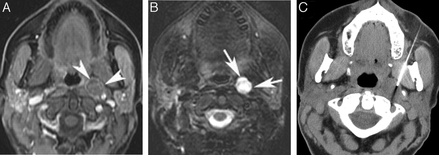
- Fig 1.
A 51-year-old woman with a history of thyroidectomy, left neck dissection, and rising thyroglobulin levels. A, Axial T1-weighted MR image shows a 12-mm solid left retropharyngeal node (asterisk) that is hyperintense relative to muscle. B, Axial T2-weighted MR image shows the left retropharyngeal lymph node. C, FDG-PET shows avid uptake in the left retropharyngeal node at the skull base, which did not concentrate radioiodine (not shown). Surgical resection confirmed metastatic papillary carcinoma. T indicates physiologic uptake in the palatine tonsils.
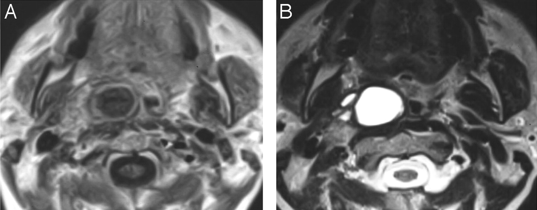
- Fig 2.
A 52-year-old woman being evaluated with MR imaging for an unrelated indication. An incidental right parapharyngeal space mass was identified. A, Gadolinium-enhanced axial T1-weighted MR image shows a multiseptate cystic rim-enhancing mass in the right parapharyngeal space. B, Axial T2-weighted MR image shows the multiseptate parapharyngeal mass. The patient went directly to surgical resection, which identified metastatic papillary cancer in a high level II lymph node in the parapharyngeal space. Subsequent thyroidectomy identified a 4-mm papillary carcinoma in the thyroid gland. No other pathologic nodes in the neck were identified on MR imaging.
- Fig 3.
A 47-year-old woman with a history of thyroidectomy, bilateral neck dissections, and rising thyroglobulin levels. A, Gadolinium-enhanced axial T1-weighted MR image shows a 1-cm complex solid and cystic lymph node metastasis in the left retropharynx (arrowheads). B, Corresponding axial T2-weighted MR image shows the complex retropharyngeal node (arrows). C, CT-guided FNA reveals metastatic papillary cancer confirmed at surgical resection.
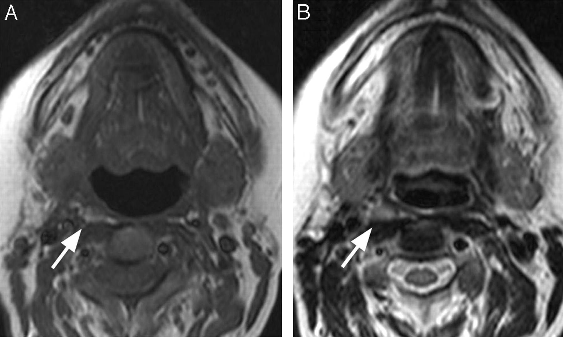
- Fig 4.
A 47-year-old woman treated with thyroidectomy and right neck dissection for papillary carcinoma presented with progressive rising thyroglobulin levels. A, Axial unenhanced T1-weighted MR image shows a 7-mm right retropharyngeal node (arrow) that is hyperintense relative to muscle. B, Axial T2-weighted MR image shows the node (arrow). Both FDG-PET and radioiodine showed increased uptake (not shown).


Tables
Characteristics of patients with retropharyngeal or parapharyngeal space nodal metastases
Age*/Sex Prior Surgery RP/PPS Node (Location/mm) MR Signal PET I-123 FNA Cytology Surgical Pathology 47/F Thyroidectomy, b/l neck dissection Left RP/10 Complex N/A N/A Papillary thyroid ca. Papillary thyroid ca. 68/F Thyroidectomy, R neck dissection Right RP/16 Solid Pos Neg Papillary thyroid ca. Papillary thyroid ca. 74/M Thyroidectomy, L neck dissection Left RP/20 Complex N/A Neg Anaplastic thyroid ca. N/A 58/F Thyroidectomy, L neck dissection Left RP/12 Solid Pos Neg No tumor Papillary thyroid ca. 49/F Thyroidectomy, L neck dissection, b/l paratracheal dissection Left RP/10 Solid Neg N/A No tumor N/A 52/F None Right PPS/25 Cystic N/A N/A N/A Papillary thyroid ca. 47/F Thyroidectomy, R neck dissection Right RP/7 Solid Neg Neg N/A N/A 47/F Thyroidectomy, b/l neck dissection, posterior neck dissection Left RP/7 Solid N/A N/A N/A N/A 56/F Thyroidectomy, R neck dissection Right RP/11 Solid N/A N/A N/A N/A Note:—RP indicates retropharyngeal node; PPS, parapharyngeal space node; b/l, bilateral; L, left; R, right; Neg, negative; Pos, positive; N/A, not applicable; ca., cancer; PET, positron-emission tomography; FNA, fine needle aspiration.
* Age at identification of pathologic lymph node on MR imaging.
In this issue


Jump to section
Related Articles
Cited By...
- No citing articles found.