Advertisement

Main menu

  • Home
  • Content
    • Current Issue
    • Accepted Manuscripts
    • Article Preview
    • Past Issue Archive
    • Video Articles
    • AJNR Case Collection
    • Case of the Week Archive
    • Case of the Month Archive
    • Classic Case Archive
  • Special Collections
    • AJNR Awards
    • Low-Field MRI
    • Alzheimer Disease
    • ASNR Foundation Special Collection
    • Photon-Counting CT
    • View All
  • Multimedia
    • AJNR Podcasts
    • AJNR SCANtastic
    • Trainee Corner
    • MRI Safety Corner
    • Imaging Protocols
  • For Authors
    • Submit a Manuscript
    • Submit a Video Article
    • Submit an eLetter to the Editor/Response
    • Manuscript Submission Guidelines
    • Statistical Tips
    • Fast Publishing of Accepted Manuscripts
    • Graphical Abstract Preparation
    • Imaging Protocol Submission
    • Author Policies
  • About Us
    • About AJNR
    • Editorial Board
    • Editorial Board Alumni
  • More
    • Become a Reviewer/Academy of Reviewers
    • Subscribers
    • Permissions
    • Alerts
    • Feedback
    • Advertisers
    • ASNR Home

User menu

  • Alerts
  • Log in

Search

  • Advanced search
American Journal of Neuroradiology

American Journal of Neuroradiology

ASHNR American Society of Functional Neuroradiology ASHNR American Society of Pediatric Neuroradiology ASSR
  • Alerts
  • Log in

Advanced Search

  • Home
  • Content
    • Current Issue
    • Accepted Manuscripts
    • Article Preview
    • Past Issue Archive
    • Video Articles
    • AJNR Case Collection
    • Case of the Week Archive
    • Case of the Month Archive
    • Classic Case Archive
  • Special Collections
    • AJNR Awards
    • Low-Field MRI
    • Alzheimer Disease
    • ASNR Foundation Special Collection
    • Photon-Counting CT
    • View All
  • Multimedia
    • AJNR Podcasts
    • AJNR SCANtastic
    • Trainee Corner
    • MRI Safety Corner
    • Imaging Protocols
  • For Authors
    • Submit a Manuscript
    • Submit a Video Article
    • Submit an eLetter to the Editor/Response
    • Manuscript Submission Guidelines
    • Statistical Tips
    • Fast Publishing of Accepted Manuscripts
    • Graphical Abstract Preparation
    • Imaging Protocol Submission
    • Author Policies
  • About Us
    • About AJNR
    • Editorial Board
    • Editorial Board Alumni
  • More
    • Become a Reviewer/Academy of Reviewers
    • Subscribers
    • Permissions
    • Alerts
    • Feedback
    • Advertisers
    • ASNR Home
  • Follow AJNR on Twitter
  • Visit AJNR on Facebook
  • Follow AJNR on Instagram
  • Join AJNR on LinkedIn
  • RSS Feeds

AJNR Awards, New Junior Editors, and more. Read the latest AJNR updates

Research ArticleHead and Neck Imaging

Graves Ophthalmopathy: The Bony Orbit in Optic Neuropathy, Its Apical Angular Capacity, and Impact on Prediction of Risk

L.-L. Chan, H.-E. Tan, S. Fook-Chong, T.-H. Teo, L.-H. Lim and L.-L. Seah
American Journal of Neuroradiology March 2009, 30 (3) 597-602; DOI: https://doi.org/10.3174/ajnr.A1413
L.-L. Chan
  • Find this author on Google Scholar
  • Find this author on PubMed
  • Search for this author on this site
H.-E. Tan
  • Find this author on Google Scholar
  • Find this author on PubMed
  • Search for this author on this site
S. Fook-Chong
  • Find this author on Google Scholar
  • Find this author on PubMed
  • Search for this author on this site
T.-H. Teo
  • Find this author on Google Scholar
  • Find this author on PubMed
  • Search for this author on this site
L.-H. Lim
  • Find this author on Google Scholar
  • Find this author on PubMed
  • Search for this author on this site
L.-L. Seah
  • Find this author on Google Scholar
  • Find this author on PubMed
  • Search for this author on this site
  • Article
  • Figures & Data
  • Info & Metrics
  • Responses
  • References
  • PDF
Loading

Article Figures & Data

Figures

  • Tables
  • Fig 1.
    • Download figure
    • Open in new tab
    • Download powerpoint
    Fig 1.

    A and B, Axial CT scans (bone window on left, soft tissue window on right) showing the orbital apex point indicated by the curved arrow (A) and the orbital rim angle (B). A, The apex point is defined as the anterolateral border of the groove in the sphenoid body formed by the intracavernous portion of the internal carotid artery (labeled 2) on the section just inferior to the anterior clinoid process. B, The orbital rim angle (43°) is measured at the level of the medial palpebral ligament (arrow). C−E, The same axial section containing the bulk of the medial and lateral rectus muscles shows the medial wall angle (C), the lateral wall angle (D), and the orbital apex angle (E). For the medial and lateral walls, the angle that best describes the widest bony point of the orbital wall around the muscular bellies is recorded. The angular change is recorded as positive if the resultant angle is wider than the orbital rim angle, and negative for narrower angles. F, The length of the lateral orbital wall is measured on the section just inferior to the anterior clinoid.
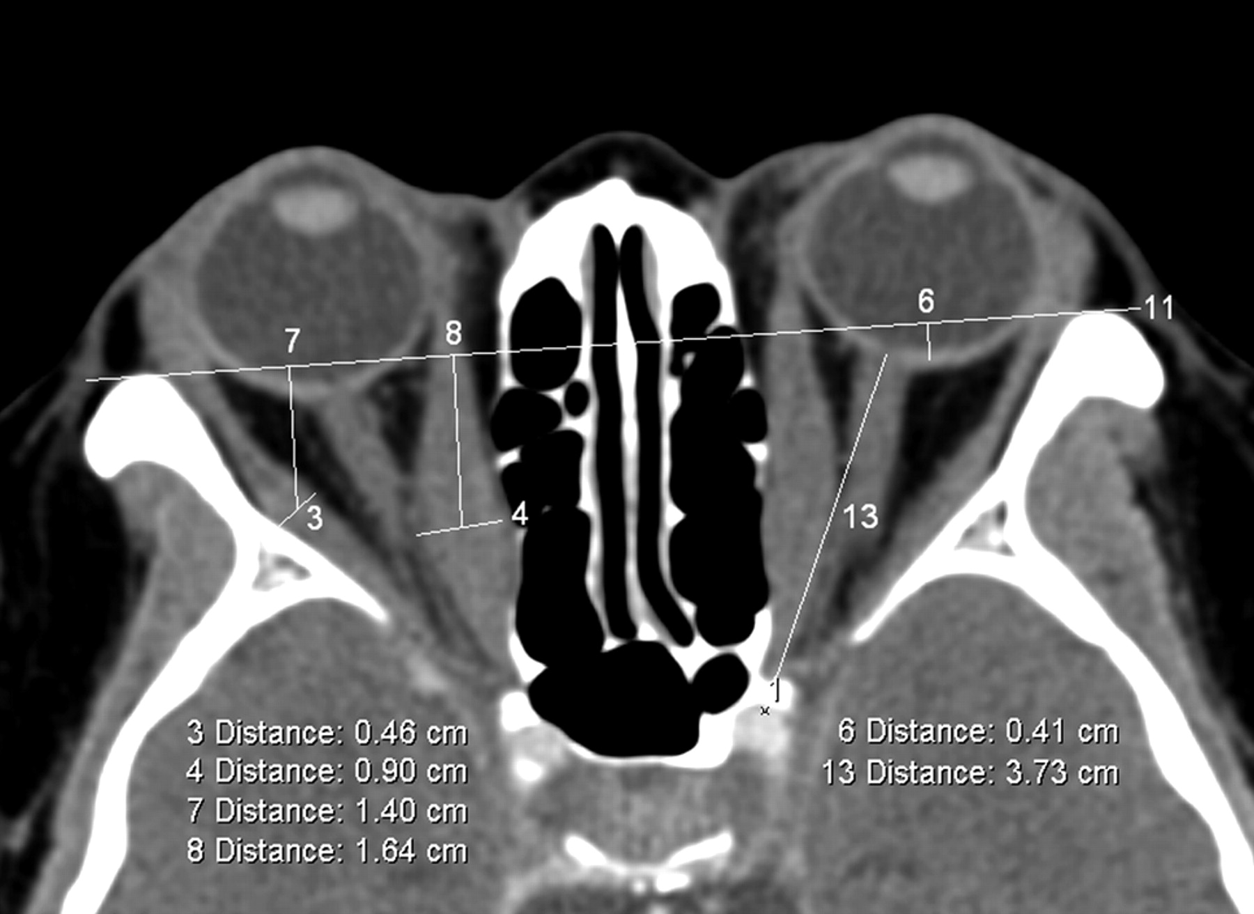
  • Fig 2.
    • Download figure
    • Open in new tab
    • Download powerpoint
    Fig 2.

    Axial section at midglobe level showing the interzygomatic line (labeled 11) and maximum horizontal diameters of the right medial and lateral rectus muscles (measurements labeled 4 and 3, respectively). The distance from the midpoint of the maximum muscular diameter of the medial (measurement 8) and lateral rectus muscles (measurement 7) to the interzygomatic line is also recorded. Proptosis of the left globe relative to the interzygomatic line is labeled as measurement 6. Left optic nerve stretch (labeled 13) is measured from the retrobulbar optic nerve to the orbital apex point (labeled 1).
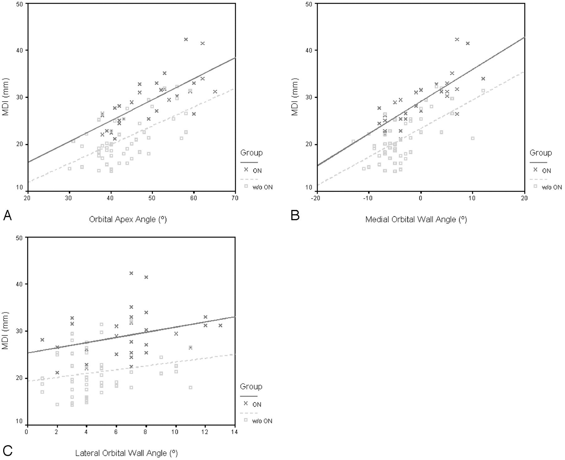
  • Fig 3.
    • Download figure
    • Open in new tab
    • Download powerpoint
    Fig 3.

    Scatterplots of the MDI and orbital apex angle (A), MDI and medial wall angle (B), and MDI and lateral wall angle (C) in patients with and without ON. Greater muscular enlargement is accompanied by wider orbital angles with or without (w/o) ON. For identical MDI and orbital angles, the orbital angles are narrower and MDI greater in patients with ON, respectively. Within the borderline MDI range of 22–30 mm in B, 15 of 26 patients with zero or negative medial angles (57.7%) had ON (P = .07), compared with 1 of 6 patients with positive medial angles (16.7%).

  • Fig 4.
    • Download figure
    • Open in new tab
    • Download powerpoint
    Fig 4.

    Scatterplot depicting the relationship between lengths of lateral orbital wall and orbital rim angles in patients with and without (w/o) ON.

Tables

  • Figures
    • View popup
    Table 1:

    Demographics, quantitative CT measurements, and categoric variables in patients with Graves ophthalmopathy, with and without ON*

    Graves OphthalmopathyONWithout ONP Value
    Demographics
        No. orbits3250
        Age55.9 ± 11.5 (39–78)44.7 ± 11.0 (28–65)
        Sex (male, female)14 M, 18 F20 M, 30 F
    Bony confines
        Bony orbital angle (degrees)
            Orbital rim42.4 ± 2.5 (39–49)42.2 ± 2.4 (37–51)<.727
            Medial wall-0.1 ± 5.7 (−8–12)−3.6 ± 5.2 (−13–12)<.006
            Lateral wall6.7 ± 3.0 (1–13)4.7 ± 2.5 (1–11)<.002
            Total angular change6.6 ± 7.5 (−3–20)1.1 ± 6.5 (−8–17)<.001
            Orbital apex49.0 ± 8.2 (38–65)43.3 ± 7.4 (30–59)<.002
        Length of lateral wall (mm)44.1 ± 2.9 (35.7–48.9)42.5 ± 3.2 (33.7–47.5)<.022
    Extraoccular muscles (mm)
        Maximum muscle diameter
            Medial rectus7.2 ± 1.8 (3.8–10.5)4.8 ± 1.8 (2.6–10.0)<.0005
            Lateral rectus4.9 ± 1.5 (2.3–8.2)3.8 ± 0.9 (2.1–6.4)<.0005
            Superior muscle group6.5 ± 1.8 (3.6–9.5)4.9 ± 1.7 (2.6–8.7)<.0005
            Inferior rectus7.1 ± 2.0 (3.2–12.8)5.0 ± 1.6 (2.6–9.5)<.0005
            Superior oblique3.0 ± 0.7 (1.8–4.5)2.8 ± 0.7 (1.8–4.4).212
            MDI29.0 ± 5.2 (21.1–42.3)21.2 ± 4.8 (14.3–32.3)<.0005
        Medial rectus bulk from IZ line14.0 ± 3.9 (5.4–21.0)12.3 ± 3.4 (6.0–21.2).049
        Lateral rectus bulk from IZ line16.0 ± 3.0 (10.8–21.2)14.8 ± 2.8 (10.7–21.1).084
    Neurovascular structures
        Proptosis from IZ line2.6 ± 2.7 (−3.5–7.0)3.5 ± 4.0 (−4.6- 12.7).200
        Optic nerve stretch40.8 ± 3.2 (35.0–47.9)39.7 ± 4.1 (30.7–46.6).243
        Optic nerve sheath
            Retrobulbar5.7 ± 0.7 (4.2–7.2)5.8 ± 0.9 (4.0–8.1).464
            Waist3.7 ± 0.6 (2.4–5.2)3.6 ± 0.7 (1.6–5.5).703
        Superior ophthalmic vein1.9 ± 0.4 (1.2–2.7)1.8 ± 0.4 (1.2–2.9).273
    Apical crowding3 (1–3)1 (0–3)<.0005†
    Presence of intracranial fat prolapse11 (34.4%)13 (26.0%).416‡
    • Note:—IZ line indicates interzygomatic line; ON, optic neuropathy; MDI, muscle diameter index.

    • * Descriptive statistics presented are mean ± SD (range) and count (%) for the presence of intracranial fat prolapse. All mean quantitative CT measurements between the 2 groups are compared using the Student 2-tailed t test. Statistical significance is defined at P < .05.

    • † The categoric variables are compared using the Mann-Whitney U test. Statistical significance is defined at P < .05.

    • ‡ The categoric variables are compared using the χ2 square test. Statistical significance is defined at P < .05.

    • View popup
    Table 2:

    Normal bony orbital measurements (24 orbits)

    Bony StructureMeasurement
    Bony orbital angle (degrees)
        Orbital rim42.5 ± 2.5 (39–47)
        Medial wall−4.6 ± 1.9 (−7 to −1)
        Lateral wall4.9 ± 2.5 (1–10)
        Total angular change0.3 ± 2.9 (−5–5)
        Orbital apex42.8 ± 4.2 (35–52)
    Length of lateral wall (mm)42.7 ± 1.9 (39.4–45.9)
    • View popup
    Table 3:

    Sensitivity, specificity, and positive and negative predictive values of using MDI alone versus MDI in combination with medial and lateral orbital angles in predicting ON in Graves ophthalmopathy

    PercentageMDI AloneMDI and Medial and Lateral Wall Angles
    Sensitivity60.0 (18/30)73.3 (22/30)
    Specificity86.0 (43/50)90.0 (45/50)
    Positive predictive value72.0 (18/25)81.5 (22/27)
    Negative predictive value78.2 (43/55)84.9 (45/53)
PreviousNext
Back to top

In this issue

American Journal of Neuroradiology: 30 (3)
American Journal of Neuroradiology
Vol. 30, Issue 3
March 2009
  • Table of Contents
  • Index by author
Advertisement
Print
Download PDF
Email Article

Thank you for your interest in spreading the word on American Journal of Neuroradiology.

NOTE: We only request your email address so that the person you are recommending the page to knows that you wanted them to see it, and that it is not junk mail. We do not capture any email address.

Enter multiple addresses on separate lines or separate them with commas.
Graves Ophthalmopathy: The Bony Orbit in Optic Neuropathy, Its Apical Angular Capacity, and Impact on Prediction of Risk
(Your Name) has sent you a message from American Journal of Neuroradiology
(Your Name) thought you would like to see the American Journal of Neuroradiology web site.
CAPTCHA
This question is for testing whether or not you are a human visitor and to prevent automated spam submissions.
Cite this article
L.-L. Chan, H.-E. Tan, S. Fook-Chong, T.-H. Teo, L.-H. Lim, L.-L. Seah
Graves Ophthalmopathy: The Bony Orbit in Optic Neuropathy, Its Apical Angular Capacity, and Impact on Prediction of Risk
American Journal of Neuroradiology Mar 2009, 30 (3) 597-602; DOI: 10.3174/ajnr.A1413

Citation Manager Formats

  • BibTeX
  • Bookends
  • EasyBib
  • EndNote (tagged)
  • EndNote 8 (xml)
  • Medlars
  • Mendeley
  • Papers
  • RefWorks Tagged
  • Ref Manager
  • RIS
  • Zotero
0 Responses
Respond to this article
Share
Bookmark this article
Graves Ophthalmopathy: The Bony Orbit in Optic Neuropathy, Its Apical Angular Capacity, and Impact on Prediction of Risk
L.-L. Chan, H.-E. Tan, S. Fook-Chong, T.-H. Teo, L.-H. Lim, L.-L. Seah
American Journal of Neuroradiology Mar 2009, 30 (3) 597-602; DOI: 10.3174/ajnr.A1413
del.icio.us logo Twitter logo Facebook logo Mendeley logo
  • Tweet Widget
  • Facebook Like
  • Google Plus One
Purchase

Jump to section

  • Article
    • Abstract
    • Materials and Methods
    • Results
    • Discussion
    • References
  • Figures & Data
  • Info & Metrics
  • Responses
  • References
  • PDF

Related Articles

  • No related articles found.
  • PubMed
  • Google Scholar

Cited By...

  • Thyroid eye disease: a Southeast Asian experience
  • Quantification of Orbital Apex Crowding for Screening of Dysthyroid Optic Neuropathy Using Multidetector CT
  • Crossref
  • Google Scholar

This article has not yet been cited by articles in journals that are participating in Crossref Cited-by Linking.

More in this TOC Section

  • Hydrops Herniation into the Semicircular Canals
  • ASL Sensitivity for Head and Neck Paraganglioma
  • Post SRS Peritumoral Hyperintense Signal of VSs
Show more Head and Neck Imaging

Similar Articles

Advertisement

Indexed Content

  • Current Issue
  • Accepted Manuscripts
  • Article Preview
  • Past Issues
  • Editorials
  • Editor's Choice
  • Fellows' Journal Club
  • Letters to the Editor
  • Video Articles

Cases

  • Case Collection
  • Archive - Case of the Week
  • Archive - Case of the Month
  • Archive - Classic Case

More from AJNR

  • Trainee Corner
  • Imaging Protocols
  • MRI Safety Corner
  • Book Reviews

Multimedia

  • AJNR Podcasts
  • AJNR Scantastics

Resources

  • Turnaround Time
  • Submit a Manuscript
  • Submit a Video Article
  • Submit an eLetter to the Editor/Response
  • Manuscript Submission Guidelines
  • Statistical Tips
  • Fast Publishing of Accepted Manuscripts
  • Graphical Abstract Preparation
  • Imaging Protocol Submission
  • Evidence-Based Medicine Level Guide
  • Publishing Checklists
  • Author Policies
  • Become a Reviewer/Academy of Reviewers
  • News and Updates

About Us

  • About AJNR
  • Editorial Board
  • Editorial Board Alumni
  • Alerts
  • Permissions
  • Not an AJNR Subscriber? Join Now
  • Advertise with Us
  • Librarian Resources
  • Feedback
  • Terms and Conditions
  • AJNR Editorial Board Alumni

American Society of Neuroradiology

  • Not an ASNR Member? Join Now

© 2025 by the American Society of Neuroradiology All rights, including for text and data mining, AI training, and similar technologies, are reserved.
Print ISSN: 0195-6108 Online ISSN: 1936-959X

Powered by HighWire