Advertisement

Main menu

  • Home
  • Content
    • Current Issue
    • Accepted Manuscripts
    • Article Preview
    • Past Issue Archive
    • Video Articles
    • AJNR Case Collection
    • Case of the Week Archive
    • Case of the Month Archive
    • Classic Case Archive
  • Special Collections
    • AJNR Awards
    • Low-Field MRI
    • Alzheimer Disease
    • ASNR Foundation Special Collection
    • Photon-Counting CT
    • View All
  • Multimedia
    • AJNR Podcasts
    • AJNR SCANtastic
    • Trainee Corner
    • MRI Safety Corner
    • Imaging Protocols
  • For Authors
    • Submit a Manuscript
    • Submit a Video Article
    • Submit an eLetter to the Editor/Response
    • Manuscript Submission Guidelines
    • Statistical Tips
    • Fast Publishing of Accepted Manuscripts
    • Graphical Abstract Preparation
    • Imaging Protocol Submission
    • Author Policies
  • About Us
    • About AJNR
    • Editorial Board
    • Editorial Board Alumni
  • More
    • Become a Reviewer/Academy of Reviewers
    • Subscribers
    • Permissions
    • Alerts
    • Feedback
    • Advertisers
    • ASNR Home

User menu

  • Alerts
  • Log in

Search

  • Advanced search
American Journal of Neuroradiology

American Journal of Neuroradiology

ASHNR American Society of Functional Neuroradiology ASHNR American Society of Pediatric Neuroradiology ASSR
  • Alerts
  • Log in

Advanced Search

  • Home
  • Content
    • Current Issue
    • Accepted Manuscripts
    • Article Preview
    • Past Issue Archive
    • Video Articles
    • AJNR Case Collection
    • Case of the Week Archive
    • Case of the Month Archive
    • Classic Case Archive
  • Special Collections
    • AJNR Awards
    • Low-Field MRI
    • Alzheimer Disease
    • ASNR Foundation Special Collection
    • Photon-Counting CT
    • View All
  • Multimedia
    • AJNR Podcasts
    • AJNR SCANtastic
    • Trainee Corner
    • MRI Safety Corner
    • Imaging Protocols
  • For Authors
    • Submit a Manuscript
    • Submit a Video Article
    • Submit an eLetter to the Editor/Response
    • Manuscript Submission Guidelines
    • Statistical Tips
    • Fast Publishing of Accepted Manuscripts
    • Graphical Abstract Preparation
    • Imaging Protocol Submission
    • Author Policies
  • About Us
    • About AJNR
    • Editorial Board
    • Editorial Board Alumni
  • More
    • Become a Reviewer/Academy of Reviewers
    • Subscribers
    • Permissions
    • Alerts
    • Feedback
    • Advertisers
    • ASNR Home
  • Follow AJNR on Twitter
  • Visit AJNR on Facebook
  • Follow AJNR on Instagram
  • Join AJNR on LinkedIn
  • RSS Feeds

AJNR Awards, New Junior Editors, and more. Read the latest AJNR updates

Research ArticleBRAIN

Topographic Analysis of the Inferior Parietal Lobule in High-Resolution 3D MR Imaging

I. Kiriyama, H. Miki, K. Kikuchi, S. Ohue, S. Matsuda and T. Mochizuki
American Journal of Neuroradiology March 2009, 30 (3) 520-524; DOI: https://doi.org/10.3174/ajnr.A1417
I. Kiriyama
  • Find this author on Google Scholar
  • Find this author on PubMed
  • Search for this author on this site
H. Miki
  • Find this author on Google Scholar
  • Find this author on PubMed
  • Search for this author on this site
K. Kikuchi
  • Find this author on Google Scholar
  • Find this author on PubMed
  • Search for this author on this site
S. Ohue
  • Find this author on Google Scholar
  • Find this author on PubMed
  • Search for this author on this site
S. Matsuda
  • Find this author on Google Scholar
  • Find this author on PubMed
  • Search for this author on this site
T. Mochizuki
  • Find this author on Google Scholar
  • Find this author on PubMed
  • Search for this author on this site
  • Article
  • Figures & Data
  • Info & Metrics
  • Responses
  • References
  • PDF
Loading

Article Figures & Data

Figures

  • Tables
  • Fig 1.
    • Download figure
    • Open in new tab
    • Download powerpoint
    Fig 1.

    VR image of the left hemisphere of a healthy volunteer. The left image and the right image are the same. The IPL locates posterior to the postCG and is the inferior component of the parietal lobe. The postCS and the IPS form the border of the IPL. The postCS connects with the anterior end of the IPS (black arrowhead) and does not connect with the posterior ascending ramus of the Sy (white arrowhead) in this case. The asterisk indicates the sulcus intermedius primus.
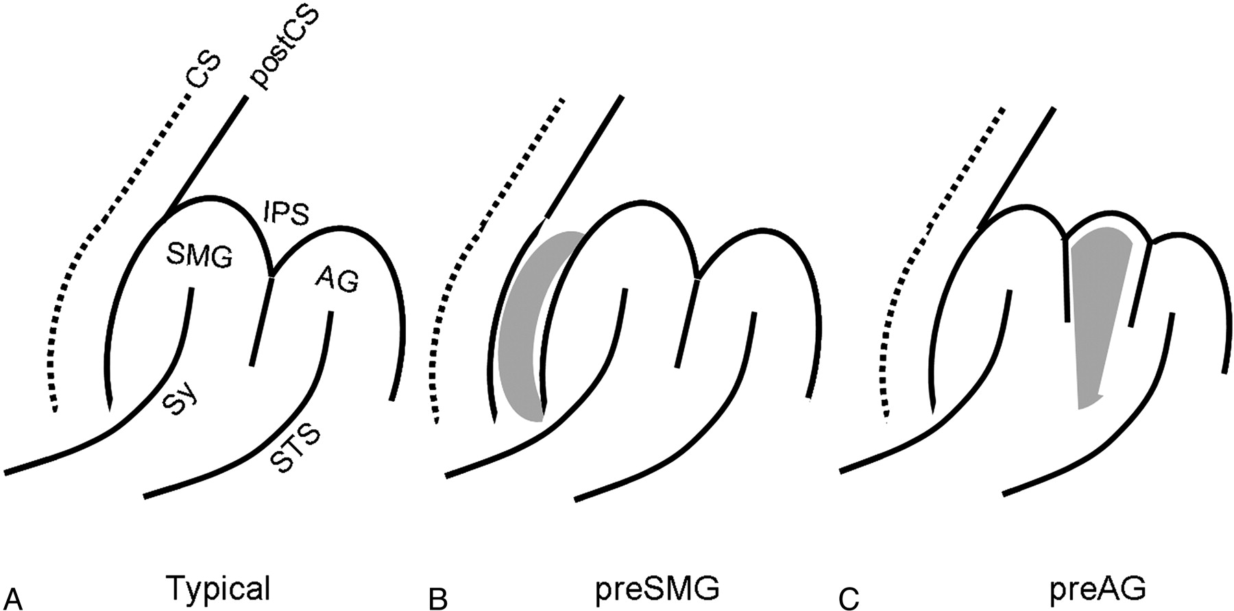
  • Fig 2.
    • Download figure
    • Open in new tab
    • Download powerpoint
    Fig 2.

    Patterns of the IPL. A, Typical pattern. There are no additional gyri. B, PreSMG pattern. The additional gyri (gray tissue) intercalated between the postCS and the SMG. C, PreAG. The additional gyri (gray tissue) positioned between the SMG and the AG.
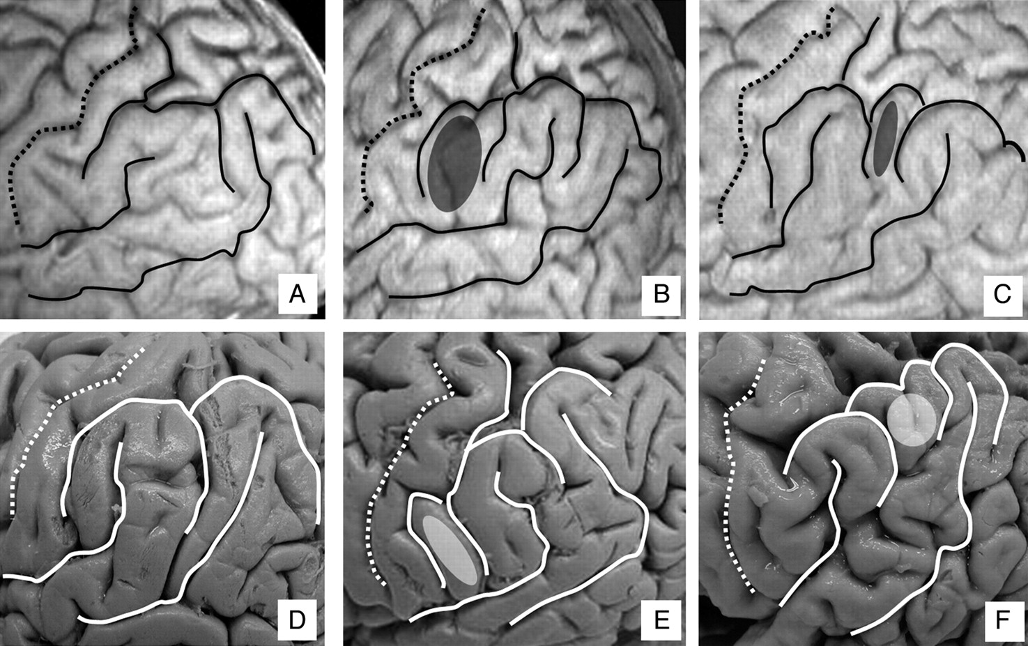
  • Fig 3.
    • Download figure
    • Open in new tab
    • Download powerpoint
    Fig 3.

    The IPL on VR images and with specimens. A and D, Typical pattern. B and E, PreSMG pattern. C and F, PreAG pattern. A through C indicate VR images; D through F, specimens. The additional gyri are colored.

  • Fig 4.
    • Download figure
    • Open in new tab
    • Download powerpoint
    Fig 4.

    Schematic (left), VR images (middle), and specimens (right) of patterns of the preSMG plus preAG pattern and the SMG plus AG pattern. A-C, PreSMG plus preAG pattern. The additional gyri (gray tissue) positioned both anterior and posterior to the SMG. D-F, SMG plus AG pattern. There is no clear boundary between the SMG and the AG.

Tables

  • Figures
    • View popup
    Table 1:

    Sulcal continuity of the inferior parietal lobule in 40 healthy brains studied with VR images and in 50 specimens studied anatomically (Ono et al.8)

    Right Hemisphere (%)Left Hemisphere (%)
    VROno et al.8 (specimens)VROno et al.8 (specimens)
    postCSContinuous47.5444048
    Discontinuous (2 segments)42.54057.548
    Discontinuous (≥3 segments)10162.54
    IPSContinuous65287072
    Discontinuous (2 segments)32.5682528
    Discontinuous (≥3 segments)2.5450
    • Note:—postCS indicates postcentral sulcus; VR, volume rendering; IPS, intraparietal sulcus.

    • View popup
    Table 2:

    Sulcal connections of the inferior parietal lobule in 40 healthy brains studied with VR images and in 50 specimens studied anatomically (Ono et al8)

    postCS Connected withRight Hemisphere (%)Left Hemisphere (%)
    VROno et al8(specimens)VROno et al8(specimens)
    IPS72.5647572
    Sy52.5683048
    • Note:—IPS indicates the anterior end of the intraparietal sulcus; Sy, Sylvian fissure (the posterior ascending ramus of the Sylvian fissure).

    • View popup
    Table 3:

    Frequency of gyral patterns of inferior parietal lobule in 40 healthy brains studied with VR images, in 20 specimens studied anatomically, and in 50 specimens studied anatomically by Naidich et al10

    Right Hemisphere (%)Left Hemisphere (%)
    VRSpecimensNaidich et al10 (specimens)VRSpecimensNaidich et al10 (specimens)
    Typical42.54548352028
    preSMG2.55417.53516
    preAG17.520160528
    Others37.53032*47.54528*
        (preSMG + preAG)(0)(15)−(2.5)(0)−
        (SMG + AG)(12.5)(10)−(37.5)(40)−
        (Pattern not identifiable)(25)(5)−(7.5)(5)−
    • Note:—preSMG indicates additional gyri intercalated between the postcentral sulcus (postCS) and the supramarginal gyrus (SMG); preAG, additional gyri positioned between the SMG and the angular gyrus (AG).

    • * Others were described as pattern not identifiable in the report by Naidich et al10.

PreviousNext
Back to top

In this issue

American Journal of Neuroradiology: 30 (3)
American Journal of Neuroradiology
Vol. 30, Issue 3
March 2009
  • Table of Contents
  • Index by author
Advertisement
Print
Download PDF
Email Article

Thank you for your interest in spreading the word on American Journal of Neuroradiology.

NOTE: We only request your email address so that the person you are recommending the page to knows that you wanted them to see it, and that it is not junk mail. We do not capture any email address.

Enter multiple addresses on separate lines or separate them with commas.
Topographic Analysis of the Inferior Parietal Lobule in High-Resolution 3D MR Imaging
(Your Name) has sent you a message from American Journal of Neuroradiology
(Your Name) thought you would like to see the American Journal of Neuroradiology web site.
CAPTCHA
This question is for testing whether or not you are a human visitor and to prevent automated spam submissions.
Cite this article
I. Kiriyama, H. Miki, K. Kikuchi, S. Ohue, S. Matsuda, T. Mochizuki
Topographic Analysis of the Inferior Parietal Lobule in High-Resolution 3D MR Imaging
American Journal of Neuroradiology Mar 2009, 30 (3) 520-524; DOI: 10.3174/ajnr.A1417

Citation Manager Formats

  • BibTeX
  • Bookends
  • EasyBib
  • EndNote (tagged)
  • EndNote 8 (xml)
  • Medlars
  • Mendeley
  • Papers
  • RefWorks Tagged
  • Ref Manager
  • RIS
  • Zotero
0 Responses
Respond to this article
Share
Bookmark this article
Topographic Analysis of the Inferior Parietal Lobule in High-Resolution 3D MR Imaging
I. Kiriyama, H. Miki, K. Kikuchi, S. Ohue, S. Matsuda, T. Mochizuki
American Journal of Neuroradiology Mar 2009, 30 (3) 520-524; DOI: 10.3174/ajnr.A1417
del.icio.us logo Twitter logo Facebook logo Mendeley logo
  • Tweet Widget
  • Facebook Like
  • Google Plus One
Purchase

Jump to section

  • Article
    • Abstract
    • Anatomy of the IPL8,10,16
    • Materials and Methods
    • Results
    • Discussion
    • Conclusions
    • References
  • Figures & Data
  • Info & Metrics
  • Responses
  • References
  • PDF

Related Articles

  • No related articles found.
  • PubMed
  • Google Scholar

Cited By...

  • Functional Subdivisions in the Left Angular Gyrus Where the Semantic System Meets and Diverges from the Default Network
  • Crossref (11)
  • Google Scholar

This article has been cited by the following articles in journals that are participating in Crossref Cited-by Linking.

  • Functional Subdivisions in the Left Angular Gyrus Where the Semantic System Meets and Diverges from the Default Network
    Mohamed L. Seghier, Elizabeth Fagan, Cathy J. Price
    The Journal of Neuroscience 2010 30 50
  • Alterations in regional homogeneity of resting-state brain activity in internet gaming addicts
    Guangheng Dong, Jie Huang, Xiaoxia Du
    Behavioral and Brain Functions 2012 8 1
  • On the Origin of Individual Functional Connectivity Variability: The Role of White Matter Architecture
    Maxime Chamberland, Gabriel Girard, Michaël Bernier, David Fortin, Maxime Descoteaux, Kevin Whittingstall
    Brain Connectivity 2017 7 8
  • The structural connectivity of the human angular gyrus as revealed by microdissection and diffusion tractography
    Laurent Petit, Kariem Mahdy Ali, François Rheault, Arnaud Boré, Sandrine Cremona, Francesco Corsini, Alessandro De Benedictis, Maxime Descoteaux, Silvio Sarubbo
    Brain Structure and Function 2023 228 1
  • Cortical and Subcortical Anatomy of the Parietal Lobe From the Neurosurgical Perspective
    Tomasz Andrzej Dziedzic, Aleksandra Bala, Andrzej Marchel
    Frontiers in Neurology 2021 12
  • The connectivity-based parcellation of the angular gyrus: fiber dissection and MR tractography study
    Fatih Yakar, Pınar Çeltikçi, Yücel Doğruel, Emrah Egemen, Abuzer Güngör
    Brain Structure and Function 2023 228 1
  • Island of Reil (Insula) in the Human Brain
    Igor Lima Maldonado, Ilyess Zemmoura, Christophe Destrieux
    2018
  • Brain Graph Interface
    Arno Klein
    Research Ideas and Outcomes 2016 2
  • Cognitive impairment assessed by static and dynamic changes of spontaneous brain activity during end stage renal disease patients on early hemodialysis
    Yunfan Wu, Rujin Li, Guihua Jiang, Ning Yang, Mengchen Liu, Yanying Chen, Zichao Chen, Kanghui Yu, Yi Yin, Shoujun Xu, Bin Xia, Shandong Meng
    Frontiers in Neurology 2025 16
  • Inferior parietal lobule gyrations in refractory epilepsy
    Javeria Nooraine, Seetharam Raghavendra
    Epileptic Disorders 2021 23 3

More in this TOC Section

  • Enhanced Axonal Metabolism during Early Natalizumab Treatment in Relapsing-Remitting Multiple Sclerosis
  • SWI or T2*: Which MRI Sequence to Use in the Detection of Cerebral Microbleeds? The Karolinska Imaging Dementia Study
  • Progression of Microstructural Damage in Spinocerebellar Ataxia Type 2: A Longitudinal DTI Study
Show more BRAIN

Similar Articles

Advertisement

Indexed Content

  • Current Issue
  • Accepted Manuscripts
  • Article Preview
  • Past Issues
  • Editorials
  • Editor's Choice
  • Fellows' Journal Club
  • Letters to the Editor
  • Video Articles

Cases

  • Case Collection
  • Archive - Case of the Week
  • Archive - Case of the Month
  • Archive - Classic Case

More from AJNR

  • Trainee Corner
  • Imaging Protocols
  • MRI Safety Corner
  • Book Reviews

Multimedia

  • AJNR Podcasts
  • AJNR Scantastics

Resources

  • Turnaround Time
  • Submit a Manuscript
  • Submit a Video Article
  • Submit an eLetter to the Editor/Response
  • Manuscript Submission Guidelines
  • Statistical Tips
  • Fast Publishing of Accepted Manuscripts
  • Graphical Abstract Preparation
  • Imaging Protocol Submission
  • Evidence-Based Medicine Level Guide
  • Publishing Checklists
  • Author Policies
  • Become a Reviewer/Academy of Reviewers
  • News and Updates

About Us

  • About AJNR
  • Editorial Board
  • Editorial Board Alumni
  • Alerts
  • Permissions
  • Not an AJNR Subscriber? Join Now
  • Advertise with Us
  • Librarian Resources
  • Feedback
  • Terms and Conditions
  • AJNR Editorial Board Alumni

American Society of Neuroradiology

  • Not an ASNR Member? Join Now

© 2025 by the American Society of Neuroradiology All rights, including for text and data mining, AI training, and similar technologies, are reserved.
Print ISSN: 0195-6108 Online ISSN: 1936-959X

Powered by HighWire