
Article Figures & Data
Figures
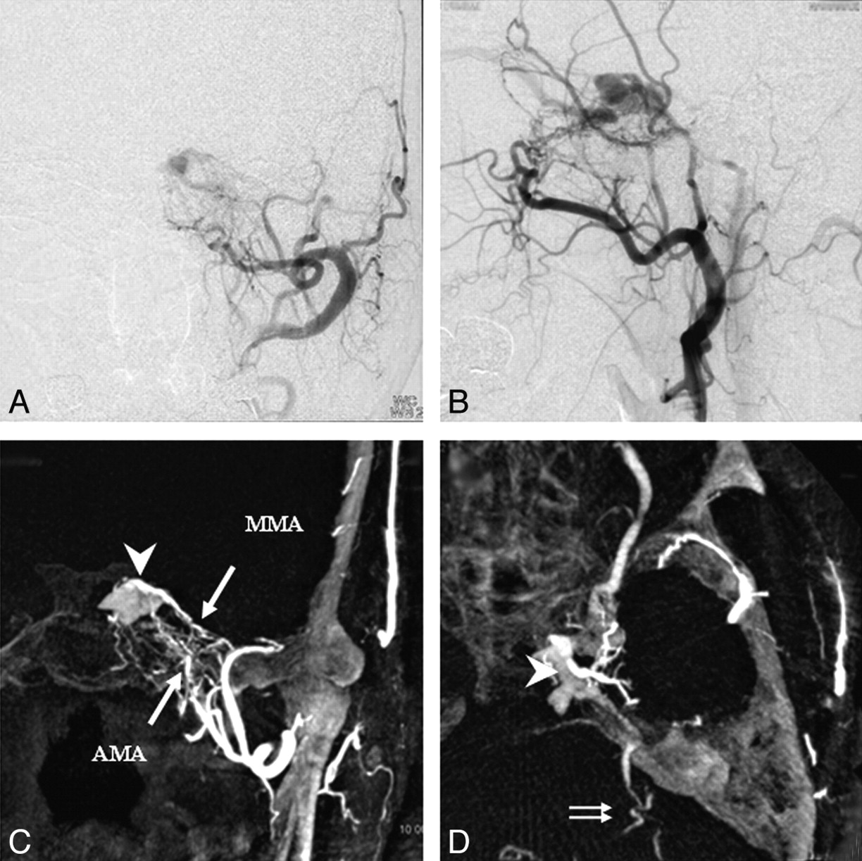
- Fig 1.
Case 4, a 68-year-old man with a left CS DAVF. A and B, 2D DSA shows the left CS DAVF draining into the SOV, the SPS, and the IPS. C (coronal image) and D (axial image), DynaCT shows a DAVF supplied by the AMA and the MMA (arrows). The fistulous point is located in the posterosuperior compartment of the left CS (arrowhead), and the left SPS drains to the left petrosal vein with cortical venous reflux (double arrows).
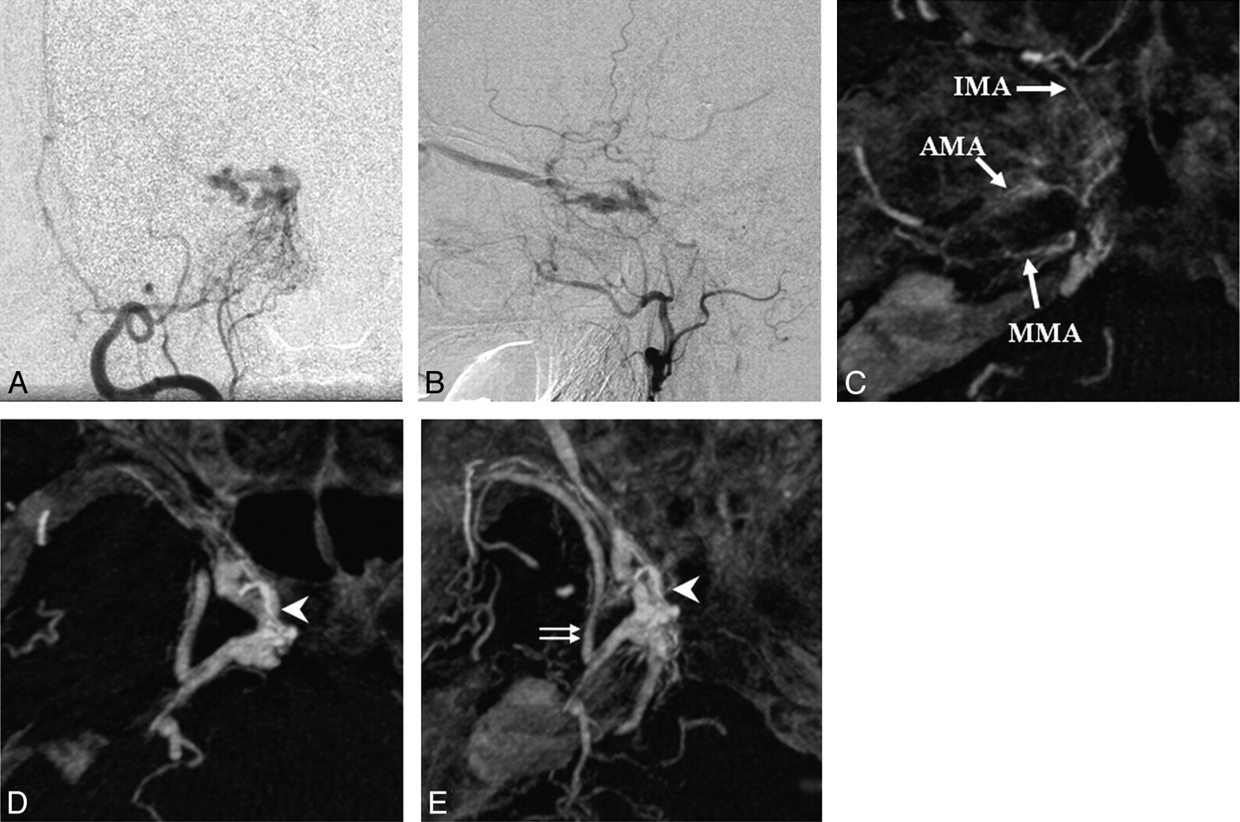
- Fig 2.
Case 7, a 73-year-old woman with a right CS DAVF. A and B, 2D DSA shows the right CS DAVF draining into the SOV, the SPS, and the IPS. C–E (axial images), DynaCT shows a DAVF supplied by the IMA, the AMA, and the MMA (arrows). The fistula is located in the medial compartment of the right CS (arrowhead), and the right SPS is connected to the superficial middle cerebral vein (ie, the sphenopetrosal sinus; double arrows).
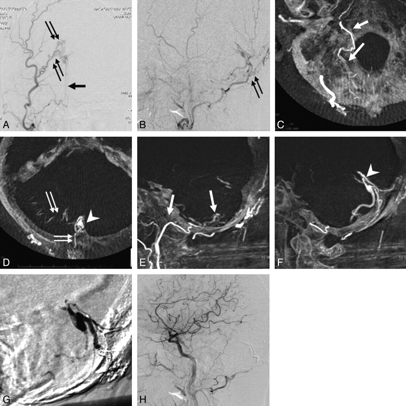
- Fig 3.
Case 10, a 58-year-old man with a tentorial DAVF. A and B, 2D DSA shows a DAVF supplied by the APA and the OA. C and D (axial images) and E and F (sagittal images), DynaCT shows a DAVF supplied by the APA (arrow) and OA (double arrows) draining into the inferior hemispheric vein. The fistula is located in the medial tentorium (arrowhead). G, Injection of n-butyl-2-cyanoacrylate into the fistula from the distal APA. H, 2D DSA shows a complete obliteration of the DAVF.


In this issue


Jump to section
Related Articles
Cited By...
- Diagnostic accuracy of three-dimensional-rotational angiography and heavily T2-weighted volumetric magnetic resonance fusion imaging for the diagnosis of spinal arteriovenous shunts
- Computational Modeling of Venous Sinus Stenosis in Idiopathic Intracranial Hypertension
- Artery of the Superior Orbital Fissure: An Undescribed Branch from the Pterygopalatine Segment of the Maxillary Artery to the Orbital Apex Connecting with the Anteromedial Branch of the Inferolateral Trunk
- Adjunctive value of intra-arterial cone beam CT angiography relative to DSA in the evaluation of cranial and spinal arteriovenous fistulas
- Angioarchitecture of Transverse-Sigmoid Sinus Dural Arteriovenous Fistulas: Evaluation of Shunted Pouches by Multiplanar Reformatted Images of Rotational Angiography
- Turn-Back Embolization Technique for Effective Transvenous Embolization of Dural Arteriovenous Fistulas
- Simultaneous Arteriovenous Shunting and Venous Congestion Identification in Dural Arteriovenous Fistulas Using Susceptibility-Weighted Imaging: Initial Experience
- CT Angiography as a Screening Tool for Dural Arteriovenous Fistula in Patients with Pulsatile Tinnitus: Feasibility and Test Characteristics
- Use of Angiographic CT Imaging in the Cardiac Catheterization Laboratory for Congenital Heart Disease
- 3D C-Arm Conebeam CT Angiography as an Adjunct in the Precise Anatomic Characterization of Spinal Dural Arteriovenous Fistulas