Advertisement

Main menu

  • Home
  • Content
    • Current Issue
    • Accepted Manuscripts
    • Article Preview
    • Past Issue Archive
    • Video Articles
    • AJNR Case Collection
    • Case of the Week Archive
    • Case of the Month Archive
    • Classic Case Archive
  • Special Collections
    • Advancing NeuroMRI with High-Relaxivity Contrast Agents
    • AJNR Awards
    • Low-Field MRI
    • Alzheimer Disease
    • ASNR Foundation Special Collection
    • Photon-Counting CT
    • View All
  • Multimedia
    • AJNR Podcasts
    • AJNR SCANtastic
    • Trainee Corner
    • MRI Safety Corner
    • Imaging Protocols
  • For Authors
    • Submit a Manuscript
    • Submit a Video Article
    • Submit an eLetter to the Editor/Response
    • Manuscript Submission Guidelines
    • Statistical Tips
    • Fast Publishing of Accepted Manuscripts
    • Graphical Abstract Preparation
    • Imaging Protocol Submission
    • Author Policies
  • About Us
    • About AJNR
    • Editorial Board
    • Editorial Board Alumni
  • More
    • Become a Reviewer/Academy of Reviewers
    • Subscribers
    • Permissions
    • Alerts
    • Feedback
    • Advertisers
    • ASNR Home

User menu

  • Alerts
  • Log in

Search

  • Advanced search
American Journal of Neuroradiology

American Journal of Neuroradiology

ASHNR American Society of Functional Neuroradiology ASHNR American Society of Pediatric Neuroradiology ASSR
  • Alerts
  • Log in

Advanced Search

  • Home
  • Content
    • Current Issue
    • Accepted Manuscripts
    • Article Preview
    • Past Issue Archive
    • Video Articles
    • AJNR Case Collection
    • Case of the Week Archive
    • Case of the Month Archive
    • Classic Case Archive
  • Special Collections
    • Advancing NeuroMRI with High-Relaxivity Contrast Agents
    • AJNR Awards
    • Low-Field MRI
    • Alzheimer Disease
    • ASNR Foundation Special Collection
    • Photon-Counting CT
    • View All
  • Multimedia
    • AJNR Podcasts
    • AJNR SCANtastic
    • Trainee Corner
    • MRI Safety Corner
    • Imaging Protocols
  • For Authors
    • Submit a Manuscript
    • Submit a Video Article
    • Submit an eLetter to the Editor/Response
    • Manuscript Submission Guidelines
    • Statistical Tips
    • Fast Publishing of Accepted Manuscripts
    • Graphical Abstract Preparation
    • Imaging Protocol Submission
    • Author Policies
  • About Us
    • About AJNR
    • Editorial Board
    • Editorial Board Alumni
  • More
    • Become a Reviewer/Academy of Reviewers
    • Subscribers
    • Permissions
    • Alerts
    • Feedback
    • Advertisers
    • ASNR Home
  • Follow AJNR on Twitter
  • Visit AJNR on Facebook
  • Follow AJNR on Instagram
  • Join AJNR on LinkedIn
  • RSS Feeds

AJNR Awards, New Junior Editors, and more. Read the latest AJNR updates


Improved Turnaround Times | Median time to first decision: 12 days

Research ArticleNeurointervention

Volume and Composition of Emboli in Neuroprotected Stenting of the Carotid Artery

P. Piñero, A. González, E. Martínez, A. Mayol, E. Rafel, J.R. González-Marcos, F. Moniche, A. Cayuela and A. Gil-Peralta
American Journal of Neuroradiology March 2009, 30 (3) 473-478; DOI: https://doi.org/10.3174/ajnr.A1407
P. Piñero
  • Find this author on Google Scholar
  • Find this author on PubMed
  • Search for this author on this site
A. González
  • Find this author on Google Scholar
  • Find this author on PubMed
  • Search for this author on this site
E. Martínez
  • Find this author on Google Scholar
  • Find this author on PubMed
  • Search for this author on this site
A. Mayol
  • Find this author on Google Scholar
  • Find this author on PubMed
  • Search for this author on this site
E. Rafel
  • Find this author on Google Scholar
  • Find this author on PubMed
  • Search for this author on this site
J.R. González-Marcos
  • Find this author on Google Scholar
  • Find this author on PubMed
  • Search for this author on this site
F. Moniche
  • Find this author on Google Scholar
  • Find this author on PubMed
  • Search for this author on this site
A. Cayuela
  • Find this author on Google Scholar
  • Find this author on PubMed
  • Search for this author on this site
A. Gil-Peralta
  • Find this author on Google Scholar
  • Find this author on PubMed
  • Search for this author on this site
  • Article
  • Figures & Data
  • Info & Metrics
  • Responses
  • References
  • PDF
Loading

Article Figures & Data

Figures

  • Tables
  • Fig 1.
    • Download figure
    • Open in new tab
    • Download powerpoint
    Fig 1.

    Volume analysis. Drops are shown with 10-, 5-, and 1-λ volumes. 1 λ is equivalent to 1 μL (0.001 mL) or 1 mm3. The measurement scale is in millimeters.
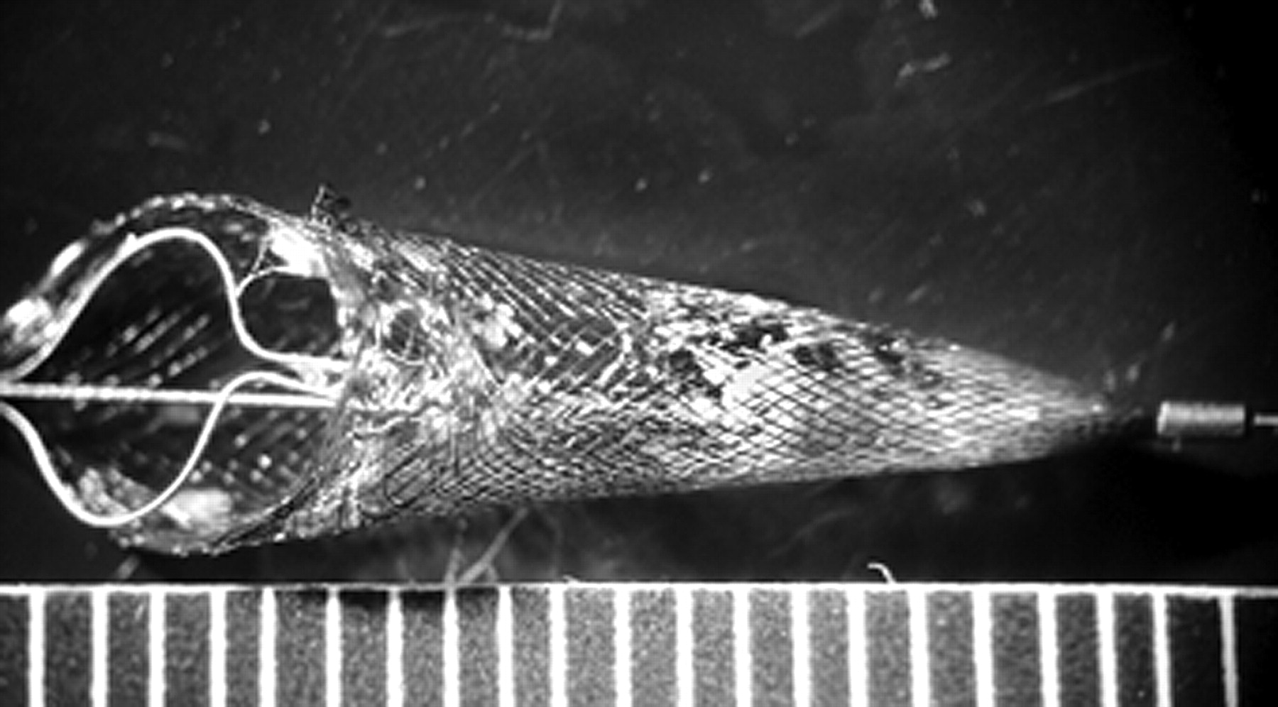
  • Fig 2.
    • Download figure
    • Open in new tab
    • Download powerpoint
    Fig 2.

    Photograph of a filter with volume content >10 λ before being cut open. The measurement scale is in millimeters.

  • Fig 3.
    • Download figure
    • Open in new tab
    • Download powerpoint
    Fig 3.

    Photographs of macroscopic views of fibrin and platelet aggregates on the outer surface of filters. A, Plugs of fibrinoplatelet material cover pores outside the filter. B, A cut-open filter shows large-volume particles inside.

  • Fig 4.
    • Download figure
    • Open in new tab
    • Download powerpoint
    Fig 4.

    Photomicrographs show ultrastructural findings. A, Small capillary. B, Endothelial cell. C, Foam cell with intracytoplasmic inclusions. D, Cholesterol crystals. E, Fragment of the vessel wall. F, Small calcification within the vessel wall. G, Collagen fibers. H, Smooth muscle cell (lead citrate and uranyl acetate, original magnification ×25,000).

Tables

  • Figures
    • View popup
    Table 1:

    Angiographic characteristics of stenosis

    CharacteristicNo. (%)
    Right ICA stenosis100 (49.8)
    Left ICA stenosis101 (50.2)
    % Stenosis (treated vessel)
        <95%144 (71.6)
        95%–98%40 (20)
        99%17 (8.45)
    % Contralateral stenosis
        <70%136 (67.5)
        70%–98%27 (13.4)
        99%4 (2)
        100%34 (17)
    Ulcerated plaque102 (50.7)
    Calcified plaque90 (45)
    Functioning AcomA159 (79.1)
    Functioning PcomA96 (45.7)
    Plaque size (cm)Mean, 1.79 cm (range, 0.1–2.4)
    Distance from carotid bulb (cm)Mean, 0.31 cm (range, 0–1.2)
    Intracranial lesion*23 (13)
    Dissection post-PTA†16 (8)
    Transient asymptomatic vasospasm59 (29.4)
    • Note:—PTA indicates percutaneous transluminal angioplasty; AcomA, anterior communicating artery; PcomA, posterior communicating artery; ICA, internal carotid artery.

    • * “Intracranial lesion” is defined as any aneurysm, stenosis, or vascular malformation discovered in the pre-CAS angiographic study.

    • † Dissection post-PTA indicates a subtle intimal irregularity shown in the angiographic post-PTA and pre-CAS examination, due to manipulation during PTA, with no clinical manifestation.

    • View popup
    Table 2:

    Anatomic study of the filter content (201 cases) after CAS

    ContentNo. (%)
    Particulate material recovered in the filter
        No84 (41.8)
        Yes117 (58.2)
    Volume of the particulate material (117 positive cases)
        ≤1 λ (0.001 mL = 1μl = 1 mm3)83 (71)
        >1 λ27 (23)
        >10 λ7 (6)
    • Note:—CAS indicates carotid angioplasty and stent placement.

    • View popup
    Table 3:

    Volume of the particulate material recovered in the distal filter and pathologic composition (117 positive cases)

    ≤1 λ (n = 83)>1 λ (n = 27)P
    No. (%)No. (%)
    Fibrin/platelet complex56 (33.5)22 (64).001
    Cell debris35 (21)24 (70.6)<.001
    Cholesterol clefts29 (17.4)21 (61.8)>.001
    Foam cells15 (9)9 (26.5).004
    Amorphous material5 (3)5 (14.7).004
    Collagen8 (4.8)1 (2.9).6
    Calcium4 (2.4)2 (5.9).2
    Smooth muscle4 (2.4)1 (2.9).8
    Capillary3 (1.8)0 (0).4
    • View popup
    Table 4:

    Association between the number of balloon dilations performed and filter content

    No. of DilationsContent in Filter (No., %)
    NoYesTotal
    123 (60.5)15 (39.5)38 (100)
    241 (38)67 (62)108 (100)
    ≥320 (36.4)35 (63.6)55 (100)
    Total84 (41.8)117 (58.2)201 (100)
PreviousNext
Back to top

In this issue

American Journal of Neuroradiology: 30 (3)
American Journal of Neuroradiology
Vol. 30, Issue 3
March 2009
  • Table of Contents
  • Index by author
Advertisement
Print
Download PDF
Email Article

Thank you for your interest in spreading the word on American Journal of Neuroradiology.

NOTE: We only request your email address so that the person you are recommending the page to knows that you wanted them to see it, and that it is not junk mail. We do not capture any email address.

Enter multiple addresses on separate lines or separate them with commas.
Volume and Composition of Emboli in Neuroprotected Stenting of the Carotid Artery
(Your Name) has sent you a message from American Journal of Neuroradiology
(Your Name) thought you would like to see the American Journal of Neuroradiology web site.
CAPTCHA
This question is for testing whether or not you are a human visitor and to prevent automated spam submissions.
Cite this article
P. Piñero, A. González, E. Martínez, A. Mayol, E. Rafel, J.R. González-Marcos, F. Moniche, A. Cayuela, A. Gil-Peralta
Volume and Composition of Emboli in Neuroprotected Stenting of the Carotid Artery
American Journal of Neuroradiology Mar 2009, 30 (3) 473-478; DOI: 10.3174/ajnr.A1407

Citation Manager Formats

  • BibTeX
  • Bookends
  • EasyBib
  • EndNote (tagged)
  • EndNote 8 (xml)
  • Medlars
  • Mendeley
  • Papers
  • RefWorks Tagged
  • Ref Manager
  • RIS
  • Zotero
0 Responses
Respond to this article
Share
Bookmark this article
Volume and Composition of Emboli in Neuroprotected Stenting of the Carotid Artery
P. Piñero, A. González, E. Martínez, A. Mayol, E. Rafel, J.R. González-Marcos, F. Moniche, A. Cayuela, A. Gil-Peralta
American Journal of Neuroradiology Mar 2009, 30 (3) 473-478; DOI: 10.3174/ajnr.A1407
del.icio.us logo Twitter logo Facebook logo Mendeley logo
  • Tweet Widget
  • Facebook Like
  • Google Plus One
Purchase

Jump to section

  • Article
    • Abstract
    • Materials and Methods
    • Results
    • Discussion
    • References
  • Figures & Data
  • Info & Metrics
  • Responses
  • References
  • PDF

Related Articles

  • No related articles found.
  • PubMed
  • Google Scholar

Cited By...

  • The association between carotid intraplaque hemorrhage and outcomes of carotid stenting: a systematic review and meta-analysis
  • Crossref
  • Google Scholar

This article has not yet been cited by articles in journals that are participating in Crossref Cited-by Linking.

More in this TOC Section

  • Connectivity for remote neurovascular procedures
  • Cavernous dural AVF treated by transfacial route
  • A Retrospective Study in Tentorial DAVFs
Show more NEUROINTERVENTION

Similar Articles

Advertisement

Indexed Content

  • Current Issue
  • Accepted Manuscripts
  • Article Preview
  • Past Issues
  • Editorials
  • Editor's Choice
  • Fellows' Journal Club
  • Letters to the Editor
  • Video Articles

Cases

  • Case Collection
  • Archive - Case of the Week
  • Archive - Case of the Month
  • Archive - Classic Case

More from AJNR

  • Trainee Corner
  • Imaging Protocols
  • MRI Safety Corner
  • Book Reviews

Multimedia

  • AJNR Podcasts
  • AJNR Scantastics

Resources

  • Turnaround Time
  • Submit a Manuscript
  • Submit a Video Article
  • Submit an eLetter to the Editor/Response
  • Manuscript Submission Guidelines
  • Statistical Tips
  • Fast Publishing of Accepted Manuscripts
  • Graphical Abstract Preparation
  • Imaging Protocol Submission
  • Evidence-Based Medicine Level Guide
  • Publishing Checklists
  • Author Policies
  • Become a Reviewer/Academy of Reviewers
  • News and Updates

About Us

  • About AJNR
  • Editorial Board
  • Editorial Board Alumni
  • Alerts
  • Permissions
  • Not an AJNR Subscriber? Join Now
  • Advertise with Us
  • Librarian Resources
  • Feedback
  • Terms and Conditions
  • AJNR Editorial Board Alumni

American Society of Neuroradiology

  • Not an ASNR Member? Join Now

© 2025 by the American Society of Neuroradiology All rights, including for text and data mining, AI training, and similar technologies, are reserved.
Print ISSN: 0195-6108 Online ISSN: 1936-959X

Powered by HighWire