Advertisement

Main menu

  • Home
  • Content
    • Current Issue
    • Accepted Manuscripts
    • Article Preview
    • Past Issue Archive
    • Video Articles
    • AJNR Case Collection
    • Case of the Week Archive
    • Case of the Month Archive
    • Classic Case Archive
  • Special Collections
    • AJNR Awards
    • Low-Field MRI
    • Alzheimer Disease
    • ASNR Foundation Special Collection
    • Photon-Counting CT
    • View All
  • Multimedia
    • AJNR Podcasts
    • AJNR SCANtastic
    • Trainee Corner
    • MRI Safety Corner
    • Imaging Protocols
  • For Authors
    • Submit a Manuscript
    • Submit a Video Article
    • Submit an eLetter to the Editor/Response
    • Manuscript Submission Guidelines
    • Statistical Tips
    • Fast Publishing of Accepted Manuscripts
    • Graphical Abstract Preparation
    • Imaging Protocol Submission
    • Author Policies
  • About Us
    • About AJNR
    • Editorial Board
    • Editorial Board Alumni
  • More
    • Become a Reviewer/Academy of Reviewers
    • Subscribers
    • Permissions
    • Alerts
    • Feedback
    • Advertisers
    • ASNR Home

User menu

  • Alerts
  • Log in

Search

  • Advanced search
American Journal of Neuroradiology

American Journal of Neuroradiology

ASHNR American Society of Functional Neuroradiology ASHNR American Society of Pediatric Neuroradiology ASSR
  • Alerts
  • Log in

Advanced Search

  • Home
  • Content
    • Current Issue
    • Accepted Manuscripts
    • Article Preview
    • Past Issue Archive
    • Video Articles
    • AJNR Case Collection
    • Case of the Week Archive
    • Case of the Month Archive
    • Classic Case Archive
  • Special Collections
    • AJNR Awards
    • Low-Field MRI
    • Alzheimer Disease
    • ASNR Foundation Special Collection
    • Photon-Counting CT
    • View All
  • Multimedia
    • AJNR Podcasts
    • AJNR SCANtastic
    • Trainee Corner
    • MRI Safety Corner
    • Imaging Protocols
  • For Authors
    • Submit a Manuscript
    • Submit a Video Article
    • Submit an eLetter to the Editor/Response
    • Manuscript Submission Guidelines
    • Statistical Tips
    • Fast Publishing of Accepted Manuscripts
    • Graphical Abstract Preparation
    • Imaging Protocol Submission
    • Author Policies
  • About Us
    • About AJNR
    • Editorial Board
    • Editorial Board Alumni
  • More
    • Become a Reviewer/Academy of Reviewers
    • Subscribers
    • Permissions
    • Alerts
    • Feedback
    • Advertisers
    • ASNR Home
  • Follow AJNR on Twitter
  • Visit AJNR on Facebook
  • Follow AJNR on Instagram
  • Join AJNR on LinkedIn
  • RSS Feeds

AJNR Awards, New Junior Editors, and more. Read the latest AJNR updates

OtherHead and Neck Imaging

Dilation of the Subarachnoid Spaces Surrounding the Cranial Nerves with Petrous Apex Cephaloceles in Usher Syndrome

T.A. Stark, A.M. McKinney, C.S. Palmer, R.H. Maisel and C.L. Truwit
American Journal of Neuroradiology February 2009, 30 (2) 434-436; DOI: https://doi.org/10.3174/ajnr.A1283
T.A. Stark
  • Find this author on Google Scholar
  • Find this author on PubMed
  • Search for this author on this site
A.M. McKinney
  • Find this author on Google Scholar
  • Find this author on PubMed
  • Search for this author on this site
C.S. Palmer
  • Find this author on Google Scholar
  • Find this author on PubMed
  • Search for this author on this site
R.H. Maisel
  • Find this author on Google Scholar
  • Find this author on PubMed
  • Search for this author on this site
C.L. Truwit
  • Find this author on Google Scholar
  • Find this author on PubMed
  • Search for this author on this site
  • Article
  • Figures & Data
  • Info & Metrics
  • Responses
  • References
  • PDF
Loading

Abstract

SUMMARY: Limited studies of brain MR imaging findings in Usher syndrome have reported atrophy with enlarged subarachnoid spaces. However, the specific appearance of the subarachnoid spaces surrounding the cranial nerves has not yet been described. Herein we describe the skull base MR imaging findings in an adult with Usher syndrome. Multiple cranial nerve exits were enlarged to the point of causing cephaloceles with bony remodeling. A combination of uncommon findings in this rare disorder raises the question of an etiologic association.

Usher syndrome, also called Graefe-Usher syndrome, is a rare, congenital, autosomal recessive disorder characterized by retinitis pigmentosa and sensorineural hearing loss, as first described by Von Graefe in 1858.1,2 In the United States, the prevalence is estimated at 4.4 per 100,000.1 Usher syndrome stems from a variety of genetic mutations for which a common mechanism is not well understood.3 This syndrome is divided into 3 clinical subtypes on the basis of the temporal course and the extent of deafness: type I patients exhibit profound hearing loss and lack of vestibular function; type II patients have moderate to severe hearing loss with preserved vestibular function; and type III patients have milder progressive hearing loss, with approximately half having vestibular deterioration.3 The CT and MR imaging literature describing Usher syndrome is sparse but describes diffuse brain atrophy and enlargement of the subarachnoid spaces with diminished cerebral and posterior fossa tissue volumes.4-6 There has not yet been a description of whether the subarachnoid space enlargement involves the transit points of the cranial nerves along the skull base.

A petrous apex cephalocele (PAC) is an uncommon lesion consisting of congenital or acquired herniation of the contents from the Meckel cave, which may cause remodeling of the adjacent petrous temporal bone.7 On pathologic examination, these have been defined as either meningoceles or arachnoid cysts.7 These lesions are usually incidental and unilateral but rarely may be symptomatic.

Here we report the case of an adult with type I Usher syndrome, in whom MR imaging demonstrated diffuse dilation of the subarachnoid spaces extending along the cranial nerves to the point of causing bony remodeling from PACs. The presence of these uncommon findings in a patient with this rare disorder raises the question of an etiologic association.

Case Report

A 58-year-old man with type I Usher syndrome (per ophthalmologic diagnosis) and baseline ocular blindness and sensorineural hearing loss presented initially with temporal pain underlying the right temporomandibular joint. An MR imaging examination was performed on a 3T scanner to exclude an intracranial cause, consisting of axial fast spin-echo (FSE) T2-weighted images (T2WI), diffusion, and fluid-attenuated inversion recovery images through the whole brain (5-mm thickness), with thinner 3D FSE T2WI images through the skull base (0.6 mm), as well as precontrast and postcontrast axial and coronal T1-weighted images of the skull base (2-mm thickness) with fat suppression.

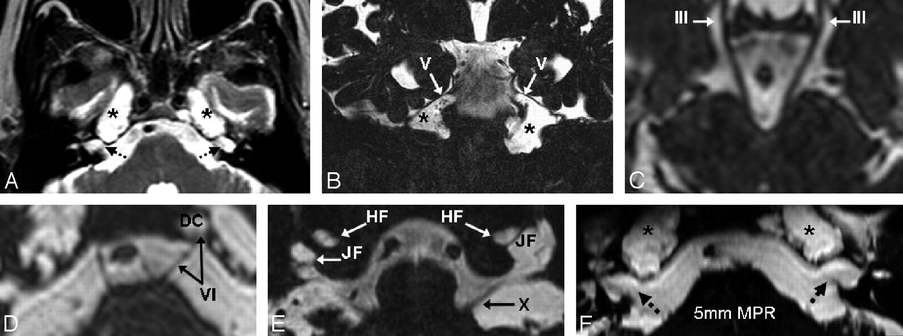
On MR imaging, there were diffusely enlarged subarachnoid spaces surrounding the exits of cranial nerves (CN) II-XII, being most prominent along the oculomotor cisterns (CN III), Meckel caves (CN V), and within the jugular foramina (CN X; Fig 1A–F). The findings were bilateral and relatively symmetric, with the exception of enlargement of the Dorello canal unilaterally surrounding CN VI. These enlarged spaces followed the signal intensity of CSF on all sequences. The most prominent enlarged spaces were 3-cm-sized lesions of the petrous apices that were considered to be PACs because of their having CSF intensity that was contiguous with CSF within the Meckel cave. There was also moderate cerebral volume loss. There was no abnormal contrast enhancement of the brain parenchyma or meninges. A temporal bone CT confirmed the enlarged foramina and the petrous apex remodeling and showed a mildly enlarged bony sellar contour (Fig 2). An MR imaging examination 1 year previously was obtained for comparison and showed an identical appearance and size of the PACs.

Fig 1.
  • Download figure
  • Open in new tab
  • Download powerpoint
Fig 1.

On MR imaging, the bilateral PACs were bright on T2WI (A) but not on diffusion or fluid-attenuated inversion recovery (not shown). Thin, 0.6-mm 3D FSE T2WI (B–E) demonstrated a communication with Meckel caves (asterisks) in Figs 1A, 1B, and 1F and depicted dilated subarachnoid spaces surrounding the cranial nerves. Shown are the oculomotor cistern (III) in Fig 1C, Dorello canal in Fig 1D (VI, DC), Meckel caves in Figs 1A, 1B, and 1F (V and asterisks), and the jugular (X, JF) and hypoglossal foramina (XII, HF) in Fig 1E. There are 5-mm-thick reconstructions from the 3D FSE T2WI that illustrate the PACs and the adjacent enlarged internal auditory canals (Fig 1F). The prominent internal auditory canals in Figs 1A and 1F (dashed arrows) did not communicate with the PACs.

Fig 2.
  • Download figure
  • Open in new tab
  • Download powerpoint
Fig 2.

A CT scan was obtained after MR imaging for confirmation of the PACs and better characterization of the extent of bony remodeling of the petrous apices (asterisks). Note the enlarged internal auditory canals as well (dashed white arrows).

The patient's symptoms were thought related to the temporomandibular joint after they resolved with nonsurgical therapy. A neuro-otologic consultation was then obtained to evaluate the petrous apex lesions; the neuro-otologist elected to observe these lesions. Nearly 2 years after presentation, the patient continues to be followed by otolaryngology should his symptoms worsen. No additional symptoms or cranial neuropathies have developed beyond his baseline blindness and sensorineural hearing loss.

Discussion

Although the underlying causes of Usher syndrome are not entirely clear, the best understood and most commonly accepted molecular mechanism in type I Usher syndrome involves the myosin VIIa protein.2,3 This protein encodes another unconventional myosin protein localized in the connecting cilium of photoreceptor cells in the retinal pigment epithelium and inner ear stereocilia.3 Reports of patients with type I Usher syndrome having bronchiectasis, chronic sinusitis, and primary ciliary aplasia suggest that the effects of these mutations are not limited to the inner ear and retina.8,9

PACs are thought to arise from increases in intracranial pressure, whether transient or sustained.10,11 Only 20 to 30 cases have been reported, 3 being bilateral, but their frequency is difficult to determine because these may have been underreported or listed under alternate entities such as meningoceles or arachnoid cysts, or even mistakenly identified as other common lesions of the petrous apex.10,11 In our patient, MR imaging ultimately excluded other lesions in the differential diagnosis of petrous apex masses, such as cholesterol granulomas (not T1-bright), epidermoid cysts (lack of decreased diffusion), bilateral glomus tumors or schwannomas (no enhancement), and petrous apicitis or effusions (no enhancement or mastoiditis).

The cause of the cystic dilations of the subarachnoid spaces in our patient is unclear, and the mechanism of formation of the PACs is unknown; however, these “cephaloceles” could result from a model similar to that proposed for arachnoid cyst formation. At approximately the 15th gestational week, the roof of the fourth ventricle opens into the cisterna magna via the foramina of Magendie and Luschka, and the CSF passing into the cisternal mesenchyme can result in arachnoid membrane splitting; this splitting may eventually lead to the formation of a CSF-containing cyst.11-14 Hence, in our patient, the PACs could result from a similar process occurring focally through the Meckel cave and the porus trigeminus as well as through the other pori associated with the other cranial nerve exits, resulting in the appearance of multiple cephaloceles.11 Also it has been postulated that intracranial hypertension or abnormal CSF pulsation can contribute to PAC enlargement.10,11

It is interesting to note that changes in intracranial pressure leading to hydrocephalus is a phenomenon that has been cited to occur in an emerging class of disorders of ciliary function, which includes Kartagener syndrome, Meckel-Gruber syndrome, Joubert syndrome, and other rare cilia dysfunctions, some of which may be associated with cephaloceles.15,16 Indeed, recent evidence from an animal model has demonstrated that in the ependymal cell layer, dysfunction of the cilia can cause aberrant ependymal flow and resultant closure of the cerebral aqueduct with subsequent hydrocephalus.16 From a theoretic standpoint, such dysfunction could have occurred to some degree in our patient, contributing to a low-level hydrocephalus and cephalocele formation. We note that our patient lacked the typical findings of acute hydrocephalus, such as enlarged temporal horns of the lateral ventricles, enlarged anterior recesses of the third ventricle, periependymal T2 hyperintensity, or enlarged ventricles relative to the cerebral sulci. On the other hand, a chronic, low-level hydrocephalus or other abnormality of CSF transmission or pulsation could not be entirely excluded, particularly given the mild bony sellar enlargement.

Another possibility is that the dilated subarachnoid spaces, the multiple cephaloceles, and the cerebral volume loss were separate, isolated findings. However, this is unlikely because PACs are quite uncommon and are especially rare to be bilateral, particularly in the setting of diffusely prominent subarachnoid spaces surrounding the cranial nerves. This combination of rare findings raises the question of a common, underlying mechanism related to abnormal CSF flow or pulsation. Although our patient did not exhibit any clear clinical findings of hydrocephalus, these signs can be nonspecific in states of chronically elevated intracranial pressure.

In the available literature on Usher syndrome, we have not found descriptions of PACs, nor have we found reports of enlarged subarachnoid spaces surrounding cranial nerve exits to the point of causing scalloping and remodeling of the skull base. The sparse literature describing the histopathologic findings in Usher syndrome focuses primarily on atrophic changes in the organ of Corti and ciliary aplasia.8,17 It is unfortunate that most reports regarding MR imaging findings in this syndrome have small patient sample sizes without long-term surveillance and have described nonspecific findings including brain atrophy and enlarged subarachnoid spaces, without mention of involvement of the cranial nerve exit sites or descriptions using thin-section MR imaging of the skull base.4-6

References

  1. ↵
    Boughman J, Vernon M, Shaver K. Usher syndrome: definition and estimate of prevalence from two high-risk populations. J Chronic Dis 1983;36:595–603
    CrossRefPubMed
  2. ↵
    Riazuddin S, Nazli S, Ahmed ZM, et al. Mutation spectrum of MYO7a and evaluation of a novel nonsyndromic deafness DFNB2 allele with residual function. Hum Mutat 2008;29:502–11
    CrossRefPubMed
  3. ↵
    Williams D. Usher syndrome: animal models, retinal function of usher proteins, and prospects for gene therapy. Vision Res 2008;48:433–41
    CrossRefPubMed
  4. ↵
    Bloom T, Fishman G, Mafee M. Usher syndrome: CNS defects determined by computed tomography. Retina 1983;3:108–13
    CrossRefPubMed
  5. Tamayo M, Maldonado C, Plaza S, et al. Neuroradiology and clinical aspects of Usher syndrome. Clin Genet 1996;50:126–32
    PubMed
  6. ↵
    Schaefer G, Bodensteiner J, Thompson J, et al. Volumetric neuroimaging in Usher syndrome: evidence of global involvement. Am J Med Genet 1998;79:1–4
    CrossRefPubMed
  7. ↵
    Moore K, Fischbein N, Harnsberger H, et al. Petrous apex cephaloceles. AJNR Am J Neuroradiology 2001;22:1867–71
    Abstract/FREE Full Text
  8. ↵
    Tosi G, de Santi M, Pradal U, et al. Usher syndrome type 1 associated with primary ciliary aplasia. Arch Ophthalmol 2003;121:407–08
    PubMed
  9. ↵
    Bonneau D, Raymond F, Kremer C, et al. Usher syndrome type I associated with bronchiectasis and immotile nasal cilia in two brothers. J Med Genet 1993;30:253–54
    Abstract/FREE Full Text
  10. ↵
    Alorainy I. Petrous apex cephalocele and empty sella: is there a relation? Eur J Radiol 2007;62:378–84
    CrossRefPubMed
  11. ↵
    Isaacson B, Coker N, Vrabec J, et al. Invasive cerebrospinal fluid cysts and cephaloceles of the petrous apex. Otol Neurotol 2006;27:1131–41
    CrossRefPubMed
  12. Abbott R. The endoscopic management of arachnoidal cysts. Neurosurg Clin N Am 2004;15:9–17
    CrossRefPubMed
  13. Starkman SP, Brown TC, Linell EA. Cerebral arachnoid cysts. J Neuropathol Exp Neurol 1958;17:484–500
    CrossRefPubMed
  14. ↵
    Cristobal R, Oghalai JS. Peripetrosal arachnoid cysts. Curr Opin Otolaryngol Head Neck Surg 2007;15:323–29
    CrossRefPubMed
  15. ↵
    Badano JL, Mitsuma N, Beales PL, et al. The ciliopathies: an emerging class of human genetic disorders. Annu Rev Genomics Hum Genet 2006;7:125–48
    CrossRefPubMed
  16. ↵
    Ibañex-Tallon I, Gorokhoba S, Heintz N. Loss of function of axonemal dynein Mdnah5 causes primary ciliary dyskinesia and hydrocephalus. Hum Mol Genet 2002;11:715–21
    Abstract/FREE Full Text
  17. ↵
    Cremers C, Delleman W. Usher's syndrome, temporal bone pathology. Int J Pediatr Otorhinolaryngol 1988;16:23–30
    CrossRefPubMed
  • Received June 29, 2008.
  • Accepted after revision July 16, 2008.
  • Copyright © American Society of Neuroradiology
View Abstract
PreviousNext
Back to top

In this issue

American Journal of Neuroradiology: 30 (2)
American Journal of Neuroradiology
Vol. 30, Issue 2
February 2009
  • Table of Contents
  • Index by author
Advertisement
Print
Download PDF
Email Article

Thank you for your interest in spreading the word on American Journal of Neuroradiology.

NOTE: We only request your email address so that the person you are recommending the page to knows that you wanted them to see it, and that it is not junk mail. We do not capture any email address.

Enter multiple addresses on separate lines or separate them with commas.
Dilation of the Subarachnoid Spaces Surrounding the Cranial Nerves with Petrous Apex Cephaloceles in Usher Syndrome
(Your Name) has sent you a message from American Journal of Neuroradiology
(Your Name) thought you would like to see the American Journal of Neuroradiology web site.
CAPTCHA
This question is for testing whether or not you are a human visitor and to prevent automated spam submissions.
Cite this article
T.A. Stark, A.M. McKinney, C.S. Palmer, R.H. Maisel, C.L. Truwit
Dilation of the Subarachnoid Spaces Surrounding the Cranial Nerves with Petrous Apex Cephaloceles in Usher Syndrome
American Journal of Neuroradiology Feb 2009, 30 (2) 434-436; DOI: 10.3174/ajnr.A1283

Citation Manager Formats

  • BibTeX
  • Bookends
  • EasyBib
  • EndNote (tagged)
  • EndNote 8 (xml)
  • Medlars
  • Mendeley
  • Papers
  • RefWorks Tagged
  • Ref Manager
  • RIS
  • Zotero
0 Responses
Respond to this article
Share
Bookmark this article
Dilation of the Subarachnoid Spaces Surrounding the Cranial Nerves with Petrous Apex Cephaloceles in Usher Syndrome
T.A. Stark, A.M. McKinney, C.S. Palmer, R.H. Maisel, C.L. Truwit
American Journal of Neuroradiology Feb 2009, 30 (2) 434-436; DOI: 10.3174/ajnr.A1283
del.icio.us logo Twitter logo Facebook logo Mendeley logo
  • Tweet Widget
  • Facebook Like
  • Google Plus One
Purchase

Jump to section

  • Article
    • Abstract
    • Case Report
    • Discussion
    • References
  • Figures & Data
  • Info & Metrics
  • Responses
  • References
  • PDF

Related Articles

  • No related articles found.
  • PubMed
  • Google Scholar

Cited By...

  • No citing articles found.
  • Crossref (23)
  • Google Scholar

This article has been cited by the following articles in journals that are participating in Crossref Cited-by Linking.

  • Lesions of the Petrous Apex: Classification and Findings at CT and MR Imaging
    Ahmed Abdel Razek, Benjamin Y. Huang
    RadioGraphics 2012 32 1
  • The association between petrous apex cephalocele and empty sella
    Dima Z. Jamjoom, Ibrahim A. Alorainy
    Surgical and Radiologic Anatomy 2015 37 10
  • ‘Do not touch’ lesions of the skull base
    Mircea C Dobre, Nancy Fischbein
    Journal of Medical Imaging and Radiation Oncology 2014 58 4
  • Imaging of Petrous Apex Lesions
    Gillian M. Potter, Rekha Siripurapu
    Neuroimaging Clinics of North America 2021 31 4
  • Imaging of Skull Base Tumors
    Bilal Battal, Carlos Zamora
    Tomography 2023 9 4
  • Cerebrospinal fluid leak with recurrent meningitis following tonsillectomy
    Shawn L. Hervey-Jumper, Ahmer K. Ghori, Douglas J. Quint, Lawrence J. Marentette, Cormac O. Maher
    Journal of Neurosurgery: Pediatrics 2010 5 3
  • Bilateral petrous apex cephaloceles: Is surgical intervention indicated?
    Ali Alkhaibary, Fahd Musawnaq, Makki Almuntashri, Abdulaziz Alarifi
    International Journal of Surgery Case Reports 2020 72
  • Analysis of petrous apex meningocele associated with meningioma: is there any relation with chronic intracranial hypertension?
    Wan-Qun Yang, Jie-Ying Feng, Hong-Jun Liu, Biao Huang, Chang-Hong Liang
    Neuroradiology 2018 60 2
  • Hipoacusia en niños con conducto auditivo interno agrandado
    Saturnino Santos, M. Jesús Domínguez, Javier Cervera, Alicia Suárez, Antonio Bueno, Margarita Bartolomé, Rafael López
    Acta Otorrinolaringológica Española 2014 65 2
  • Hearing loss in a child with cystic dilated internal auditory canal
    Muath Alsabih, Khalid Alosaimi, Roa Halawani, Farid Alzhrani
    Indian Journal of Otology 2019 25 3

More in this TOC Section

  • ASL Sensitivity for Head and Neck Paraganglioma
  • Post SRS Peritumoral Hyperintense Signal of VSs
  • Contrast Enhanced Pituitary CISS/FIESTA
Show more Head and Neck Imaging

Similar Articles

Advertisement

Indexed Content

  • Current Issue
  • Accepted Manuscripts
  • Article Preview
  • Past Issues
  • Editorials
  • Editor's Choice
  • Fellows' Journal Club
  • Letters to the Editor
  • Video Articles

Cases

  • Case Collection
  • Archive - Case of the Week
  • Archive - Case of the Month
  • Archive - Classic Case

More from AJNR

  • Trainee Corner
  • Imaging Protocols
  • MRI Safety Corner
  • Book Reviews

Multimedia

  • AJNR Podcasts
  • AJNR Scantastics

Resources

  • Turnaround Time
  • Submit a Manuscript
  • Submit a Video Article
  • Submit an eLetter to the Editor/Response
  • Manuscript Submission Guidelines
  • Statistical Tips
  • Fast Publishing of Accepted Manuscripts
  • Graphical Abstract Preparation
  • Imaging Protocol Submission
  • Evidence-Based Medicine Level Guide
  • Publishing Checklists
  • Author Policies
  • Become a Reviewer/Academy of Reviewers
  • News and Updates

About Us

  • About AJNR
  • Editorial Board
  • Editorial Board Alumni
  • Alerts
  • Permissions
  • Not an AJNR Subscriber? Join Now
  • Advertise with Us
  • Librarian Resources
  • Feedback
  • Terms and Conditions
  • AJNR Editorial Board Alumni

American Society of Neuroradiology

  • Not an ASNR Member? Join Now

© 2025 by the American Society of Neuroradiology All rights, including for text and data mining, AI training, and similar technologies, are reserved.
Print ISSN: 0195-6108 Online ISSN: 1936-959X

Powered by HighWire