
Article Figures & Data
Figures
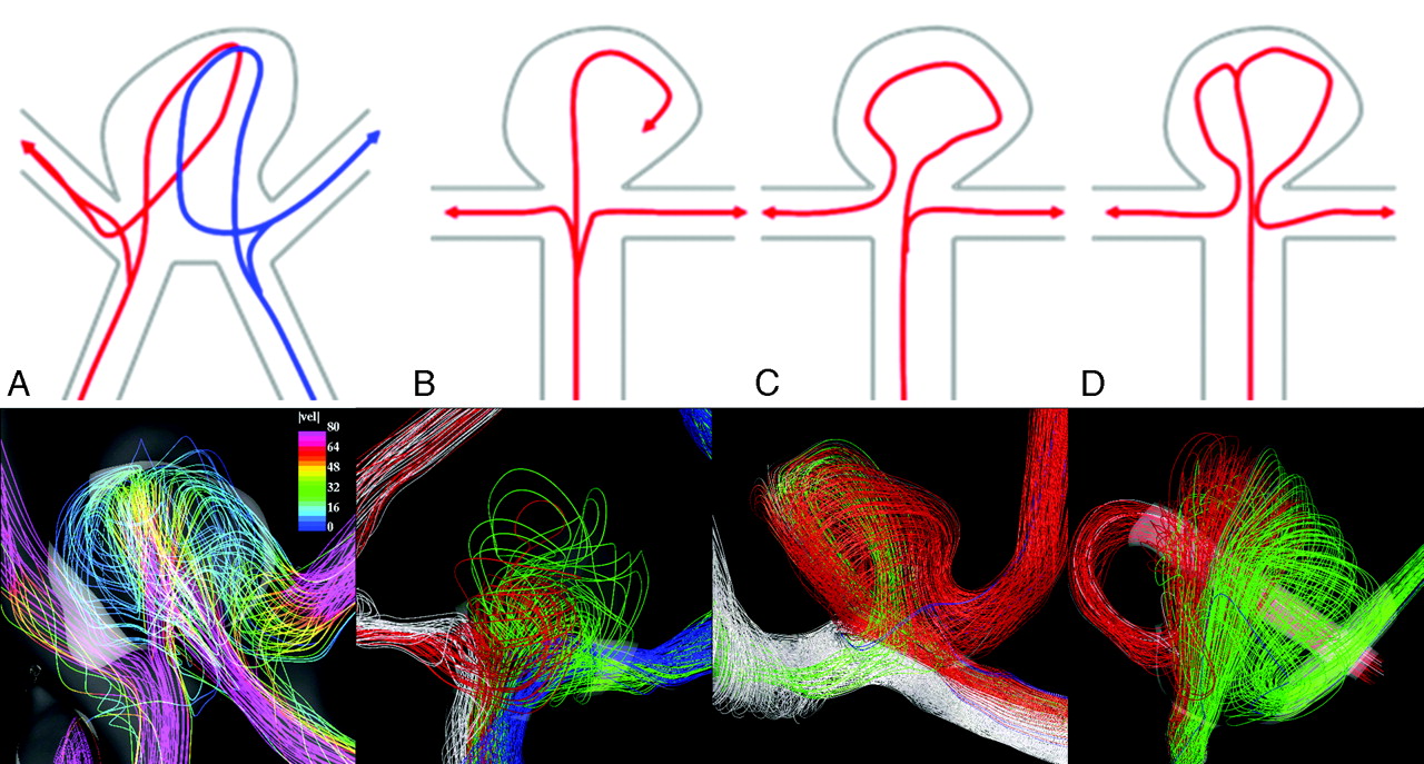
- Fig 1.
A–D, Different flow patterns found in AcomA aneurysms are shown in the top row. Pictures of the streamlines colored by the magnitude of the velocity and their path from the A1 segment to the A2 segments in 4 selected aneurysms for each flow type are shown in the bottom row.
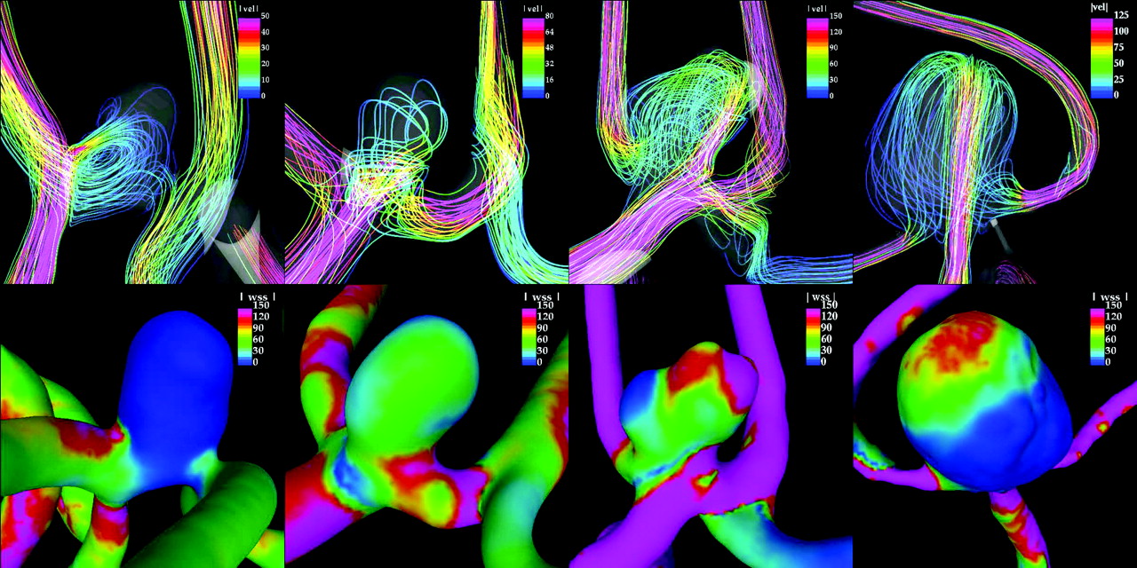
- Fig 2.
Streamlines colored with the velocity magnitude (top row) and instantaneous WSS distributions (bottom row) at the peak systole for 4 selected aneurysms with different flow types (from left to right: flow types A, B, C, and D) whose MWSS is of 10, 60, 200, and 150 dyne/cm2, respectively. The same color scale of 150 dyne/cm2 is used for all the WSS images. Only flow type C aneurysm is ruptured.


Tables
- Table 1:
Number and percentage of ruptured and unruptured aneurysms in each flow type based on velocity magnitude maps at selected cut planes
Flow Type Ruptured (%) Unruptured (%) Total I 3 (75) 1 (25) 4 II 6 (75) 2 (25) 8 III 1 (100) 0 (0) 1 IV 8 (62) 5 (38) 13 Total 18 (70) 8 (30) 26 - Table 2:
Number, percentage, and average peak WSS of ruptured and unruptured aneurysms in each flow type group
Flow Type Ruptured Unruptured Total No. (%) WSS (dyne/cm2) No. (%) WSS (dyne/cm2) A 1 (25) 700 3 (75) 63 4 B 2 (50) 78 2 (50) 60 4 C 13 (87) 283 2 (13) 225 15 D 2 (67) 160 1 (33) 150 3 Total 18 (70) 271 8 (30) 114 26 Note:—WSS indicates wall shear stress.
In this issue


Jump to section
Related Articles
Cited By...
- Fluid-structure interaction simulation of tissue degradation and its effects on intra-aneurysm hemodynamics
- Impact of A1 Asymmetry on the Woven EndoBridge Device in Anterior Communicating Artery Aneurysms
- Residual Flow Inside the Woven EndoBridge Device at Follow-Up: Potential Predictors of the Bicetre Occlusion Scale Score 1 Phenomenon
- Inflow Jet Patterns of Unruptured Cerebral Aneurysms Based on the Flow Velocity in the Parent Artery: Evaluation Using 4D Flow MRI
- Hemodynamic characteristics of large unruptured internal carotid artery aneurysms prior to rupture: a case control study
- Morphologic and hemodynamic analysis of paraclinoid aneurysms: ruptured versus unruptured
- CFD: Computational Fluid Dynamics or Confounding Factor Dissemination? The Role of Hemodynamics in Intracranial Aneurysm Rupture Risk Assessment
- Effects of Circle of Willis Anatomic Variations on Angiographic and Clinical Outcomes of Coiled Anterior Communicating Artery Aneurysms
- High WSS or Low WSS? Complex Interactions of Hemodynamics with Intracranial Aneurysm Initiation, Growth, and Rupture: Toward a Unifying Hypothesis
- Quantification of speed-up and accuracy of multi-CPU computational flow dynamics simulations of hemodynamics in a posterior communicating artery aneurysm of complex geometry
- Intracranial Aneurysm Neck Size Overestimation with 3D Rotational Angiography: The Impact on Intra-Aneurysmal Hemodynamics Simulated with Computational Fluid Dynamics
- Comparison of Phase-Contrast MR Imaging and Endovascular Sonography for Intracranial Blood Flow Velocity Measurements
- Counterpoint: Realizing the Clinical Utility of Computational Fluid Dynamics--Closing the Gap
- Wall Shear Stress Distribution Inside Growing Cerebral Aneurysm
- Patient-Specific Computational Hemodynamics of Intracranial Aneurysms from 3D Rotational Angiography and CT Angiography: An In Vivo Reproducibility Study
- Hemodynamic-Morphologic Discriminants for Intracranial Aneurysm Rupture
- Potent Risk Factor for Aneurysm Formation: Termination Aneurysms of the Anterior Communicating Artery and Detection of A1 Vessel Asymmetry by Flow Dilution
- Blood-Flow Characteristics in a Terminal Basilar Tip Aneurysm Prior to Its Fatal Rupture