Advertisement
  • Main menu
  • User menu
  • Search
  • English ▼
    • English
    • Afrikaans
    • Albanian
    • Amharic
    • Arabic
    • Armenian
    • Azerbaijani
    • Basque
    • Belarusian
    • Bengali
    • Bosnian
    • Bulgarian
    • Catalan
    • Cebuano
    • Chichewa
    • Chinese (Simplified)
    • Chinese (Traditional)
    • Corsican
    • Croatian
    • Czech
    • Danish
    • Dutch
    • Esperanto
    • Estonian
    • Filipino
    • Finnish
    • French
    • Frisian
    • Galician
    • Georgian
    • German
    • Greek
    • Gujarati
    • Haitian Creole
    • Hausa
    • Hawaiian
    • Hebrew
    • Hindi
    • Hmong
    • Hungarian
    • Icelandic
    • Igbo
    • Indonesian
    • Irish
    • Italian
    • Japanese
    • Javanese
    • Kannada
    • Kazakh
    • Khmer
    • Korean
    • Kurdish (Kurmanji)
    • Kyrgyz
    • Lao
    • Latin
    • Latvian
    • Lithuanian
    • Luxembourgish
    • Macedonian
    • Malagasy
    • Malay
    • Malayalam
    • Maltese
    • Maori
    • Marathi
    • Mongolian
    • Myanmar (Burmese)
    • Nepali
    • Norwegian
    • Pashto
    • Persian
    • Polish
    • Portuguese
    • Punjabi
    • Romanian
    • Russian
    • Samoan
    • Scottish Gaelic
    • Serbian
    • Sesotho
    • Shona
    • Sindhi
    • Sinhala
    • Slovak
    • Slovenian
    • Somali
    • Spanish
    • Sudanese
    • Swahili
    • Swedish
    • Tajik
    • Tamil
    • Telugu
    • Thai
    • Turkish
    • Ukrainian
    • Urdu
    • Uzbek
    • Vietnamese
    • Welsh
    • Xhosa
    • Yiddish
    • Yoruba
    • Zulu

Main menu

  • Home
  • Content
    • Current Issue
    • Accepted Manuscripts
    • Article Preview
    • Past Issue Archive
    • Video Articles
    • AJNR Case Collection
    • Case of the Week Archive
    • Case of the Month Archive
    • Classic Case Archive
  • Special Collections
    • Advancing NeuroMRI with High-Relaxivity Contrast Agents
    • AJNR Awards
    • Low-Field MRI
    • Alzheimer Disease
    • ASNR Foundation Special Collection
    • Photon-Counting CT
    • View All
  • Multimedia
    • AJNR Podcasts
    • AJNR SCANtastic
    • Trainee Corner
    • MRI Safety Corner
    • Imaging Protocols
  • For Authors
    • Submit a Manuscript
    • Submit a Video Article
    • Submit an eLetter to the Editor/Response
    • Manuscript Submission Guidelines
    • Statistical Tips
    • Fast Publishing of Accepted Manuscripts
    • Graphical Abstract Preparation
    • Imaging Protocol Submission
    • Author Policies
  • About Us
    • About AJNR
    • Editorial Board
    • Editorial Board Alumni
  • More
    • Become a Reviewer/Academy of Reviewers
    • Subscribers
    • Permissions
    • Alerts
    • Feedback
    • Advertisers
    • ASNR Home

User menu

  • Alerts
  • Log in

Search

  • Advanced search
American Journal of Neuroradiology

American Journal of Neuroradiology

ASHNR American Society of Functional Neuroradiology ASHNR American Society of Pediatric Neuroradiology ASSR
  • Alerts
  • Log in

Advanced Search

English ▼
  • English
  • Afrikaans
  • Albanian
  • Amharic
  • Arabic
  • Armenian
  • Azerbaijani
  • Basque
  • Belarusian
  • Bengali
  • Bosnian
  • Bulgarian
  • Catalan
  • Cebuano
  • Chichewa
  • Chinese (Simplified)
  • Chinese (Traditional)
  • Corsican
  • Croatian
  • Czech
  • Danish
  • Dutch
  • Esperanto
  • Estonian
  • Filipino
  • Finnish
  • French
  • Frisian
  • Galician
  • Georgian
  • German
  • Greek
  • Gujarati
  • Haitian Creole
  • Hausa
  • Hawaiian
  • Hebrew
  • Hindi
  • Hmong
  • Hungarian
  • Icelandic
  • Igbo
  • Indonesian
  • Irish
  • Italian
  • Japanese
  • Javanese
  • Kannada
  • Kazakh
  • Khmer
  • Korean
  • Kurdish (Kurmanji)
  • Kyrgyz
  • Lao
  • Latin
  • Latvian
  • Lithuanian
  • Luxembourgish
  • Macedonian
  • Malagasy
  • Malay
  • Malayalam
  • Maltese
  • Maori
  • Marathi
  • Mongolian
  • Myanmar (Burmese)
  • Nepali
  • Norwegian
  • Pashto
  • Persian
  • Polish
  • Portuguese
  • Punjabi
  • Romanian
  • Russian
  • Samoan
  • Scottish Gaelic
  • Serbian
  • Sesotho
  • Shona
  • Sindhi
  • Sinhala
  • Slovak
  • Slovenian
  • Somali
  • Spanish
  • Sudanese
  • Swahili
  • Swedish
  • Tajik
  • Tamil
  • Telugu
  • Thai
  • Turkish
  • Ukrainian
  • Urdu
  • Uzbek
  • Vietnamese
  • Welsh
  • Xhosa
  • Yiddish
  • Yoruba
  • Zulu
  • Home
  • Content
    • Current Issue
    • Accepted Manuscripts
    • Article Preview
    • Past Issue Archive
    • Video Articles
    • AJNR Case Collection
    • Case of the Week Archive
    • Case of the Month Archive
    • Classic Case Archive
  • Special Collections
    • Advancing NeuroMRI with High-Relaxivity Contrast Agents
    • AJNR Awards
    • Low-Field MRI
    • Alzheimer Disease
    • ASNR Foundation Special Collection
    • Photon-Counting CT
    • View All
  • Multimedia
    • AJNR Podcasts
    • AJNR SCANtastic
    • Trainee Corner
    • MRI Safety Corner
    • Imaging Protocols
  • For Authors
    • Submit a Manuscript
    • Submit a Video Article
    • Submit an eLetter to the Editor/Response
    • Manuscript Submission Guidelines
    • Statistical Tips
    • Fast Publishing of Accepted Manuscripts
    • Graphical Abstract Preparation
    • Imaging Protocol Submission
    • Author Policies
  • About Us
    • About AJNR
    • Editorial Board
    • Editorial Board Alumni
  • More
    • Become a Reviewer/Academy of Reviewers
    • Subscribers
    • Permissions
    • Alerts
    • Feedback
    • Advertisers
    • ASNR Home
  • Follow AJNR on Twitter
  • Visit AJNR on Facebook
  • Follow AJNR on Instagram
  • Join AJNR on LinkedIn
  • RSS Feeds

AJNR Awards, New Junior Editors, and more. Read the latest AJNR updates


Improved Turnaround Times | Median time to first decision: 12 days

Research ArticlePHYSICS REVIEW
Open Access

Susceptibility-Weighted Imaging: Technical Aspects and Clinical Applications, Part 2

S. Mittal, Z. Wu, J. Neelavalli and E.M. Haacke
American Journal of Neuroradiology February 2009, 30 (2) 232-252; DOI: https://doi.org/10.3174/ajnr.A1461
S. Mittal
  • Find this author on Google Scholar
  • Find this author on PubMed
  • Search for this author on this site
Z. Wu
  • Find this author on Google Scholar
  • Find this author on PubMed
  • Search for this author on this site
J. Neelavalli
  • Find this author on Google Scholar
  • Find this author on PubMed
  • Search for this author on this site
E.M. Haacke
  • Find this author on Google Scholar
  • Find this author on PubMed
  • Search for this author on this site
  • Article
  • Figures & Data
  • Info & Metrics
  • Responses
  • References
  • PDF
Loading

Article Figures & Data

Figures

  • Fig 1.
    • Download figure
    • Open in new tab
    • Download powerpoint
    Fig 1.

    A 38-year-old woman who sustained severe TBI following a motor vehicle crash and lapsed into a coma. CT and MR images were obtained on the second day of hospitalization. A, Nonenhanced CT scan shows suspicious low attenuations in the pons and bilateral brachium pontis. B, T1-weighted image shows similar hypointense lesions in the pons, right brachium pontis, and right hemisphere of the cerebellum. C, T2-weighted image identifies multiple hyperintense lesions in the pons, bilateral brachium pontis, and right cerebellar hemisphere. D, SWI demonstrates multiple hypointense lesions in the left brachium pontis and both hemispheres of the cerebellum. There are more lesions shown on SWI, and they are also larger than those shown on CT and conventional MR imaging sequences. The shape of hemorrhagic lesions in the right cerebellar hemisphere reflects damage caused by shearing effect, indicating diffuse axonal injury. Courtesy of Dr Lei Jing, Tianjing Huanhu Hospital, Tianjing, China.
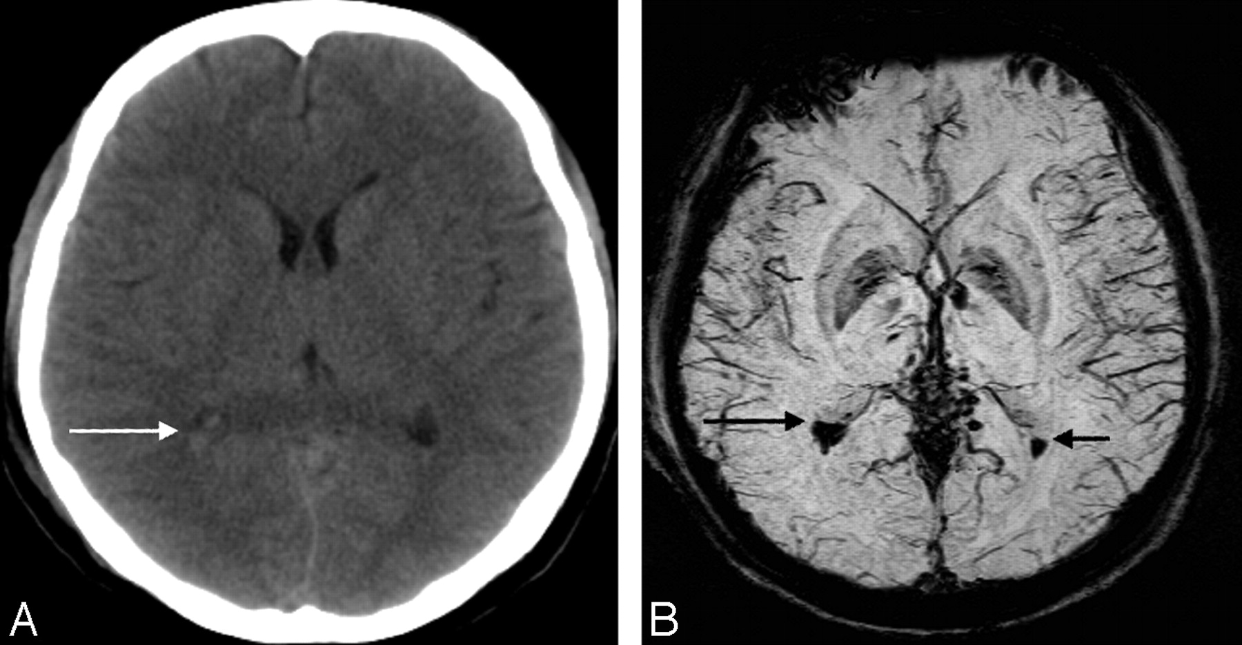
  • Fig 2.
    • Download figure
    • Open in new tab
    • Download powerpoint
    Fig 2.

    A, CT scan shows slightly higher attenuation (white arrow) in the posterior horn of right lateral ventricle. B, The SWI data clearly show hemorrhage (black arrows) inside both posterior horns of the lateral ventricle. Courtesy of Dr Lei Jing, Tianjing Huanhu Hospital, Tianjing, China.
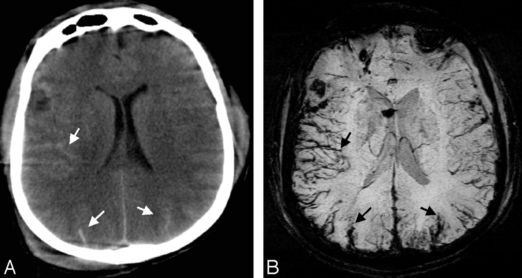
  • Fig 3.
    • Download figure
    • Open in new tab
    • Download powerpoint
    Fig 3.

    A, CT scan shows high-attenuation signals within the sulci of the cerebrum (white arrows), indicating traumatic subarachnoid hemorrhage. B, SWI clearly shows hemorrhage within the sulci of the cerebrum (black arrows), in agreement with the results of CT, but with a sharper contrast. Courtesy of Dr Lei Jing, Tianjing Huanhu Hospital, Tianjing, China.
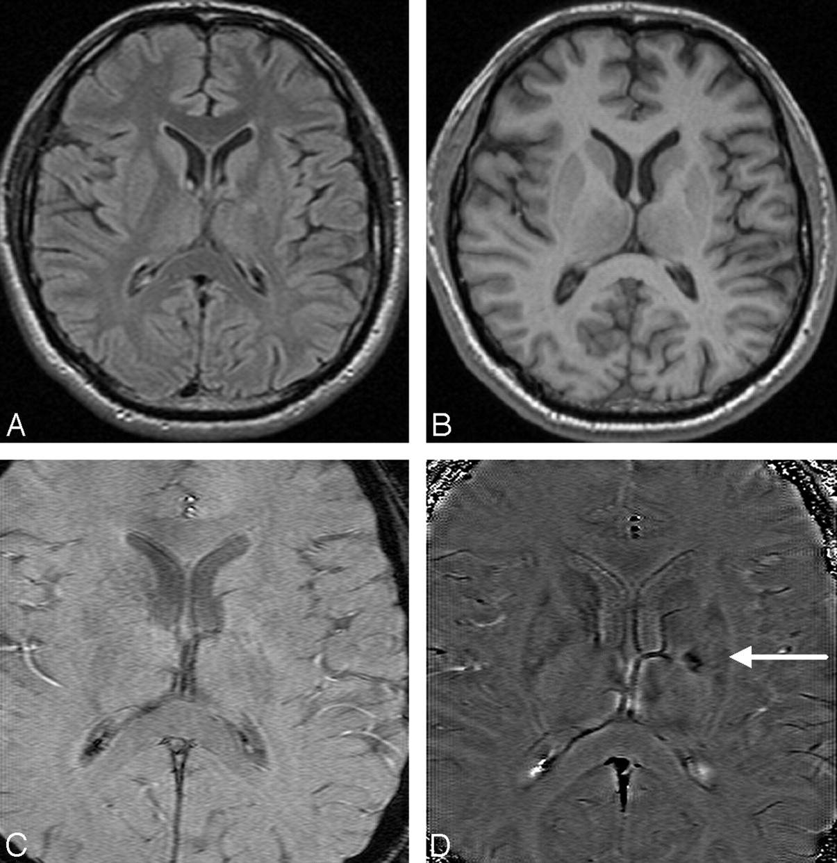
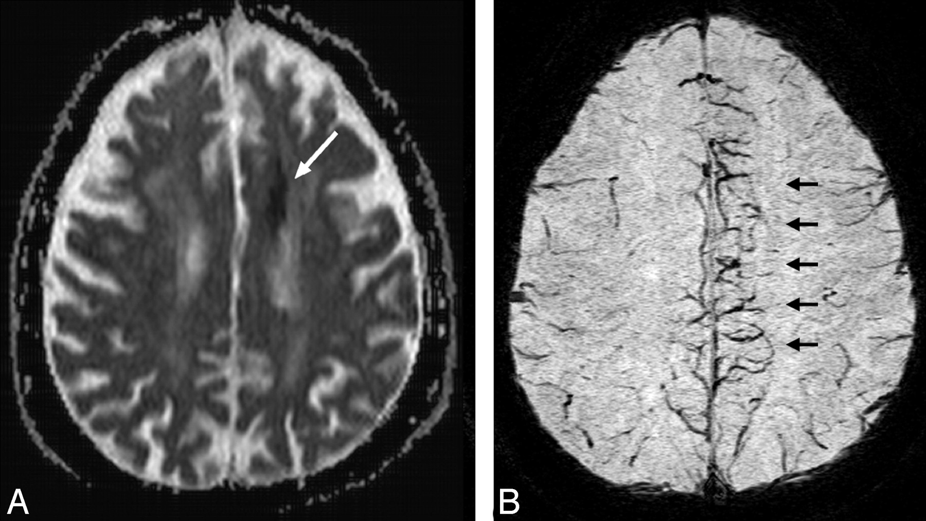
  • Fig 4.
    • Download figure
    • Open in new tab
    • Download powerpoint
    Fig 4.

    A patient who had sudden onset of aphasia and right paresthesias 5 years earlier and who partially recovered neurologic function after treatment of the acute stroke. A and B, Findings of follow-up MR images are almost normal on the FLAIR (A) and T1-weighted images (B). C and D, The original TE = 40 ms magnitude image (C) does not show any abnormality; however, the SWI phase image (D) shows a small hemorrhagic lesion (white arrow) in the genu of the internal capsule and internal globus pallidus.
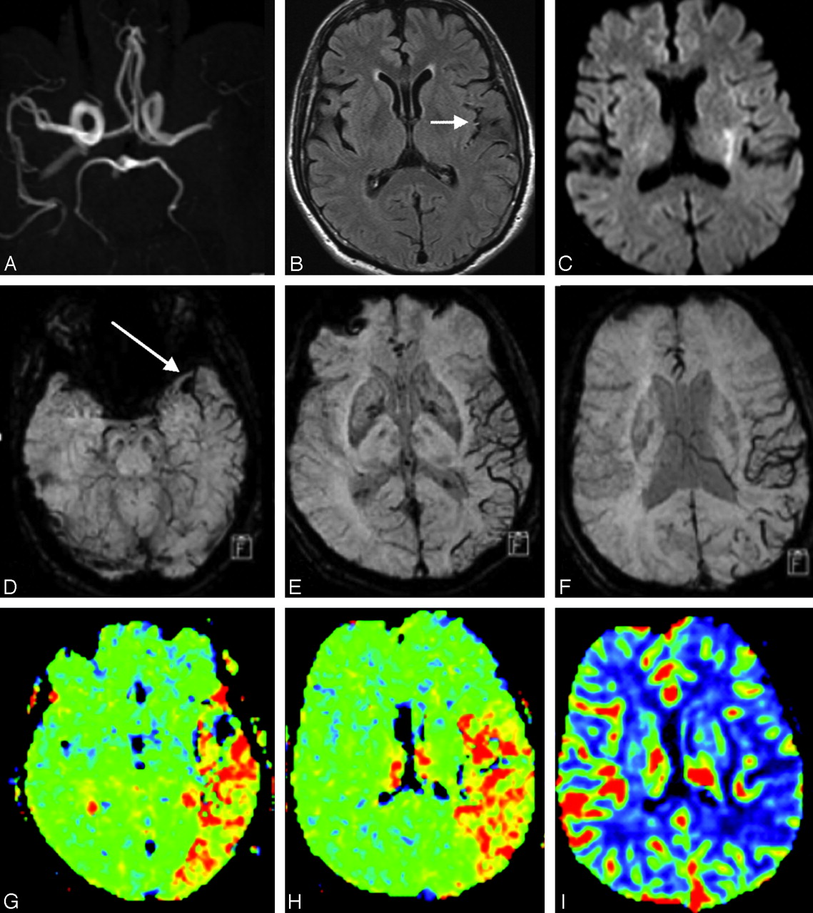
  • Fig 5.
    • Download figure
    • Open in new tab
    • Download powerpoint
    Fig 5.

    Cardioembolic stroke in the left MCA, 2 hours after onset. A, MRA shows lack of time-of-flight signal intensity in the distal M1 segment of the left MCA. B, FLAIR image displays intra-arterial signal intensity (arrow) of the left MCA branches along the lateral sulcus, indicating acute occlusion. C, DWI shows a small hyperintense lesion within the territory of the left MCA. D, SWI reveals localized hyposignal (arrow) in the left M1 segment, representing the acute thromboembolus itself. E and F, SWI demonstrates prominently hypointense cortical veins within the left MCA territory, suggesting relatively increased deoxyhemoglobin in the draining veins within the acutely ischemic region. G and H, The MTT map shows delayed transit times in the left MCA territory, matching SWI well. I, The rCBF is moderately reduced. Courtesy of Dr Masahiro Ida, Ebara Hospital, Tokyo, Japan.

  • Fig 6.
    • Download figure
    • Open in new tab
    • Download powerpoint
    Fig 6.

    Multiple microbleeds in CAA. A and B, T1-weighted (A) and T2-weighted (B) images do not reveal significant abnormalities except for the lesion in the left temporoparietal area. C, MRA shows normal brain vascular structure. D, SWI demonstrates, in addition to hemorrhage in the left temporoparietal region, multiple microbleeds distributed along the gray/white matter interface in the whole brain, strongly suggesting CAA.

  • Fig 7.
    • Download figure
    • Open in new tab
    • Download powerpoint
    Fig 7.

    An acute stroke patient with severe stenosis of the left internal carotid artery. The apparent diffusion coefficient map (A) shows an acute infarction as a low signal intensity (white arrow) together with old infarctions (which appear bright). The SWI data (B) shows prominently hypointense cortical veins reflecting the reduced oxygenation levels in the left anterior cerebral artery territory (arrows). Courtesy of Jonathon Grynspan, McMaster University, Hamilton, Ontario, Canada.
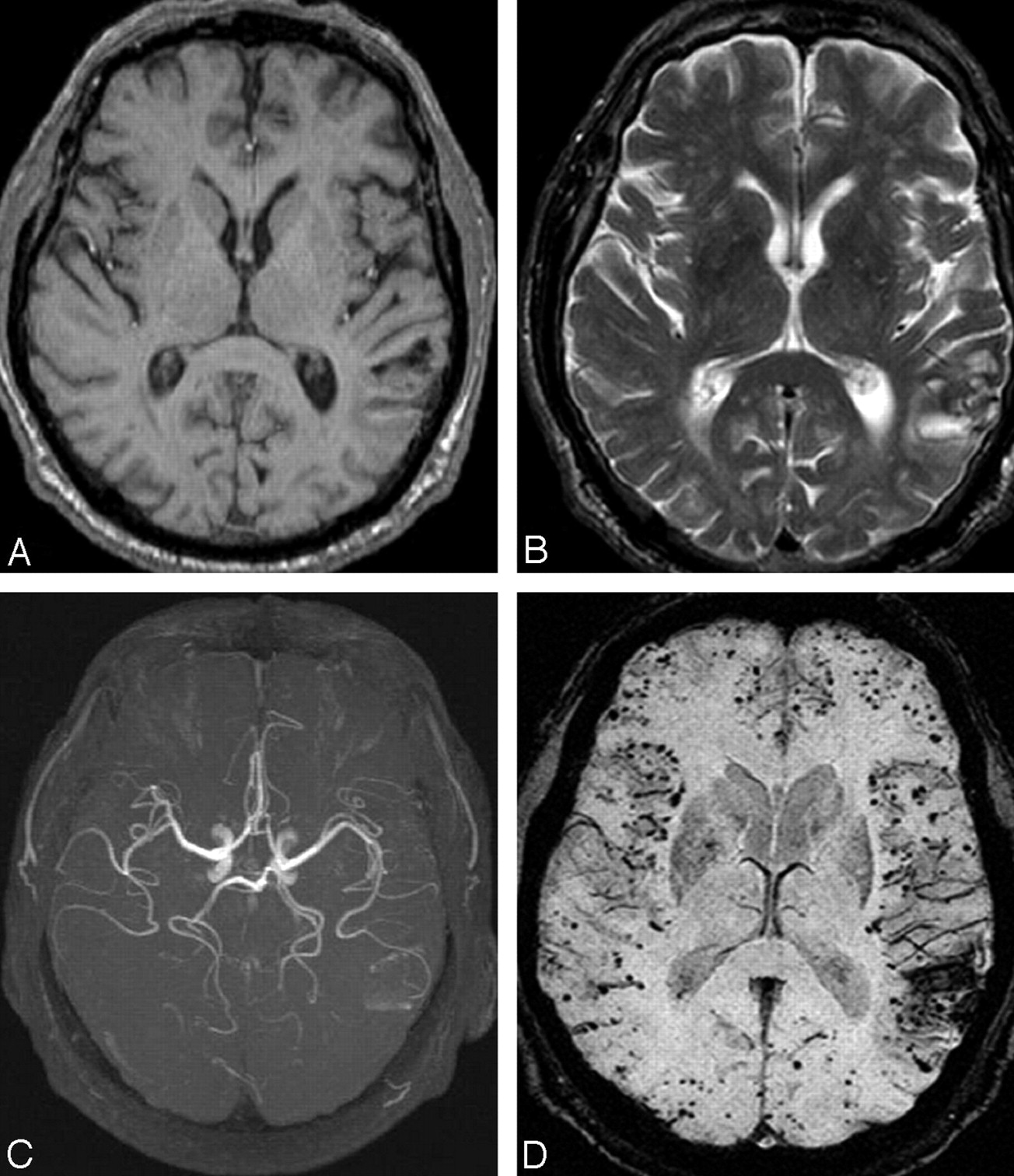
  • Fig 8.
    • Download figure
    • Open in new tab
    • Download powerpoint
    Fig 8.

    A, The SWI processed data for this 57-year-old patient with CADASIL shows 2 small bleeds in the anterior thalamus (arrows) and increased iron content in the basal ganglia. B, The SWI processed image for a healthy 53-year-old volunteer shows significant iron only in the globus pallidus. C and D, The T2 (C) and T1 (D) spin-echo images of the patient with CADASIL show diffuse white matter hyperintensities.
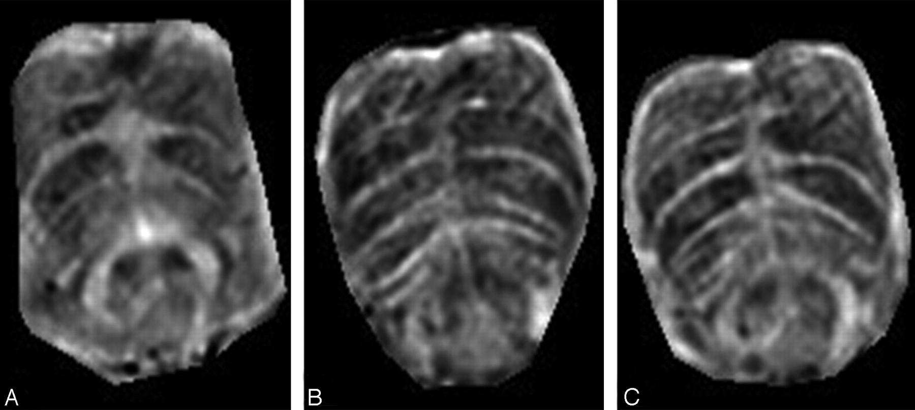
  • Fig 9.
    • Download figure
    • Open in new tab
    • Download powerpoint
    Fig 9.

    SWI data for 3 slices through the pons of a progressive MS patient with severe iron build up in various parts of the brain.

  • Fig 10.
    • Download figure
    • Open in new tab
    • Download powerpoint
    Fig 10.

    Different lesion distributions on SWI phase images and conventional MR images at 4T. A and B, Two lesions (long arrows) shown on the SWI phase image (A) are barely seen on FLAIR (B). C and D, On the other hand, lesions (short arrows) along the lateral ventricles shown on FLAIR (C) are not obvious on the SWI phase image (D).

  • Fig 11.
    • Download figure
    • Open in new tab
    • Download powerpoint
    Fig 11.

    In a patient with MS, iron builds up not only in the basal ganglia but also in the pulvinar nucleus of the thalamus. A, The SWI image for the patient shows the areas of increased iron content, including the pulvinar. B, In the healthy age-matched volunteer, there is little iron deposition in the pulvinar nucleus.

  • Fig 12.
    • Download figure
    • Open in new tab
    • Download powerpoint
    Fig 12.

    SWI data in a patient with progressive MS exquisitely demonstrate the location of iron deposition in the deep gray matter. The second scan (B) was acquired 1.5 years after the first scan (A) and shows an increase in the iron content in the globus pallidus as demonstrated by the increased phase values and aliasing seen in B.
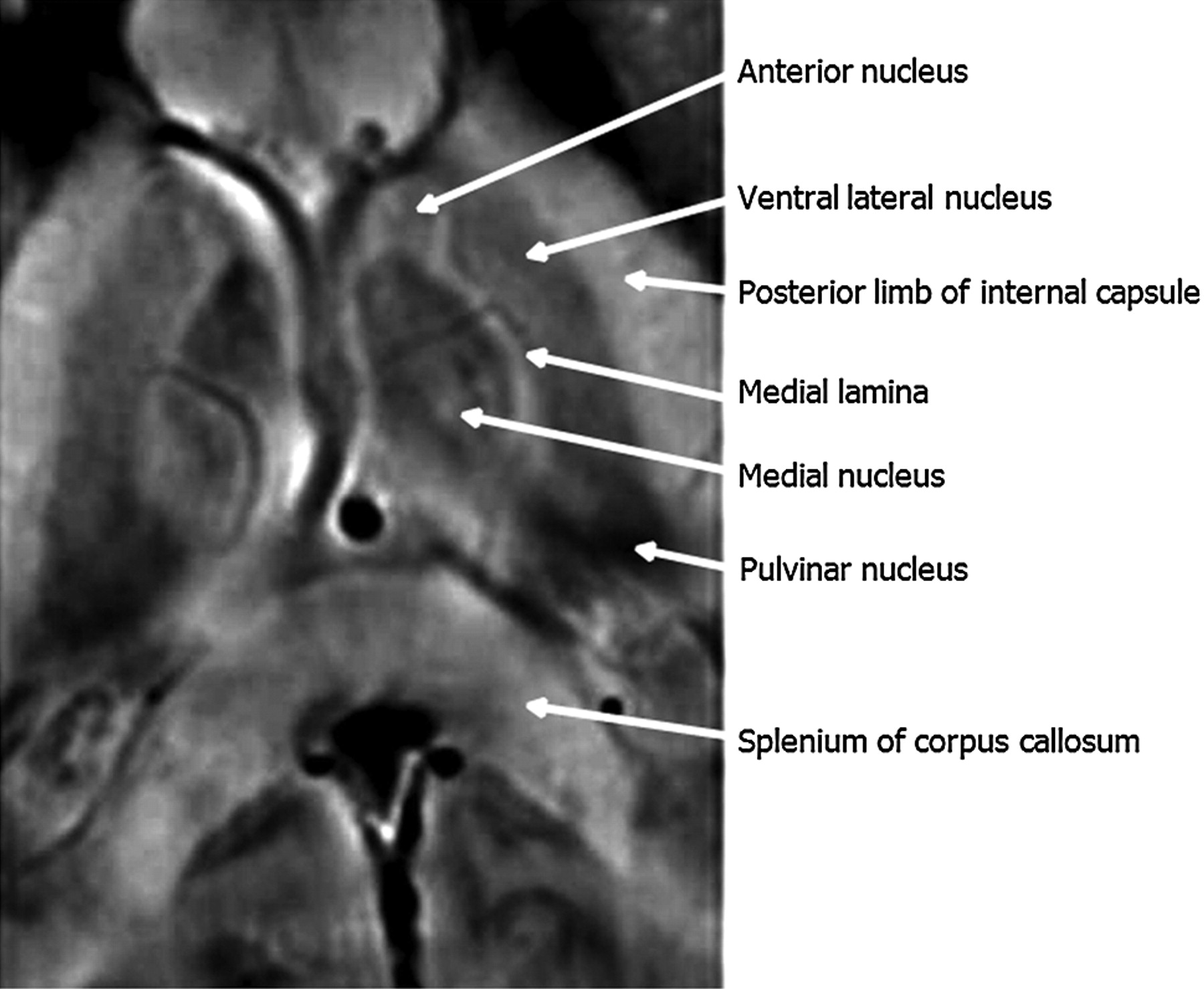
  • Fig 13.
    • Download figure
    • Open in new tab
    • Download powerpoint
    Fig 13.

    Detailed visualization of the thalamic structures by using SWI filtered-phase images in a patient with MS.

  • Fig 14.
    • Download figure
    • Open in new tab
    • Download powerpoint
    Fig 14.

    Another MS case shows a ringlike structure in the SWI filtered-phase image. The lesion of interest is highlighted by the either a white or black arrow. A−D, The T2 (A), T1 (B), original magnitude (C), and SWI filtered-phase (D) images show different lesion contrast. Iron appears in a ring structure with a smaller amount of iron scattered inside the lesion.
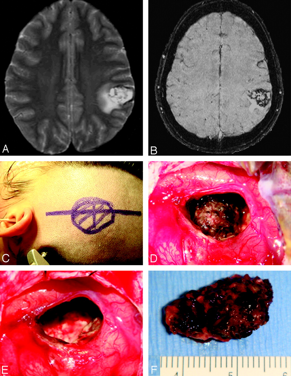
  • Fig 15.
    • Download figure
    • Open in new tab
    • Download powerpoint
    Fig 15.

    A 24-year-old Hispanic woman presenting with partial sensorimotor seizures. A, Axial T2-weighted FSE image shows a left parietal cavernous malformation. B, Because of its exquisite sensitivity in identifying venous structures and blood products, SWI provides a more complete evaluation of patients with CCM. C−F, Intraoperative photographs show circumferential dissection and complete resection of the left parietal cavernous malformation by using microsurgical techniques.

  • Fig 16.
    • Download figure
    • Open in new tab
    • Download powerpoint
    Fig 16.

    A 25-year-old man evaluated for long-term migraine problems. A, T2-weighted image shows a small vascular flow void (arrow) surrounded by an ill-defined region of mixed intensities in the left superior frontal gyrus. B, SWI depicts numerous fine well-delineated anomalous medullary veins that converge into a dilated transcortical collector vein. This forms the classic “caput medusa.” C, Postcontrast 3D T1-weighted image reveals the dilated medullary veins and an enlarged collector vein in excellent agreement with SWI.

  • Fig 17.
    • Download figure
    • Open in new tab
    • Download powerpoint
    Fig 17.

    SWS in a 5-year-old girl. A, The postcontrast T1-weighted image shows enhancement of the leptomeninges (white arrowhead) and periventricular veins (arrow). B, SWI shows extensive hypointensity along the gyri (arrowhead and dotted arrow) due to calcification of the gyri. The abnormal periventricular veins (arrow) shows better detail than the postcontrast T1-weighted image (A).

  • Fig 18.
    • Download figure
    • Open in new tab
    • Download powerpoint
    Fig 18.

    Cerebral venous sinus thrombosis. A and B, Precontrast T1-weighted (A) and T2-weighted (B) images show normal cerebral parenchyma except in the sagittal sinus (arrow), where a hyperintense (T1) and isointense (T2) thrombus is replacing the normal low signal intensity of flowing blood. C, SWI shows significant engorgement of the venous system of the whole brain because of venous hypertension secondary to CVST. D, Three months later (after thrombolytic therapy), SWI shows a return to a normal-appearing venous system. Courtesy of Dr Guangbin Wang, Shandong Medical Imaging Research Institute, Jinan, China.
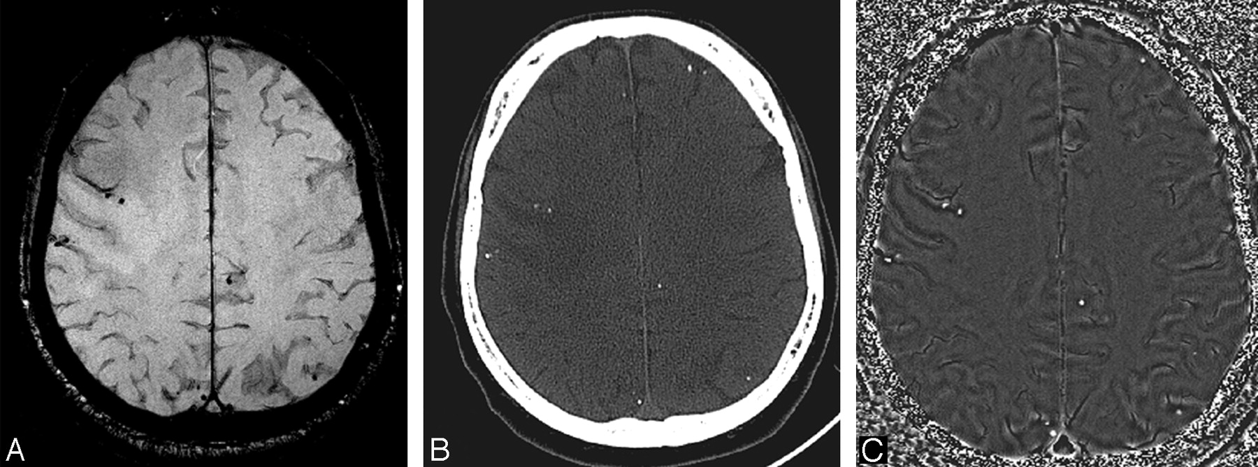
  • Fig 19.
    • Download figure
    • Open in new tab
    • Download powerpoint
    Fig 19.

    Conventional GE magnitude images show both calcifications and hemorrhage as areas of hypointense signal intensity. A, This is equally true for the SWI magnitude image with a TE of 40 ms at 1.5T. B, Usually, a CT scan is used to identify calcium. Here, the CT scan reveals many small calcifications. The SWI filtered-phase image is a means to differentiate paramagnetic from diamagnetic substances because the former will have a negative phase and the latter a positive phase (for a right-handed system). C, The phase image associated with the magnitude image in A shows a 1-to-1 correspondence of the positive phase with the calcifications in the CT scan (B).

  • Fig 20.
    • Download figure
    • Open in new tab
    • Download powerpoint
    Fig 20.

    Left temporo-occipital high-grade glioma. SWI contrast-enhanced image (C) shows similar boundary information and considerably more detail of the internal architecture of the tumor compared with the T1-weighted precontrast (A) or postcontrast image (B).

  • Fig 21.
    • Download figure
    • Open in new tab
    • Download powerpoint
    Fig 21.

    Ill-defined right temporal tumor histologically proved to be a glioblastoma. Compared with contrast-enhanced T1 (A), precontrast T1 (B), and FSE T2 (D) images, SWI (C) and phase images (E and F) are more sensitive in outlining areas of hemorrhage as well as the venous structures and thus can be a useful adjunct in defining the tumor characteristics. The phase images are shown for left-handed (E) and right-handed systems (F).

  • Fig 22.
    • Download figure
    • Open in new tab
    • Download powerpoint
    Fig 22.

    A−C, Left temporal glioblastoma. High-grade gliomas are well depicted on usual diagnostic sequences. D−F, However, SWI and phase images provide valuable additional information on the internal vascular architecture and presence of microhemorrhages within the tumor; F, phase image for a left-handed system.

  • Fig 23.
    • Download figure
    • Open in new tab
    • Download powerpoint
    Fig 23.

    Left thalamic glioma. The size, location, and surrounding edema are well shown on the T1-weighted (A) and T2-weighted images (C). B, However, SWI delineates the interior structure and hemorrhages inside the tumors. D, The SWI phase image shows that the thalamostriate veins are compressed to the right by the tumor.

  • Fig 24.
    • Download figure
    • Open in new tab
    • Download powerpoint
    Fig 24.

    A meningioma located in the left posterior horn of the lateral ventricle. A, The tumor is diffusely enhanced in the postcontrast T1-weighted image. B, SWI delineates the veins around the tumor.
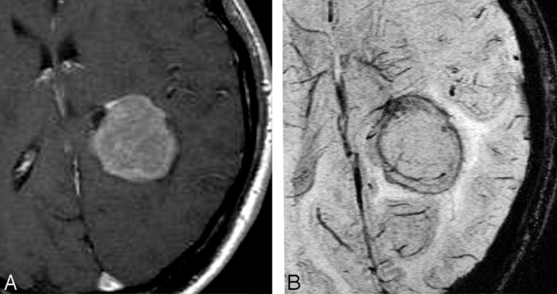
  • Fig 25.
    • Download figure
    • Open in new tab
    • Download powerpoint
    Fig 25.

    A tumor located in the posterior horn of the left lateral ventricle. A, Postcontrast T1-weighted image shows enhancement of the tumor periphery. B, Precontrast SWI shows a small hemorrhage inside the tumor (arrow). C, In postcontrast SWI, a new hypointense signal intensity appears after contrast agent injection, which indicates the presence of intratumoral vessels; the signal intensity from the hemorrhage inside the tumor remains unchanged (arrow).

  • Fig 26.
    • Download figure
    • Open in new tab
    • Download powerpoint
    Fig 26.

    An oligodendroglioma in the right frontoinsular region. A, CT scan shows patchy calcification inside the tumor. B, SWI magnitude image shows hypointense signal intensity inside the tumor but cannot identify whether the hypointensity is hemorrhage or calcification. C, On the SWI phase image, calcification is identified and matches the CT results well.

  • Fig 27.
    • Download figure
    • Open in new tab
    • Download powerpoint
    Fig 27.

    Bilateral periventricular metastases from renal cell carcinoma treated with gamma knife radiosurgery. SWI (A) effectively identifies areas of microhemorrhages consistent with radiation necrosis, compared with the contrast-enhanced T1-weighted image (B).

PreviousNext
Back to top

In this issue

American Journal of Neuroradiology: 30 (2)
American Journal of Neuroradiology
Vol. 30, Issue 2
February 2009
  • Table of Contents
  • Index by author
Advertisement
Print
Download PDF
Email Article
Cite this article
0 Responses
Respond to this article
Share
Bookmark this article
Susceptibility-Weighted Imaging: Technical Aspects and Clinical Applications, Part 2
S. Mittal, Z. Wu, J. Neelavalli, E.M. Haacke
American Journal of Neuroradiology Feb 2009, 30 (2) 232-252; DOI: 10.3174/ajnr.A1461
del.icio.us logo Twitter logo Facebook logo Mendeley logo
  • Tweet Widget
Purchase

Jump to section

  • Article
    • Abstract
    • TBI: Diffuse Axonal Injury
    • Stroke
    • Neurodegenerative Disorders
    • MS
    • Vascular Malformations and Venous Disease
    • Neurocysticercosis
    • Brain Tumors
    • Future Directions
    • Acknowledgments
    • Footnotes
    • References
  • Figures & Data
  • Info & Metrics
  • Responses
  • References
  • PDF

Related Articles

  • No related articles found.
  • PubMed
  • Google Scholar

Cited By...

  • The relationship of white matter tract orientation to vascular geometry in the human brain
  • Know your stroke mimics
  • Brain Alterations in COVID Recovered Revealed by Susceptibility-Weighted Magnetic Resonance Imaging
  • CT-like images in MRI improve specificity of erosion detection in patients with hand arthritis: a diagnostic accuracy study with CT as standard of reference
  • Adapting the UK Biobank brain imaging protocol and analysis pipeline for the C-MORE multi-organ study of COVID-19 survivors
  • Susceptibility vessel sign as a predictor for recanalization and clinical outcome in acute ischaemic stroke: A Systematic Review and Meta-analysis
  • Medium-term effects of SARS-CoV-2 infection on multiple vital organs, exercise capacity, cognition, quality of life and mental health, post-hospital discharge
  • Black Dipole or White Dipole: Using Susceptibility Phase Imaging to Differentiate Cerebral Microbleeds from Intracranial Calcifications
  • The index vein pointing to the origin of the migraine aura symptom: A case series
  • Direct thromboaspiration efficacy for mechanical thrombectomy is related to the angle of interaction between the aspiration catheter and the clot
  • Frequency, Extent, and Correlates of Superficial Siderosis and Ependymal Siderosis in Premature Infants with Germinal Matrix Hemorrhage: An SWI Study
  • Validation of Highly Accelerated Wave-CAIPI SWI Compared with Conventional SWI and T2*-Weighted Gradient Recalled-Echo for Routine Clinical Brain MRI at 3T
  • Increased Diameters of the Internal Cerebral Veins and the Basal Veins of Rosenthal Are Associated with White Matter Hyperintensity Volume
  • Gadolinium-Enhanced Susceptibility-Weighted Imaging in Multiple Sclerosis: Optimizing the Recognition of Active Plaques for Different MR Imaging Sequences
  • SWI filtered-phase imaging in calcific cerebral embolism secondary to cardiac myxoma
  • Engorgement of Deep Medullary Veins in Neurosarcoidosis: A Common-Yet-Underrecognized Cerebrovascular Finding on SWI
  • The Bone Does Not Predict the Brain in Sturge-Weber Syndrome
  • Quantitative Susceptibility Mapping after Sports-Related Concussion
  • Spatiotemporal evolution of venous narrowing in acute MS lesions
  • In Vivo Imaging of Venous Side Cerebral Small-Vessel Disease in Older Adults: An MRI Method at 7T
  • Arterial Spin Labeling Perfusion Magnetic Resonance Imaging Performed in Acute Perinatal Stroke Reveals Hyperperfusion Associated With Ischemic Injury
  • Prominence of Medullary Veins on Susceptibility-Weighted Images Provides Prognostic Information in Patients with Subacute Stroke
  • Susceptibility-weighted imaging in parenchymal neurosyphilis: identification of a new MRI finding
  • Susceptibility-weighted imaging in Todd paralysis
  • SWI or T2*: Which MRI Sequence to Use in the Detection of Cerebral Microbleeds? The Karolinska Imaging Dementia Study
  • Susceptibility-Weighted Imaging in Pediatric Arterial Ischemic Stroke: A Valuable Alternative for the Noninvasive Evaluation of Altered Cerebral Hemodynamics
  • Persistent Cerebrovascular Damage After Stroke in Type Two Diabetic Rats Measured by Magnetic Resonance Imaging
  • Double Inversion Recovery MR Sequence for the Detection of Subacute Subarachnoid Hemorrhage
  • MR Quantitative Susceptibility Imaging for the Evaluation of Iron Loading in the Brains of Patients with {beta}-Thalassemia Major
  • Improved T2* Imaging without Increase in Scan Time: SWI Processing of 2D Gradient Echo
  • Susceptibility-Weighted Imaging is More Reliable Than T2*-Weighted Gradient-Recalled Echo MRI for Detecting Microbleeds
  • Current differential diagnoses and treatment options of vascular occlusions presenting as bilateral thalamic infarcts: a review of the literature
  • The venous angioarchitecture of sporadic cerebral cavernous malformations: a susceptibility weighted imaging study at 7 T MRI
  • Progressive Brain Iron Accumulation in Neuroferritinopathy Measured by the Thalamic T2* Relaxation Rate
  • Differentiation of Pyogenic Brain Abscesses from Necrotic Glioblastomas with Use of Susceptibility-Weighted Imaging
  • Developmental venous anomaly coexisting with a true arteriovenous malformation: a rare clinical entity
  • Susceptibility-Weighted Imaging in Patients with Pyogenic Brain Abscesses at 1.5T: Characteristics of the Abscess Capsule
  • Detection of Intratumoral Calcification in Oligodendrogliomas by Susceptibility-Weighted MR Imaging
  • Sporadic cerebral amyloid angiopathy revisited: recent insights into pathophysiology and clinical spectrum
  • Simultaneous Arteriovenous Shunting and Venous Congestion Identification in Dural Arteriovenous Fistulas Using Susceptibility-Weighted Imaging: Initial Experience
  • The Agfa Mayneord lecture: MRI of short and ultrashort T2 and T2* components of tissues, fluids and materials using clinical systems
  • Evaluation of Parenchymal Neuro-Behcet Disease by Using Susceptibility-Weighted Imaging
  • Early Evaluation of Tumoral Response to Antiangiogenic Therapy by Arterial Spin Labeling Perfusion Magnetic Resonance Imaging and Susceptibility Weighted Imaging in a Patient With Recurrent Glioblastoma Receiving Bevacizumab
  • Suspicious Neuroimaging Pattern of Thrombotic Microangiopathy
  • Susceptibility-Weighted MR Phase Imaging Can Demonstrate Retrograde Leptomeningeal Venous Drainage in Patients with Dural Arteriovenous Fistula
  • Cerebral Microhemorrhages Detected by Susceptibility-Weighted Imaging in Amateur Boxers
  • Susceptibility-Weighted Imaging in Migraine with Aura
  • Distinguishing Recurrent Primary Brain Tumor from Radiation Injury: A Preliminary Study Using a Susceptibility-Weighted MR Imaging-Guided Apparent Diffusion Coefficient Analysis Strategy
  • Familial versus Sporadic Cavernous Malformations: Differences in Developmental Venous Anomaly Association and Lesion Phenotype
  • Localization of the Subthalamic Nucleus: Optimization with Susceptibility-Weighted Phase MR Imaging
  • Added Value and Diagnostic Performance of Intratumoral Susceptibility Signals in the Differential Diagnosis of Solitary Enhancing Brain Lesions: Preliminary Study
  • Semiquantitative Assessment of Intratumoral Susceptibility Signals Using Non-Contrast-Enhanced High-Field High-Resolution Susceptibility-Weighted Imaging in Patients with Gliomas: Comparison with MR Perfusion Imaging
  • Hemorrhage in Posterior Reversible Encephalopathy Syndrome: Imaging and Clinical Features
  • Crossref
  • Google Scholar

This article has not yet been cited by articles in journals that are participating in Crossref Cited-by Linking.

More in this TOC Section

  • Theoretic Basis and Technical Implementations of CT Perfusion in Acute Ischemic Stroke, Part 2: Technical Implementations
  • Theoretic Basis and Technical Implementations of CT Perfusion in Acute Ischemic Stroke, Part 1: Theoretic Basis
Show more PHYSICS REVIEW

Similar Articles

Advertisement

Indexed Content

  • Current Issue
  • Accepted Manuscripts
  • Article Preview
  • Past Issues
  • Editorials
  • Editor's Choice
  • Fellows' Journal Club
  • Letters to the Editor
  • Video Articles

Cases

  • Case Collection
  • Archive - Case of the Week
  • Archive - Case of the Month
  • Archive - Classic Case

More from AJNR

  • Trainee Corner
  • Imaging Protocols
  • MRI Safety Corner
  • Book Reviews

Multimedia

  • AJNR Podcasts
  • AJNR Scantastics

Resources

  • Turnaround Time
  • Submit a Manuscript
  • Submit a Video Article
  • Submit an eLetter to the Editor/Response
  • Manuscript Submission Guidelines
  • Statistical Tips
  • Fast Publishing of Accepted Manuscripts
  • Graphical Abstract Preparation
  • Imaging Protocol Submission
  • Evidence-Based Medicine Level Guide
  • Publishing Checklists
  • Author Policies
  • Become a Reviewer/Academy of Reviewers
  • News and Updates

About Us

  • About AJNR
  • Editorial Board
  • Editorial Board Alumni
  • Alerts
  • Permissions
  • Not an AJNR Subscriber? Join Now
  • Advertise with Us
  • Librarian Resources
  • Feedback
  • Terms and Conditions
  • AJNR Editorial Board Alumni

American Society of Neuroradiology

  • Not an ASNR Member? Join Now

© 2025 by the American Society of Neuroradiology All rights, including for text and data mining, AI training, and similar technologies, are reserved.
Print ISSN: 0195-6108 Online ISSN: 1936-959X

Powered by HighWire
Email this Article

Thank you for your interest in spreading the word on American Journal of Neuroradiology.

NOTE: We only request your email address so that the person you are recommending the page to knows that you wanted them to see it, and that it is not junk mail. We do not capture any email address.

Enter multiple addresses on separate lines or separate them with commas.
Susceptibility-Weighted Imaging: Technical Aspects and Clinical Applications, Part 2
(Your Name) has sent you a message from American Journal of Neuroradiology
(Your Name) thought you would like to see the American Journal of Neuroradiology web site.
CAPTCHA
This question is for testing whether or not you are a human visitor and to prevent automated spam submissions.
Cite this article
S. Mittal, Z. Wu, J. Neelavalli, E.M. Haacke
Susceptibility-Weighted Imaging: Technical Aspects and Clinical Applications, Part 2
American Journal of Neuroradiology Feb 2009, 30 (2) 232-252; DOI: 10.3174/ajnr.A1461

Citation Manager Formats

  • BibTeX
  • Bookends
  • EasyBib
  • EndNote (tagged)
  • EndNote 8 (xml)
  • Medlars
  • Mendeley
  • Papers
  • RefWorks Tagged
  • Ref Manager
  • RIS
  • Zotero

We use cookies on this site to enhance your user experience. By clicking any link on this page you are giving your consent for us to set cookies.