Advertisement
  • Main menu
  • User menu
  • Search
  • English ▼
    • English
    • Afrikaans
    • Albanian
    • Amharic
    • Arabic
    • Armenian
    • Azerbaijani
    • Basque
    • Belarusian
    • Bengali
    • Bosnian
    • Bulgarian
    • Catalan
    • Cebuano
    • Chichewa
    • Chinese (Simplified)
    • Chinese (Traditional)
    • Corsican
    • Croatian
    • Czech
    • Danish
    • Dutch
    • Esperanto
    • Estonian
    • Filipino
    • Finnish
    • French
    • Frisian
    • Galician
    • Georgian
    • German
    • Greek
    • Gujarati
    • Haitian Creole
    • Hausa
    • Hawaiian
    • Hebrew
    • Hindi
    • Hmong
    • Hungarian
    • Icelandic
    • Igbo
    • Indonesian
    • Irish
    • Italian
    • Japanese
    • Javanese
    • Kannada
    • Kazakh
    • Khmer
    • Korean
    • Kurdish (Kurmanji)
    • Kyrgyz
    • Lao
    • Latin
    • Latvian
    • Lithuanian
    • Luxembourgish
    • Macedonian
    • Malagasy
    • Malay
    • Malayalam
    • Maltese
    • Maori
    • Marathi
    • Mongolian
    • Myanmar (Burmese)
    • Nepali
    • Norwegian
    • Pashto
    • Persian
    • Polish
    • Portuguese
    • Punjabi
    • Romanian
    • Russian
    • Samoan
    • Scottish Gaelic
    • Serbian
    • Sesotho
    • Shona
    • Sindhi
    • Sinhala
    • Slovak
    • Slovenian
    • Somali
    • Spanish
    • Sudanese
    • Swahili
    • Swedish
    • Tajik
    • Tamil
    • Telugu
    • Thai
    • Turkish
    • Ukrainian
    • Urdu
    • Uzbek
    • Vietnamese
    • Welsh
    • Xhosa
    • Yiddish
    • Yoruba
    • Zulu

Main menu

  • Home
  • Content
    • Current Issue
    • Accepted Manuscripts
    • Article Preview
    • Past Issue Archive
    • Video Articles
    • AJNR Case Collection
    • Case of the Week Archive
    • Case of the Month Archive
    • Classic Case Archive
  • Special Collections
    • Advancing NeuroMRI with High-Relaxivity Contrast Agents
    • AJNR Awards
    • Low-Field MRI
    • Alzheimer Disease
    • ASNR Foundation Special Collection
    • Photon-Counting CT
    • View All
  • Multimedia
    • AJNR Podcasts
    • AJNR SCANtastic
    • Trainee Corner
    • MRI Safety Corner
    • Imaging Protocols
  • For Authors
    • Submit a Manuscript
    • Submit a Video Article
    • Submit an eLetter to the Editor/Response
    • Manuscript Submission Guidelines
    • Statistical Tips
    • Fast Publishing of Accepted Manuscripts
    • Graphical Abstract Preparation
    • Imaging Protocol Submission
    • Author Policies
  • About Us
    • About AJNR
    • Editorial Board
    • Editorial Board Alumni
  • More
    • Become a Reviewer/Academy of Reviewers
    • Subscribers
    • Permissions
    • Alerts
    • Feedback
    • Advertisers
    • ASNR Home

User menu

  • Alerts
  • Log in

Search

  • Advanced search
American Journal of Neuroradiology

American Journal of Neuroradiology

ASHNR American Society of Functional Neuroradiology ASHNR American Society of Pediatric Neuroradiology ASSR
  • Alerts
  • Log in

Advanced Search

English ▼
  • English
  • Afrikaans
  • Albanian
  • Amharic
  • Arabic
  • Armenian
  • Azerbaijani
  • Basque
  • Belarusian
  • Bengali
  • Bosnian
  • Bulgarian
  • Catalan
  • Cebuano
  • Chichewa
  • Chinese (Simplified)
  • Chinese (Traditional)
  • Corsican
  • Croatian
  • Czech
  • Danish
  • Dutch
  • Esperanto
  • Estonian
  • Filipino
  • Finnish
  • French
  • Frisian
  • Galician
  • Georgian
  • German
  • Greek
  • Gujarati
  • Haitian Creole
  • Hausa
  • Hawaiian
  • Hebrew
  • Hindi
  • Hmong
  • Hungarian
  • Icelandic
  • Igbo
  • Indonesian
  • Irish
  • Italian
  • Japanese
  • Javanese
  • Kannada
  • Kazakh
  • Khmer
  • Korean
  • Kurdish (Kurmanji)
  • Kyrgyz
  • Lao
  • Latin
  • Latvian
  • Lithuanian
  • Luxembourgish
  • Macedonian
  • Malagasy
  • Malay
  • Malayalam
  • Maltese
  • Maori
  • Marathi
  • Mongolian
  • Myanmar (Burmese)
  • Nepali
  • Norwegian
  • Pashto
  • Persian
  • Polish
  • Portuguese
  • Punjabi
  • Romanian
  • Russian
  • Samoan
  • Scottish Gaelic
  • Serbian
  • Sesotho
  • Shona
  • Sindhi
  • Sinhala
  • Slovak
  • Slovenian
  • Somali
  • Spanish
  • Sudanese
  • Swahili
  • Swedish
  • Tajik
  • Tamil
  • Telugu
  • Thai
  • Turkish
  • Ukrainian
  • Urdu
  • Uzbek
  • Vietnamese
  • Welsh
  • Xhosa
  • Yiddish
  • Yoruba
  • Zulu
  • Home
  • Content
    • Current Issue
    • Accepted Manuscripts
    • Article Preview
    • Past Issue Archive
    • Video Articles
    • AJNR Case Collection
    • Case of the Week Archive
    • Case of the Month Archive
    • Classic Case Archive
  • Special Collections
    • Advancing NeuroMRI with High-Relaxivity Contrast Agents
    • AJNR Awards
    • Low-Field MRI
    • Alzheimer Disease
    • ASNR Foundation Special Collection
    • Photon-Counting CT
    • View All
  • Multimedia
    • AJNR Podcasts
    • AJNR SCANtastic
    • Trainee Corner
    • MRI Safety Corner
    • Imaging Protocols
  • For Authors
    • Submit a Manuscript
    • Submit a Video Article
    • Submit an eLetter to the Editor/Response
    • Manuscript Submission Guidelines
    • Statistical Tips
    • Fast Publishing of Accepted Manuscripts
    • Graphical Abstract Preparation
    • Imaging Protocol Submission
    • Author Policies
  • About Us
    • About AJNR
    • Editorial Board
    • Editorial Board Alumni
  • More
    • Become a Reviewer/Academy of Reviewers
    • Subscribers
    • Permissions
    • Alerts
    • Feedback
    • Advertisers
    • ASNR Home
  • Follow AJNR on Twitter
  • Visit AJNR on Facebook
  • Follow AJNR on Instagram
  • Join AJNR on LinkedIn
  • RSS Feeds

AJNR Awards, New Junior Editors, and more. Read the latest AJNR updates


Improved Turnaround Times | Median time to first decision: 12 days

Research ArticleBrain
Open Access

Metachromatic Leukodystrophy: A Scoring System for Brain MR Imaging Observations

F. Eichler, W. Grodd, E. Grant, M. Sessa, A. Biffi, A. Bley, A. Kohlschuetter, D.J. Loes and I. Kraegeloh-Mann
American Journal of Neuroradiology November 2009, 30 (10) 1893-1897; DOI: https://doi.org/10.3174/ajnr.A1739
F. Eichler
  • Find this author on Google Scholar
  • Find this author on PubMed
  • Search for this author on this site
W. Grodd
  • Find this author on Google Scholar
  • Find this author on PubMed
  • Search for this author on this site
E. Grant
  • Find this author on Google Scholar
  • Find this author on PubMed
  • Search for this author on this site
M. Sessa
  • Find this author on Google Scholar
  • Find this author on PubMed
  • Search for this author on this site
A. Biffi
  • Find this author on Google Scholar
  • Find this author on PubMed
  • Search for this author on this site
A. Bley
  • Find this author on Google Scholar
  • Find this author on PubMed
  • Search for this author on this site
A. Kohlschuetter
  • Find this author on Google Scholar
  • Find this author on PubMed
  • Search for this author on this site
D.J. Loes
  • Find this author on Google Scholar
  • Find this author on PubMed
  • Search for this author on this site
I. Kraegeloh-Mann
  • Find this author on Google Scholar
  • Find this author on PubMed
  • Search for this author on this site
  • Article
  • Figures & Data
  • Info & Metrics
  • Responses
  • References
  • PDF
Loading

Article Figures & Data

Figures

  • Tables
  • Fig 1.
    • Download figure
    • Open in new tab
    • Download powerpoint
    Fig 1.

    Scoring details of the metachromatic leukodystrophy (MLD) MR imaging severity score. Differences between scoring faint (1 point) versus dense appearance (2 points) are shown. A, Periventricular and central involvement in the frontal white matter is categorized as faint (1 point, thin arrow), whereas myelination is preserved in the subcortical U-fibers (0 points, thick arrow). B, Periventricular and central involvement in the frontal white matter is categorized as dense (2 points, thin arrow), and there are areas that lack subcortical U-fiber myelination (2 points, thick arrow). C, Inner atrophy as measured in the third ventricle (arrow).
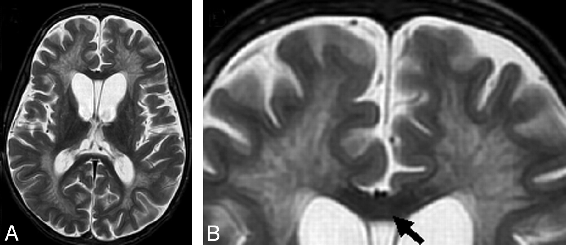
  • Fig 2.
    • Download figure
    • Open in new tab
    • Download powerpoint
    Fig 2.

    Midline of the corpus callosum in advanced stages of MLD. T2 lesion hyperintensities in the midline are reduced compared with the adjacent supratentorial white matter lesion signal intensity (arrow).

  • Fig 3.
    • Download figure
    • Open in new tab
    • Download powerpoint
    Fig 3.

    Correlation of age and MR imaging severity score in patients with MLD. The greatest variability in MR imaging lesion severity is seen in the first decade of life. Thereafter, most patients have a severity score of >20. Patients with serial scans have scores with connecting lines.

Tables

  • Figures
    • View popup
    Table 1:

    Brain MR imaging scoring system for MLD

    Brain AreasScore*Maximum per Area
    Frontal WM6†
        Periventricular012
        Central012
        U-fibers012
    Parieto-occipital WM6†
        Periventricular012
        Central012
        U-fibers012
    Temporal WM6†
        Periventricular012
        Central012
        U-fibers012
    Corpus callosum4†
        Genu012
        Splenium012
    Projection fibers6†
        Internal capsule posterior limb012
        Internal capsule anterior limb012
        Midline pons012
    Cerebral atrophy0122†
    Thalamus011†
    Basal ganglia011†
    Cerebellum2†
        WM01
        Atrophy01
    Total34†
    • Note:—MLD indicates metachromatic leukodystrophy; WM, white matter.

    • * 0 indicates normal; 1, faint hyperintensity; 2, dense hyperintensity.

    • † XXX.

    • View popup
    Table 2:

    Subtypes and initial MR imaging severity scores of patients with MLD

    Patient No.Phenotype*Age at MRIDeep Gray NucleiProjection FibersCerebellumAtrophyMRI Severity Score
    1Late infantile1.92NoNoNoNo1
    2Late infantile2.08YesNoNoNo17
    3Late infantile2.5NoNoNoNo4
    4Late infantile2.5NoNoNoNo4
    5Late infantile2.5NoNoNoNo4
    6Late infantile3NoNoNoNo6
    7Late infantile2.08NoNoNoNo0
    8Late infantile3.66YesNoNoNo21
    9Late infantile7.16YesYesYesYes30
    10Late infantile9.6YesYesYesYes32
    11Juvenile4.84NoNoNoNo12
    12Juvenile4.84NoNoNoNo12
    13Juvenile8.08YesNoNoNo13
    14Juvenile10.25NoNoNoNo3
    15Juvenile3NoNoNoNo2
    16Juvenile4.17NoNoNoNo6
    17Juvenile4.17NoNoNoNo10
    18Juvenile5.66YesYesNoYes26
    19Juvenile6.25YesYesNoYes27
    20Juvenile7NoNoNoNo18
    21Juvenile7.75NoNoNoYes19
    22Juvenile8.25YesYesYesYes30
    23Juvenile9.4NoNoNoNo12
    24Juvenile13.08YesYesNoYes26
    25Juvenile18.42YesYesYesYes28
    26Juvenile27NoYesNoYes24
    27Adult34.5YesYesNoYes22
    28Adult46YesYesNoYes26
    • Note:—MRI indicates MR imaging.

    • * The individual phenotypes, age as well as the MR imaging severity score with its subscores at initial MR imaging are provided.

PreviousNext
Back to top

In this issue

American Journal of Neuroradiology: 30 (10)
American Journal of Neuroradiology
Vol. 30, Issue 10
1 Nov 2009
  • Table of Contents
  • Index by author
Advertisement
Print
Download PDF
Email Article
Cite this article
0 Responses
Respond to this article
Share
Bookmark this article
Metachromatic Leukodystrophy: A Scoring System for Brain MR Imaging Observations
F. Eichler, W. Grodd, E. Grant, M. Sessa, A. Biffi, A. Bley, A. Kohlschuetter, D.J. Loes, I. Kraegeloh-Mann
American Journal of Neuroradiology Nov 2009, 30 (10) 1893-1897; DOI: 10.3174/ajnr.A1739
del.icio.us logo Twitter logo Facebook logo Mendeley logo
  • Tweet Widget
Purchase

Jump to section

  • Article
    • Abstract
    • Materials and Methods
    • Results
    • Discussion
    • Conclusions
    • Acknowledgments
    • Footnotes
    • References
  • Figures & Data
  • Info & Metrics
  • Responses
  • References
  • PDF

Related Articles

  • No related articles found.
  • PubMed
  • Google Scholar

Cited By...

  • AAV-mediated ARSA replacement for the treatment of Metachromatic Leukodystrophy
  • Clinical Reasoning: A 23-Year-Old Woman Presenting With Cognitive Impairment and Gait Disturbance
  • Association of Age at Onset and First Symptoms With Disease Progression in Patients With Metachromatic Leukodystrophy
  • Quantitative MR spectroscopic imaging in metachromatic leukodystrophy: value for prognosis and treatment
  • Efficacy of hematopoietic cell transplantation in metachromatic leukodystrophy: the Dutch experience
  • Cerebral gray and white matter changes and clinical course in metachromatic leukodystrophy
  • Toward a Better Understanding of Brain Lesions during Metachromatic Leukodystrophy Evolution
  • MRI characteristics and scoring in HDLS due to CSF1R gene mutations
  • Crossref
  • Google Scholar

This article has not yet been cited by articles in journals that are participating in Crossref Cited-by Linking.

More in this TOC Section

  • Predictors of Reperfusion in Patients with Acute Ischemic Stroke
  • Enhanced Axonal Metabolism during Early Natalizumab Treatment in Relapsing-Remitting Multiple Sclerosis
  • Progression of Microstructural Damage in Spinocerebellar Ataxia Type 2: A Longitudinal DTI Study
Show more Brain

Similar Articles

Advertisement

Indexed Content

  • Current Issue
  • Accepted Manuscripts
  • Article Preview
  • Past Issues
  • Editorials
  • Editor's Choice
  • Fellows' Journal Club
  • Letters to the Editor
  • Video Articles

Cases

  • Case Collection
  • Archive - Case of the Week
  • Archive - Case of the Month
  • Archive - Classic Case

More from AJNR

  • Trainee Corner
  • Imaging Protocols
  • MRI Safety Corner
  • Book Reviews

Multimedia

  • AJNR Podcasts
  • AJNR Scantastics

Resources

  • Turnaround Time
  • Submit a Manuscript
  • Submit a Video Article
  • Submit an eLetter to the Editor/Response
  • Manuscript Submission Guidelines
  • Statistical Tips
  • Fast Publishing of Accepted Manuscripts
  • Graphical Abstract Preparation
  • Imaging Protocol Submission
  • Evidence-Based Medicine Level Guide
  • Publishing Checklists
  • Author Policies
  • Become a Reviewer/Academy of Reviewers
  • News and Updates

About Us

  • About AJNR
  • Editorial Board
  • Editorial Board Alumni
  • Alerts
  • Permissions
  • Not an AJNR Subscriber? Join Now
  • Advertise with Us
  • Librarian Resources
  • Feedback
  • Terms and Conditions
  • AJNR Editorial Board Alumni

American Society of Neuroradiology

  • Not an ASNR Member? Join Now

© 2025 by the American Society of Neuroradiology All rights, including for text and data mining, AI training, and similar technologies, are reserved.
Print ISSN: 0195-6108 Online ISSN: 1936-959X

Powered by HighWire
Email this Article

Thank you for your interest in spreading the word on American Journal of Neuroradiology.

NOTE: We only request your email address so that the person you are recommending the page to knows that you wanted them to see it, and that it is not junk mail. We do not capture any email address.

Enter multiple addresses on separate lines or separate them with commas.
Metachromatic Leukodystrophy: A Scoring System for Brain MR Imaging Observations
(Your Name) has sent you a message from American Journal of Neuroradiology
(Your Name) thought you would like to see the American Journal of Neuroradiology web site.
CAPTCHA
This question is for testing whether or not you are a human visitor and to prevent automated spam submissions.
Cite this article
F. Eichler, W. Grodd, E. Grant, M. Sessa, A. Biffi, A. Bley, A. Kohlschuetter, D.J. Loes, I. Kraegeloh-Mann
Metachromatic Leukodystrophy: A Scoring System for Brain MR Imaging Observations
American Journal of Neuroradiology Nov 2009, 30 (10) 1893-1897; DOI: 10.3174/ajnr.A1739

Citation Manager Formats

  • BibTeX
  • Bookends
  • EasyBib
  • EndNote (tagged)
  • EndNote 8 (xml)
  • Medlars
  • Mendeley
  • Papers
  • RefWorks Tagged
  • Ref Manager
  • RIS
  • Zotero

We use cookies on this site to enhance your user experience. By clicking any link on this page you are giving your consent for us to set cookies.