
Article Figures & Data
Figures
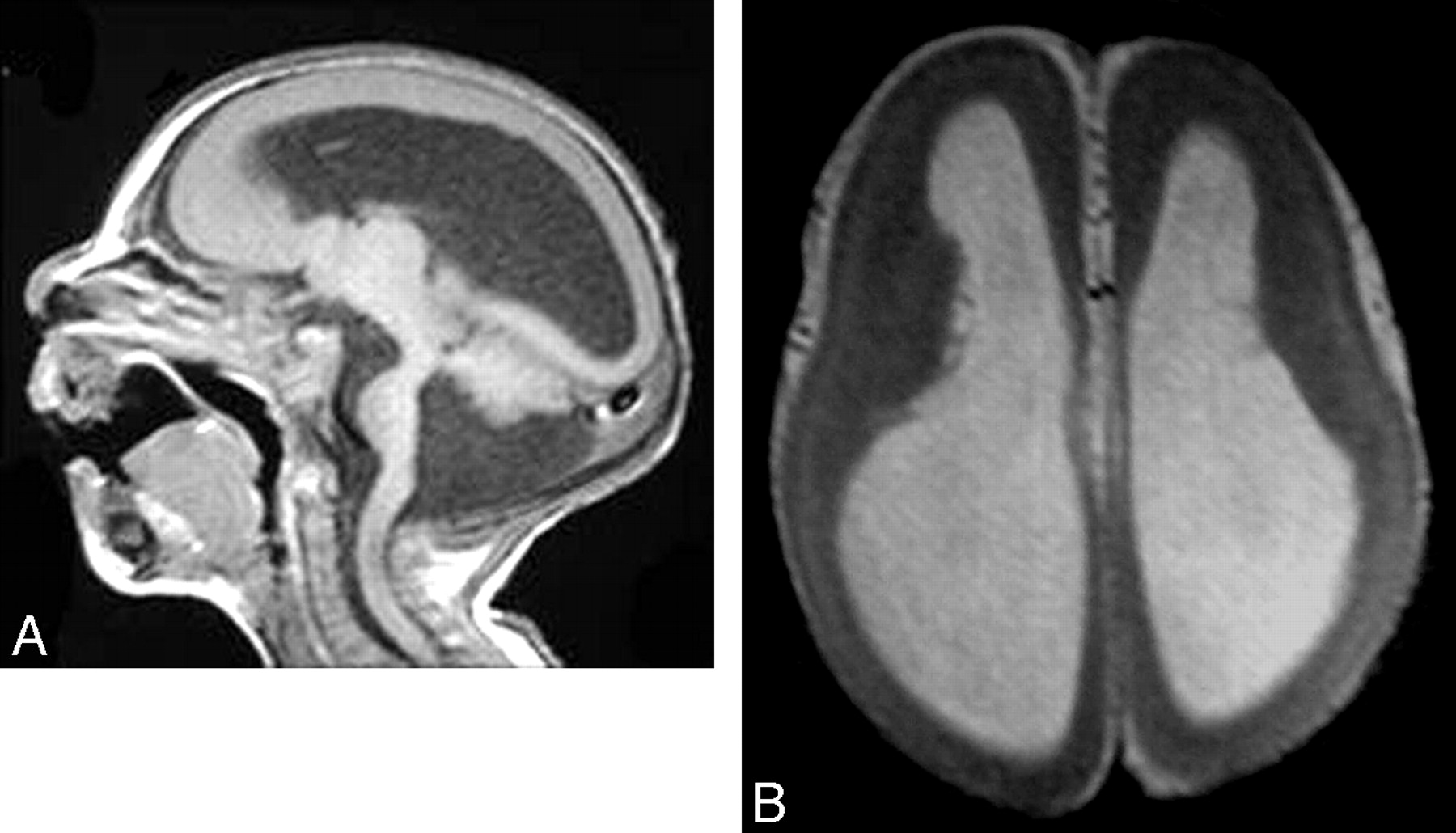
- Fig 1.
Microlissencephaly. A, Sagittal T1-weighted image shows no sulcation in the cerebrum, a hypoplastic cerebellum, and a small brain stem with Dandy-Walker syndrome. B, Axial T1-weighted image shows agyria of the cortex.
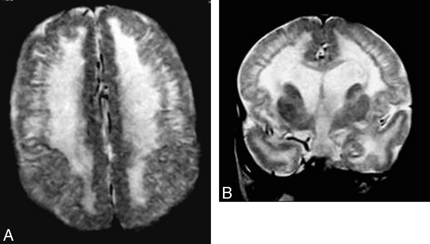
- Fig 2.
Hemimegalencephaly. A and B, Coronal T2-weighted images show an enlarged left cerebral hemisphere, a dilated left lateral ventricle, and a thickened cerebral cortex. Courtesy of Dr R. Zimmerman, Philadelphia.
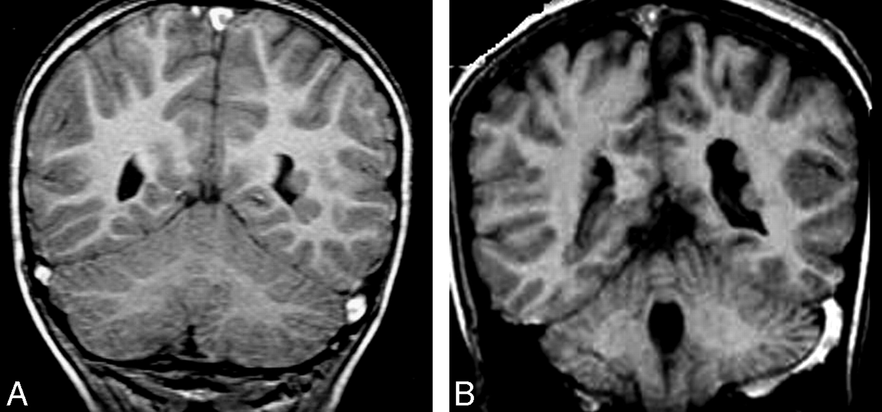
- Fig 3.
Focal cortical dysplasia. A and B, Axial and coronal T2-weighted images show focal cortical thickening in the right frontal lobe. Courtesy of Dr R. Zimmerman, Philadelphia.
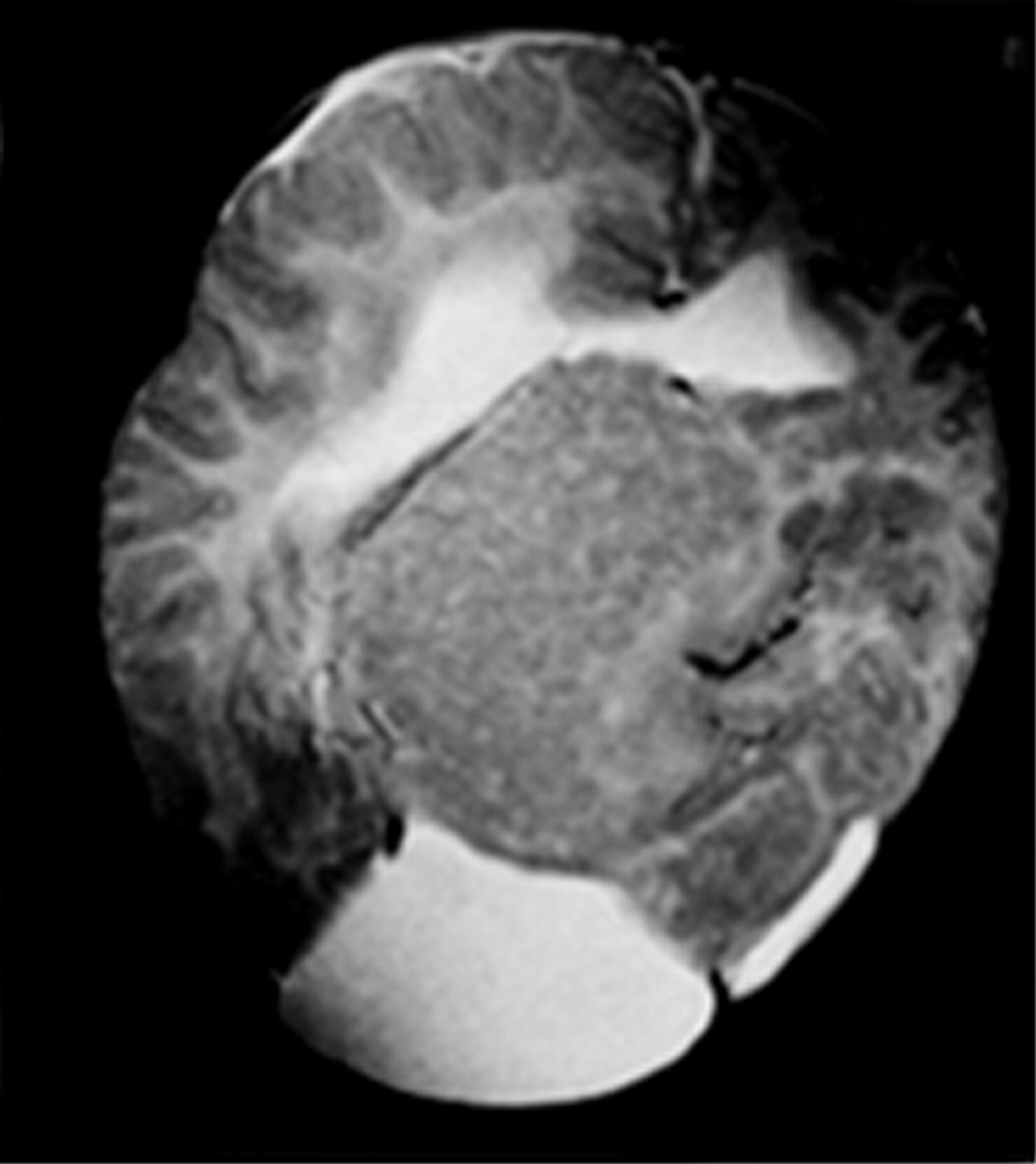
- Fig 4.
Complete lissencephaly. Axial T2-weighted image shows complete absence of sulci with a thick cortex, a shallow Sylvian fissure, and a circumferential band of high signal intensity in the parietooccipital cortex.
- Fig 5.
Cobblestone lissencephaly. A and B, Axial and coronal T2-weighted images show an irregular nodular cortex with hypomyelination of the white matter.
- Fig 6.
WWS. A, Axial T2-weighted image shows a cobblestone cerebral cortex with hypomyelination of the white matter. B, Sagittal T1-weighted image shows hydrocephalus, an occipital encephalocele, a pontomedullary kink, and a small pons.
- Fig 7.
MEB syndrome. A, Axial T2-weighted image shows undersulcation of the frontal lobe, hypomyelination of the frontal white matter, and dilated lateral ventricles. B, Axial T2-weighted image shows multiple small cerebellar cysts, with a small pons and bilateral temporal arachnoid cysts.
- Fig 8.
PVH. A and B, Coronal T1-weighted images show a few small periventricular nodules, isointense to the gray matter, along the lateral ventricular wall.
- Fig 9.
Curvilinear SCH. Axial T2-weighted MR image shows curvilinear heterotopias in the right cerebral hemisphere that are associated with its decrease in size.
- Fig 10.
Mixed SCH. Axial T2-weighted MR image shows a large nodule adjacent to the lateral ventricle with a signal intensity similar to that of the gray matter and curvilinear convolutions in the superficial part.
- Fig 11.
PMG. Axial T2-weighted image shows microgyria with normal cortical thickness that is associated with the high signal intensity of the white matter.
- Fig 12.
Schizencephaly. A, Coronal T2-weighted image shows a closed-lip right-sided schizencephalic defect lined by pachygyria. (Courtesy of Dr R. Zimmerman, Philadelphia.) B, Coronal T2-weighted image shows a wide CSF-filled cleft connecting the left lateral ventricle with the subarachnoid space, which is lined with gray matter parenchyma.


Tables
Disorders of cortical formation according to stages
Stage Cause Disorder Proliferative Decreased proliferation Microlissencephaly Increased proliferation Hemimegalencephaly Abnormal proliferation Focal cortical dysplasia Migration Undermigration Complete (classic) lissencephaly Overmigration Congenital muscular dystrophy Ectopic migration Heterotopia Organization Deranged organization Polymicrogyria Schizencephaly
In this issue


Jump to section
- Article
- Abstract
- Embryology
- Classification
- Microlissencephaly/Microcephaly with a Simplified Gyral Pattern
- Hemimegalencephaly
- FCD
- Classic (Type I) Lissencephaly (4-Layer Lissencephaly)
- Cobblestone (Type II) Lissencephaly (Congenital Muscular Dystrophy)
- Heterotopia
- Periventricular (Subependymal) Heterotopias
- Subcortical Heterotopias
- Band (Laminar) Heterotopia
- PMG
- Schizencephaly
- Conclusion
- Acknowledgments
- Footnotes
- References
- Figures & Data
- Info & Metrics
- Responses
- References
Related Articles
Cited By...
- A Unified Imaging-Histology Framework for Superficial White Matter Architecture Studies in the Human Brain
- Cortical malformation adjacent to a large pial arteriovenous fistula
- Using Correlative Properties of Neighboring Pixels to Improve Gray-White Differentiation in Pediatric Head CT Images
- Prenatal Diagnostic Challenges and Pitfalls for Schizencephaly