
Article Figures & Data
Figures
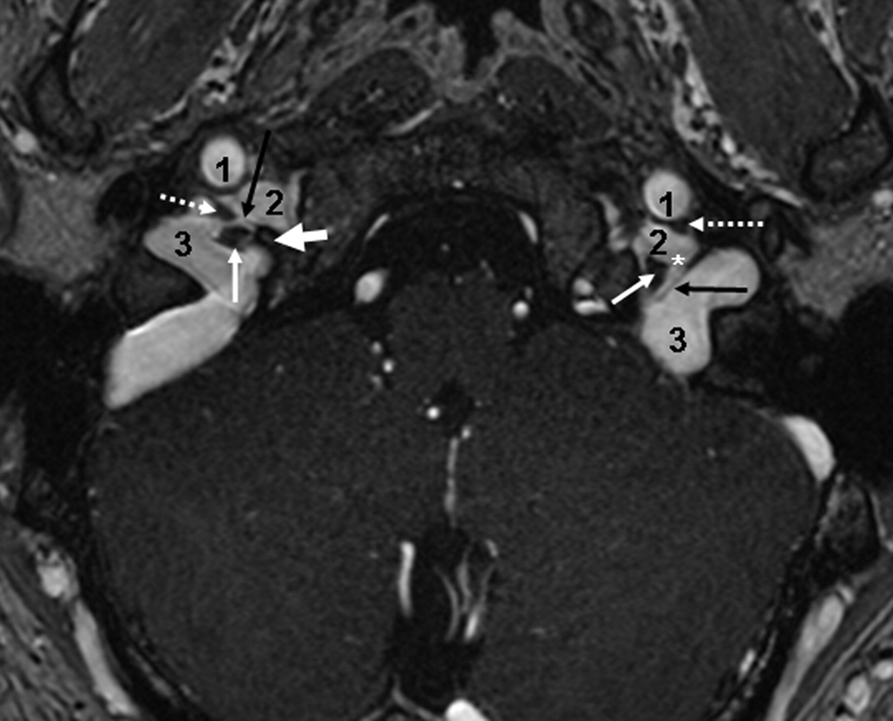
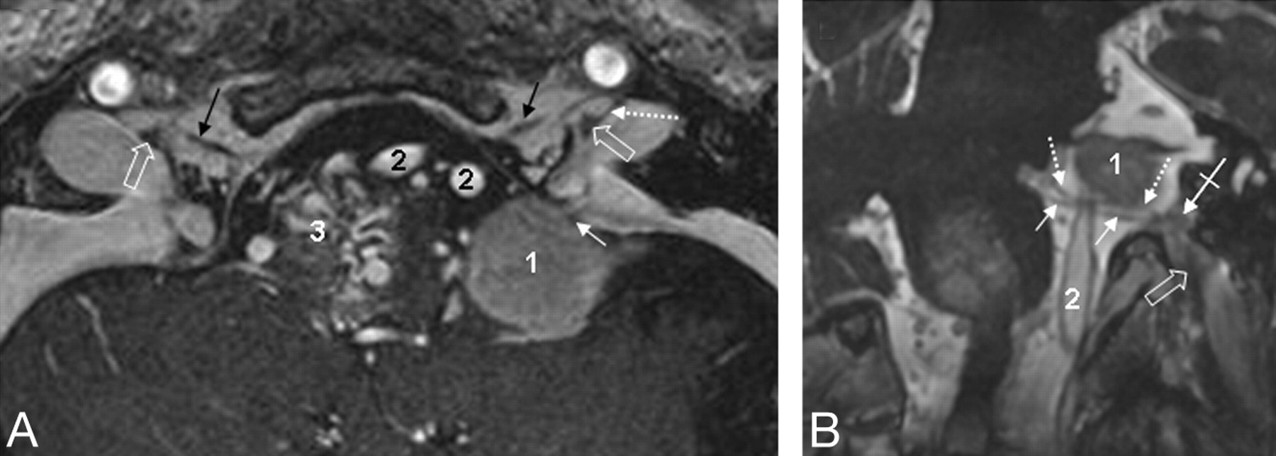
- Fig 1.
Petrosal and sigmoid parts of the JF. Axial CE-MRA image demonstrates the petrosal and sigmoid parts of the JF as well as the cranial nerves within the intrajugular compartment. The thick white arrow marks the right interjugular process of the occipital bone; the black arrow depicts the dural septum between the petrosal and sigmoid part of the JF. Asterisks mark the drainage of the inferior petrosal sinus into the jugular bulb between CNIX anterolaterally (dotted arrows) and the CNX/XI complex at the level of the supCNX posteromedially (thin white arrow). 1 indicates the ICA; 2, the inferior petrosal sinus; 3, the sigmoid sinus.
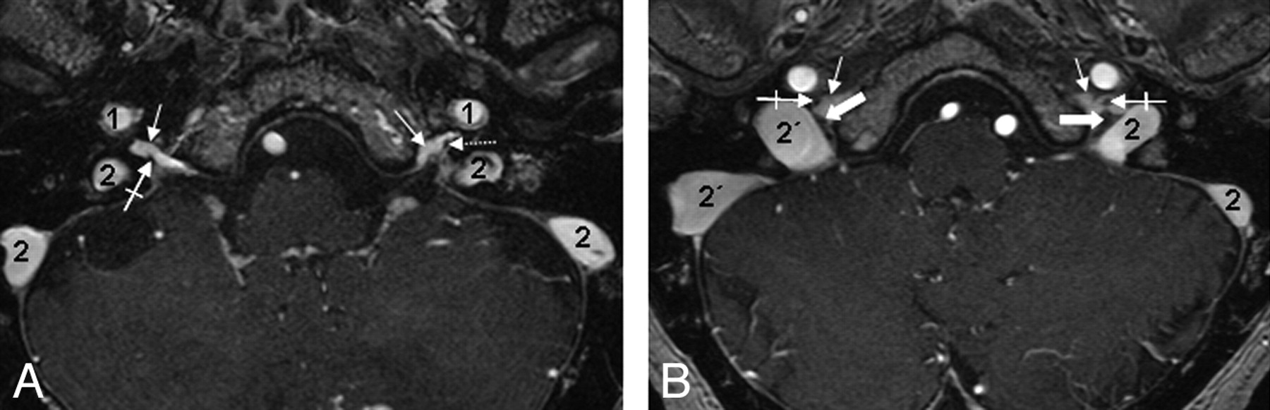
- Fig 2.
The cranial nerves and their ganglia within the JF. Consecutive axial CE-MRA images from the cranial-to-caudal direction depict the CNIX and the CNX/XI complex within the JF. A, The most cranial section is located on the level of the right supCNIX and the left infCNIX (crossed arrow). Note that in this case, no circumscript thickening of the nerve is identifiable. Thin white arrows indicate the inferior petrosal sinus; 1, the ICA; 2, the sigmoid sinus. B, Section at the level of the supCNIX (thick arrows). Crossed arrows indicate CNIX within the JF; thin white arrows, the inferior petrosal sinus; 1, the ICA; 2, the sigmoid sinus. Note that the right sigmoid part (2`) is significantly larger than the left one (2) in this case.
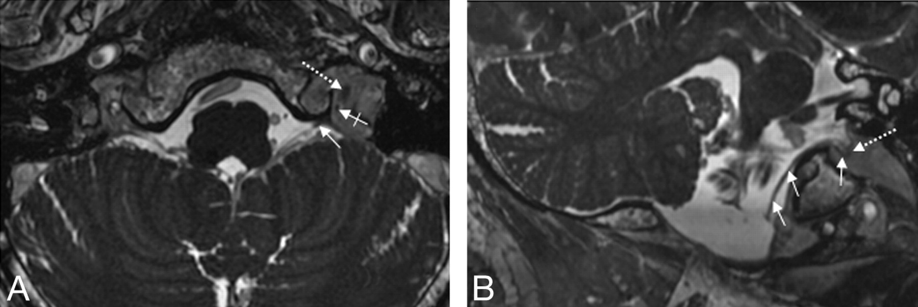
- Fig 3.
A and B, Depiction of the spCNXI on FIESTA MR imaging. The spCNXI (thin white arrows) is visualized on axial (A) and oblique coronal (B) FIESTA MR images in its cisternal course (thin arrows) and after its entrance into the JF (crossed arrow). The dotted arrow indicates the CNX/XI complex.
- Fig 4.
Visualization of the supCNIX. Axial (A) and oblique sagittal (B) 3D-FIESTA MR images. B, The oblique sagittal image is reconstructed parallel to the cisternal course of the lower CNIXs. Nerve root bundles of CNIX (thin white arrows), CNX (crossed white arrows), crCNXI (black arrow), and CNXII (black arrowhead) and the glossopharyngeal meatus (white arrowhead), vagal meatus (thick white arrow), and the supCNXI (dotted white arrows) are depicted.
- Fig 5.
A and B, CNXII within the hypoglossal canal. Axial FIESTA MR image (A) and CE-MRA image (B) clearly demonstrate the canalicular segment of CNXII. Dotted arrows indicate borders of the hypoglossal canal; crossed arrows, the canalicular segment of CNXII; thin white arrows, the CNX/XI complex within the JF; 1, the ICA; 2, the jugular bulb.
- Fig 6.
Demonstration of a bilaterally duplicated hypoglossal canal. Oblique reconstructed FIESTA images of a patient with a duplicated hypoglossal canal on both sides. Thumbnails indicate the reconstruction planes. Open thick arrows indicate borders of the caudal part of the duplicated canal; crossed arrows, the canalicular segments of the hypoglossal nerves. Note that 2 distinct canalicular nerve root bundles can be identified, both of which are demonstrated simultaneously in the images (crossed arrows). B, Dotted arrow indicates the bony septum between both the cranial and the caudal parts of the duplicated hypoglossal canal.
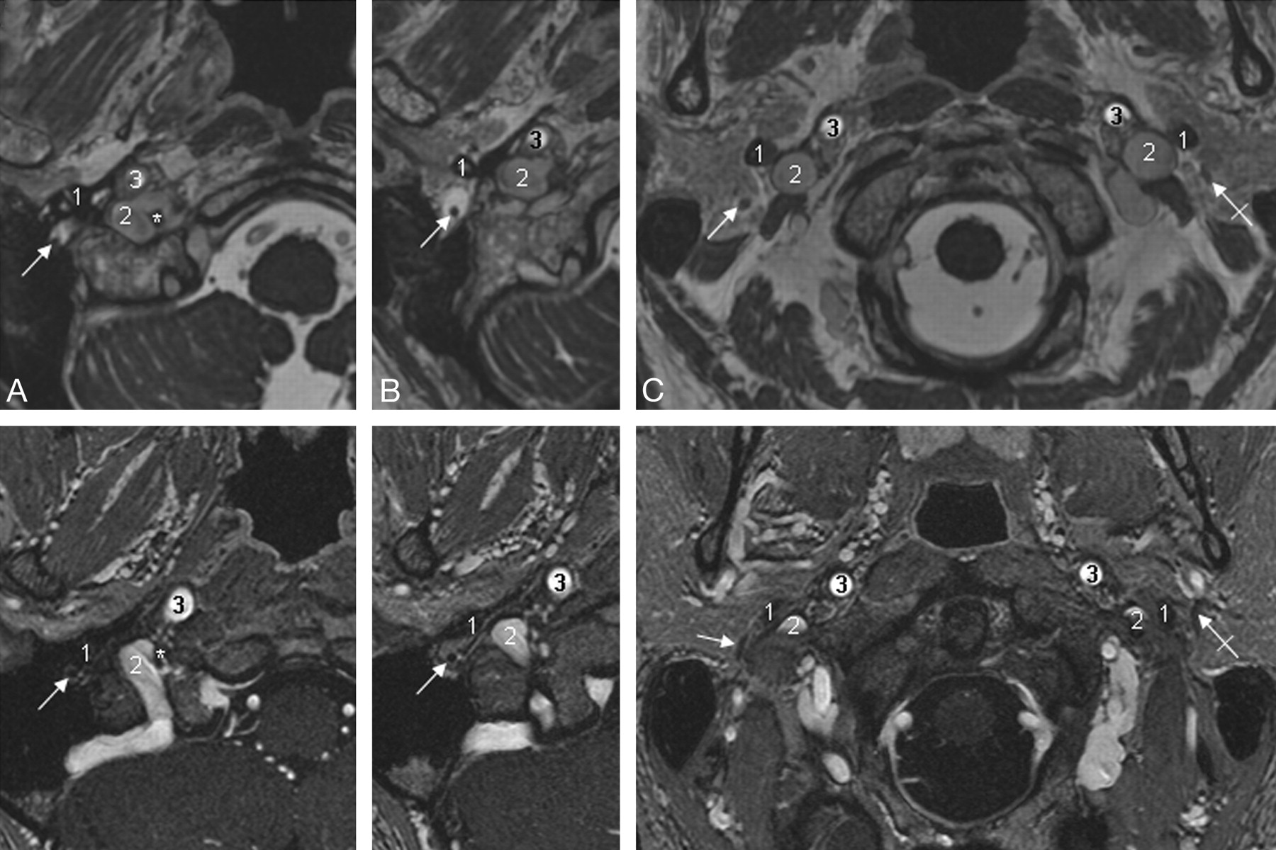
- Fig 7.
Depiction of CNVII. Axial FIESTA (upper row) and CE-MRA images demonstrate CNVII (thin white arrows indicate the right CNVII; crossed arrows, the left CNVII) within the stylomastoid foramen (A) and in its extracranial course after leaving the temporal bone via the stylomastoid foramen (B and C). 1 indicates the styloid process of the temporal bone; 2, the sigmoid sinus (A) and jugular vein (B and C), respectively; 3, the ICA.
- Fig 8.
Illustrative clinical case. A 52-year-old male patient with a left-sided meningioma of the petrosellar ligament (marked as 1). A and B, The meningioma extends throughout the lateral cerebellomedullary cistern as far caudal as the level of the hypoglossal canal. CE-MRA (A) and FIESTA (B) images allow the detailed depiction of the lower cranial nerves and their ganglia with respect to the tumor extension. A, Axial CE-MRA image shows the meningioma (1) which spreads around the spCNXI (thin white arrow). Black arrows indicate the hypoglossal nerve within the hypoglossal canal; open thick white arrow, supCNX; dotted white arrow, the dural septum between the petrosal and jugular part of the JF; 2, vertebral arteries. Note that a brain stem arteriovenous malformation is also present as an incidental finding in this patient (marked as 3). B, FIESTA images reconstructed in a considerably oblique plane aligned precisely along the anatomic course of the cisternal segment of the glossopharyngeal nerve root bundle (dotted white arrows) and the vagal nerve root bundle (thin white arrows). The crossed arrows mark supCNIX, whereas the thick open arrows delineate the supCNX. 1 indicates a meningeoma; 2, the vertebral artery.


Tables
CE-FIESTA CE-MRA TR (s) 4.5 6.6 TE (s) 1.8 2.3 FOV (mm) 160 180 Flip angle (°) 50 20 Matrix (mm) 256 × 256 450 × 450 Section thickness (mm) 0.6 0.8 NEX 2 1.15 Resolution (mm) 0.6 × 0.6 × 0.6 0.4 × 0.4 × 0.8 Duration (min:s) 7:45 7:15 Note:—CE-FIESTA indicates contrast-enhanced fast imaging employing steady-state acquisition; CE-MRA, contrast-enhanced MR angiography.
- Table 2:
Identification of anatomic structures within and adjacent to the jugular foramen*
Anatomic Structure (49 sides) CE-FIESTA CE-MRA 2 1 0 Total† 2 1 0 Total† Dural septum 0 (0) 21 (43) 28 (0) 21 (43) 29 (59.1) 15 (30.6) 5 (10.2) 44 (89.9) Cranial nerves CNIX 36 (73.5) 8 (16.3) 5 (10.2) 44 (90) 49 (100) 0 (0) 0 (0) 49 (100) CNX/XI complex‡ 39 (79.6) 7 (14.3) 3 (6.1) 46 (94) 49 (100) 0 (0) 0 (0) 49 (100) spCNXI 10 (40) 6 (24) 9 (36) 25 (51) 0 (0) 0 (0) 49 (100) 0 (0) Cochlear aqueduct 49 (100) 0 (0) 0 (0) 49 (100) 33 (67) 16 (33) 0 (0) 49 (100) Ganglia supCNIX 12 (24.5) 32 (65.3) 5 (10.2) 44 (89.9) 13 (26.6) 30 (61.2) 6 (12.2) 43 (87.8) infCNIX 0 (0) 36 (73) 13 (26.5) 36 (73) 24 (0) 25 (100) 0 (0) 49 (100) supCNX 0 (0) 48 (98) 2 (4) 48 (98) 0 (0) 49 (100) 0 (0) 49 (100) Adjacent cranial nerve CNVII 33 (67.3) 10 (20.4) 6 (12.2) 43 (87.6) 20 (40.8) 8 (16.3) 21 (42.9) 28 (57.1) Hypoglossal canal 49 (100) 0 (0) 0 (0) 49 (100) 49 (100) 0 (0) 0 (0) 49 (100) CNXII within canal 46 (93.9) 3 (6.1) 0 (0) 49 (100) 49 (100) 0 (0) 0 (0) 49 (100) Note:—sup indicates superior; inf, inferior.
* Data are numbers of sides (percentage).
† Total number of sides (percentage) on which the respective anatomic structure was identified (score of 1 or 2).
‡ The cranial nerve rootlets of CNXI intermingle with CNX inside the JF, and thus these nerves are referred to as the CNX/XI complex.
Right Side (mm) Left Side (mm) Length 6.3 (3.6–9.5) 6.4 (3.8–8.7) Width of external opening 2.9 (1.2–5.2) 2.8 (2–5.1) * Data are mean (range) measured on CE-FIESTA.
In this issue


Jump to section
Related Articles
Cited By...
- 3D Double-Echo Steady-State with Water Excitation MR Imaging of the Intraparotid Facial Nerve at 1.5T: A Pilot Study
- Detailed MR Imaging Anatomy of the Cisternal Segments of the Glossopharyngeal, Vagus, and Spinal Accessory Nerves in the Posterior Fossa: The Use of 3D Balanced Fast-Field Echo MR Imaging