Advertisement

Main menu

  • Home
  • Content
    • Current Issue
    • Accepted Manuscripts
    • Article Preview
    • Past Issue Archive
    • Video Articles
    • AJNR Case Collection
    • Case of the Week Archive
    • Case of the Month Archive
    • Classic Case Archive
  • Special Collections
    • AJNR Awards
    • Low-Field MRI
    • Alzheimer Disease
    • ASNR Foundation Special Collection
    • Photon-Counting CT
    • View All
  • Multimedia
    • AJNR Podcasts
    • AJNR SCANtastic
    • Trainee Corner
    • MRI Safety Corner
    • Imaging Protocols
  • For Authors
    • Submit a Manuscript
    • Submit a Video Article
    • Submit an eLetter to the Editor/Response
    • Manuscript Submission Guidelines
    • Statistical Tips
    • Fast Publishing of Accepted Manuscripts
    • Graphical Abstract Preparation
    • Imaging Protocol Submission
    • Author Policies
  • About Us
    • About AJNR
    • Editorial Board
    • Editorial Board Alumni
  • More
    • Become a Reviewer/Academy of Reviewers
    • Subscribers
    • Permissions
    • Alerts
    • Feedback
    • Advertisers
    • ASNR Home

User menu

  • Alerts
  • Log in

Search

  • Advanced search
American Journal of Neuroradiology

American Journal of Neuroradiology

ASHNR American Society of Functional Neuroradiology ASHNR American Society of Pediatric Neuroradiology ASSR
  • Alerts
  • Log in

Advanced Search

  • Home
  • Content
    • Current Issue
    • Accepted Manuscripts
    • Article Preview
    • Past Issue Archive
    • Video Articles
    • AJNR Case Collection
    • Case of the Week Archive
    • Case of the Month Archive
    • Classic Case Archive
  • Special Collections
    • AJNR Awards
    • Low-Field MRI
    • Alzheimer Disease
    • ASNR Foundation Special Collection
    • Photon-Counting CT
    • View All
  • Multimedia
    • AJNR Podcasts
    • AJNR SCANtastic
    • Trainee Corner
    • MRI Safety Corner
    • Imaging Protocols
  • For Authors
    • Submit a Manuscript
    • Submit a Video Article
    • Submit an eLetter to the Editor/Response
    • Manuscript Submission Guidelines
    • Statistical Tips
    • Fast Publishing of Accepted Manuscripts
    • Graphical Abstract Preparation
    • Imaging Protocol Submission
    • Author Policies
  • About Us
    • About AJNR
    • Editorial Board
    • Editorial Board Alumni
  • More
    • Become a Reviewer/Academy of Reviewers
    • Subscribers
    • Permissions
    • Alerts
    • Feedback
    • Advertisers
    • ASNR Home
  • Follow AJNR on Twitter
  • Visit AJNR on Facebook
  • Follow AJNR on Instagram
  • Join AJNR on LinkedIn
  • RSS Feeds

AJNR Awards, New Junior Editors, and more. Read the latest AJNR updates

OtherSPINE

Intradural Hemangiopericytoma of the Lumbar Spine: A Rare Entity

D. Fitzpatrick, J. Mahajan, M. Lewkowitz, K. Black, A. Setton and R. Woldenberg
American Journal of Neuroradiology January 2009, 30 (1) 152-154; DOI: https://doi.org/10.3174/ajnr.A1262
D. Fitzpatrick
  • Find this author on Google Scholar
  • Find this author on PubMed
  • Search for this author on this site
J. Mahajan
  • Find this author on Google Scholar
  • Find this author on PubMed
  • Search for this author on this site
M. Lewkowitz
  • Find this author on Google Scholar
  • Find this author on PubMed
  • Search for this author on this site
K. Black
  • Find this author on Google Scholar
  • Find this author on PubMed
  • Search for this author on this site
A. Setton
  • Find this author on Google Scholar
  • Find this author on PubMed
  • Search for this author on this site
R. Woldenberg
  • Find this author on Google Scholar
  • Find this author on PubMed
  • Search for this author on this site
  • Article
  • Figures & Data
  • Info & Metrics
  • Responses
  • References
  • PDF
Loading

Abstract

SUMMARY: We report a case of a patient with an intradural hemangiopericytoma of the lumbar spine and the unusual MR angiography (MRA) and spinal angiography findings of arteriovenous shunting with spinal venous congestion. We highlight the concordance of the unusual MRA and angiographic findings and their relationship to combined endovascular and surgical therapy.

Hemangiopericytomas, initially described by Stout and Murray,1 are vascular tumors derived from pericytes, the smooth muscle cells that control the diameter of the capillaries. The mass itself is composed of numerous capillaries with an intact basement membrane, with the neoplastic pericytes in the extravascular space.2 Hemangiopericytomas may be considered benign or malignant, have been identified in all age groups, and demonstrate no sexual predisposition.3 Primary spinal hemangiopericytomas are rare, with only 39 reported cases in the literature, those being primarily extradural.

We describe the unique presentation of a lumbar intradural hemangiopericytoma that produced imaging findings consistent with an arteriovenous shunt, with congested spinal veins and concordant spinal MR angiography (MRA) and conventional angiographic findings. Endovascular embolization of the hemangiopericytoma was performed to treat the arteriovenous shunt and prevent excessive intraoperative blood loss. In addition, the enlarged radicular vein was ligated during surgery to definitively prevent any myelopathic complications.

Case Report

A 54-year-old man presented to our emergency department with left paraspinal, buttock, and anterolateral thigh pain that radiated down the anterior shin and left foot. Initial noncontrast MR examination revealed a lesion in the left L4-L5 neural foramen, which was soft tissue signal intensity on all sequences. Serpiginous, intradural signal intensity voids were identified in the lumbar spinal canal without change in signal intensity in the cord (Fig 1). Contrast-enhanced spinal MRA and postcontrast images were recommended to elucidate the nature of the intradural flow voids and better define the neuroforaminal mass.

Fig 1.
  • Download figure
  • Open in new tab
  • Download powerpoint
Fig 1.

Sagittal T2-weighted images of the lumbar spine demonstrating multiple intradural flow voids (left, white oval), which appear on the surface of the conus (right, white arrows). No change in signal intensity was identified within the cord.

The L4-L5 neuroforaminal mass enhanced with contrast (Fig 2). MRA (Fig 3, left) of the lumbar spine demonstrated prominent intradural vessels contiguous with the mass with congested pial veins, compatible with a spinal arteriovenous fistula or arteriovenous malformation. Spinal angiographic examination was recommended both to further elucidate the relationship between the soft tissue mass and the abnormal surface veins and for possible perioperative intervention.

Fig 2.
  • Download figure
  • Open in new tab
  • Download powerpoint
Fig 2.

Precontrast (left) and postcontrast (right) axial T1-weighted images demonstrate an enhancing mass in the left L4 neural foramen (white arrows). The low signal intensity within the posterior aspect of the vertebral body enhanced after contrast administration, consistent with atypical hemangiomas.

Fig 3.
  • Download figure
  • Open in new tab
  • Download powerpoint
Fig 3.

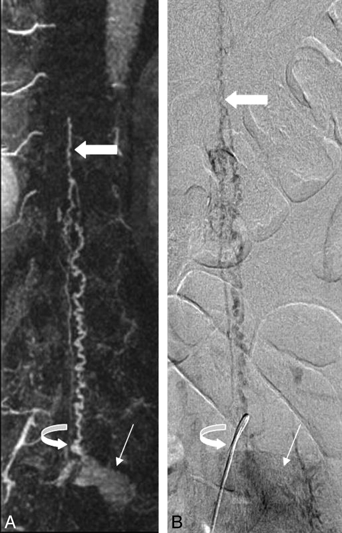
Correlative coronal contrast-enhanced spinal MRA maximum intensity projection image (left) and late-phase anteroposterior conventional spinal angiogram (right) demonstrate a hypervascular mass (thin arrow) with radicular venous drainage (curved arrow). Prominent intradural veins are present, which ascend to the level of the conus, where prominent anterior and posterior pial veins (thick arrow) are visualized.

Spinal angiography (Fig 3, right) revealed a hypervascular mass in the left L4 recess with radicular venous drainage and filling of congested intradural veins ascending to the level of the conus, where congested anterior and posterior spinal veins were apparent. Devascularization of the tumor was performed without residual tumor blush and disappearance of the apparent arteriovenous shunting accounting for spinal venous congestion. At surgery, the L4-L5 nerve sheath was incised, revealing a soft tumor that was adherent to both the overlying dura and the subjacent L4 nerve root. The large radicular draining vein, identified on the angiogram, was coagulated to prevent spinal venous hypertension.

Pathologic examination revealed a monomorphous tumor with closely packed, randomly oriented tumor cells, demonstrating numerous slitlike vessels, consistent with a hemangiopericytoma. The patient completed outpatient radiation therapy and made an uneventful recovery. Follow-up MRA demonstrated resolution of the spinal venous congestion.

Discussion

Intradural hemangiopericytomas are rare and do not demonstrate specific imaging features in the central nervous system. Typical imaging findings are a lobular, dural-based mass with heterogeneous signal intensity on T1-weighted and T2-weighted images. Hemangiopericytomas are hypervascular tumors that demonstrate marked enhancement with prominent flow voids on MR imaging. Like MR imaging, there are no specific findings of hemangiopericytoma on spinal angiography. Conventional angiographic findings typically reported for hemangiopericytomas are a hypervascular mass with irregular, “corkscrew” vessels with a prolonged tumor blush and slow circulation time.4 It is not surprising that, because hemangiopericytomas consist of dilated vascular channels, arteriovenous shunting with hemangiopericytomas on conventional angiography has been described.3

The presence of congested intradural veins, identified on spinal MRA, suggested the presence of a vascular shunt. Hemangiopericytomas are composed of capillary vascular spaces with an intact basement membrane and neoplastic pericytes within the interstitium. When present within the intradural space with multiple arterial feeders, the presence of a hemangiopericytoma may result in arteriovenous shunting from a lack of an intervening capillary bed within the mass. This results in venous hypertension, the presence of arterialized perimedullary veins, and ultimately myelopathy.

Because a hypervascular mass with dilated spinal surface veins was identified on the MRA images, preoperative embolization was suggested to both devascularize the hypervascular tumor (possibly preventing significant intraoperative blood loss) and treat the apparent arteriovenous shunting and spinal venous congestion to prevent myelopathy. The visualization of radicular venous drainage to the conus on conventional angiography predicated surgical ligation of the vein to definitively treat the venous hypertension.

Preoperative angiographic embolization of spinal hemangiopericytomas has been described in a small case series.4 Embolization was performed after significant intraoperative blood loss was identified in 2 cases of the series, which allowed for improved surgical therapy. The presence of intradural perimedullary vessels and arteriovenous shunting were not described. The diagnosis in this case, in addition to the other case reports of intradural hemangiopericytomas,5–7 was presumed to be a meningioma or neurofibroma intraoperatively, suggesting that no abnormal intradural vasculature was identified. Similar to this case, several other cases reported a primary pain component to the presentation of the tumor with lower extremity paresthesias, bladder dysfunction, and no defined motor weakness.

Conclusions

We present the case of a patient with a rare intradural mass, a lumbar hemangiopericytoma, which presented in a highly unusual fashion with markedly dilated spinal surface veins presumably from arteriovenous shunting through the lesion. Subsequent diagnostic conventional angiography with endovascular treatment of the mass and surgical resection with radicular vein ligation relieved the patient's radiculopathy and allowed for resolution of prominent spinal surface vasculature, thus avoiding the potential complication of myelopathy.

Footnotes

  • Case previously presented at: Annual Meeting of the American Society of Neuroradiology, June 5, 2008; New Orleans, La.

References

  1. ↵
    Stout AP, Murray MR. Hemangiopericytoma: a vascular tumor featuring Zimmerman's pericytes. Ann Surg 1942;116:26–32
    PubMed
  2. ↵
    Harris DJ, Fornasier VL, Livingston KE. Hemangiopericytoma of the spinal canal: report of three cases. J Neurosurg49:914–20
  3. ↵
    Stella L, Cappabianca P, Pettinato, G, et al. Hemangiopericytoma of the meninges: report of two cases. Acta Neurologica4:185–95
  4. ↵
    Muraszko KM, Antunes JL, Hilal SK, et al. Hemangiopericytomas of the spine. Neurosurgery 1982;10:473–79
    PubMed
  5. ↵
    Ijiri K, Yuasa S, Yone K, et al. Primary epidural hemangiopericytoma of the lumbar spine. Spine 2002;27:E189–92
    PubMed
  6. Betchen S, Schwartz A, Black C, et al. Intradural hemangiopericytoma of the lumbar spine: case report. Neurosurgery 2002;50:654–57
    PubMed
  7. ↵
    Ecker RD, Marsh WR, Pollock BE, et al. Hemangiopericytoma in the central nervous system: treatment, pathologic features, and long-term follow-up in 38 patients. J Neurosurg 2003;98:1182–97
    PubMed
  • Received June 1, 2008.
  • Accepted after revision June 19, 2008.
  • Copyright © American Society of Neuroradiology
View Abstract
PreviousNext
Back to top

In this issue

American Journal of Neuroradiology: 30 (1)
American Journal of Neuroradiology
Vol. 30, Issue 1
January 2009
  • Table of Contents
  • Index by author
Advertisement
Print
Download PDF
Email Article

Thank you for your interest in spreading the word on American Journal of Neuroradiology.

NOTE: We only request your email address so that the person you are recommending the page to knows that you wanted them to see it, and that it is not junk mail. We do not capture any email address.

Enter multiple addresses on separate lines or separate them with commas.
Intradural Hemangiopericytoma of the Lumbar Spine: A Rare Entity
(Your Name) has sent you a message from American Journal of Neuroradiology
(Your Name) thought you would like to see the American Journal of Neuroradiology web site.
CAPTCHA
This question is for testing whether or not you are a human visitor and to prevent automated spam submissions.
Cite this article
D. Fitzpatrick, J. Mahajan, M. Lewkowitz, K. Black, A. Setton, R. Woldenberg
Intradural Hemangiopericytoma of the Lumbar Spine: A Rare Entity
American Journal of Neuroradiology Jan 2009, 30 (1) 152-154; DOI: 10.3174/ajnr.A1262

Citation Manager Formats

  • BibTeX
  • Bookends
  • EasyBib
  • EndNote (tagged)
  • EndNote 8 (xml)
  • Medlars
  • Mendeley
  • Papers
  • RefWorks Tagged
  • Ref Manager
  • RIS
  • Zotero
0 Responses
Respond to this article
Share
Bookmark this article
Intradural Hemangiopericytoma of the Lumbar Spine: A Rare Entity
D. Fitzpatrick, J. Mahajan, M. Lewkowitz, K. Black, A. Setton, R. Woldenberg
American Journal of Neuroradiology Jan 2009, 30 (1) 152-154; DOI: 10.3174/ajnr.A1262
del.icio.us logo Twitter logo Facebook logo Mendeley logo
  • Tweet Widget
  • Facebook Like
  • Google Plus One
Purchase

Jump to section

  • Article
    • Abstract
    • Case Report
    • Discussion
    • Conclusions
    • Footnotes
    • References
  • Figures & Data
  • Info & Metrics
  • Responses
  • References
  • PDF

Related Articles

  • No related articles found.
  • PubMed
  • Google Scholar

Cited By...

  • Intracranial Meningeal Hemangiopericytomas in Children and Adolescents: CT and MR Imaging Findings
  • Crossref (16)
  • Google Scholar

This article has been cited by the following articles in journals that are participating in Crossref Cited-by Linking.

  • Hemangiopericytomas in the Spine
    Huan-guang Liu, An-chao Yang, Ning Chen, Jun Yang, Xiao-guang Qiu, Jian-guo Zhang
    Neurosurgery 2013 72 1
  • Intracranial Meningeal Hemangiopericytomas in Children and Adolescents: CT and MR Imaging Findings
    Q. Chen, X.-Z. Chen, J.-M. Wang, S.-W. Li, T. Jiang, J.-P. Dai
    American Journal of Neuroradiology 2012 33 1
  • Surgical management of primary spinal hemangiopericytomas: an institutional case series and review of the literature
    Ali Shirzadi, Doniel Drazin, Marcus Gates, Neda Shirzadi, Sergei Banykh, Xuemo Fan, Leonel Hunt, Eli M. Baron, Wesley A. King, Terrence T. Kim, J. Patrick Johnson
    European Spine Journal 2013 22 S3
  • Spinal hemangiopericytoma: an institutional experience and review of literature
    Amitabha Das, Pankaj Kumar Singh, Vaishali Suri, Mukund N. Sable, Bhawani Shankar Sharma
    European Spine Journal 2015 24 S4
  • An Institutional Review of 10 Cases of Spinal Hemangiopericytoma/Solitary Fibrous Tumor
    Raghav Singla, PankajK Singh, Gaurav Khanna, Vaishali Suri, Deepak Agarwal, PS Chandra, SS Kale, AK Mahapatra
    Neurology India 2020 68 2
  • Intradural Extramedullary Hemangiopericytoma of the Thoracic Spine Infiltrating a Nerve Root
    Samuel Moscovici, Fernando Ramirez-DeNoriega, Yakov Fellig, Guy Rosenthal, José E. Cohen, Eyal Itshayek
    Spine 2011 36 23
  • Spinal Hemangiopericytoma Which Needed Intraoperative Embolization due to Unexpected Bleeding
    Chang-Hyun Lee, Ki-Jeong Kim, Tae-Ahn Jahng, Hyun-Jib Kim
    Journal of Korean Neurosurgical Society 2013 54 3
  • Dumbbell-type hemangiopericytoma in the cervical spine: a case report and review
    Hiroaki Nakashima, Shiro Imagama, Yoshito Sakai, Hiroshi Nakamura, Yoshito Katayama, Zenya Ito, Norimitsu Wakao, Naoki Ishiguro, Yukihiro Matsuyama
    Journal of Orthopaedic Science 2013 18 5
  • Primary endodermal hemangiopericytoma/solitary fibrous tumor of the cervical spine: a case report and literature review
    Liyun Wang, Jianbo Yu, Dongping Shu, Bin Huang, Yumin Wang, Luyuan Zhang
    BMC Surgery 2021 21 1
  • Surgical Treatment of Intraspinal Angiomatous Meningiomas from a Single Center
    Liang WU, Tao YANG, Chenlong YANG, Xiaofeng DENG, Jingyi FANG, Yulun XU
    Neurologia medico-chirurgica 2015 55 4

More in this TOC Section

  • Bern Score Validity for SIH
  • MP2RAGE 7T in MS Lesions of the Cervical Spine
  • Resisted Inspiration for CSF-Venous Fistula
Show more Spine

Similar Articles

Advertisement

Indexed Content

  • Current Issue
  • Accepted Manuscripts
  • Article Preview
  • Past Issues
  • Editorials
  • Editor's Choice
  • Fellows' Journal Club
  • Letters to the Editor
  • Video Articles

Cases

  • Case Collection
  • Archive - Case of the Week
  • Archive - Case of the Month
  • Archive - Classic Case

More from AJNR

  • Trainee Corner
  • Imaging Protocols
  • MRI Safety Corner
  • Book Reviews

Multimedia

  • AJNR Podcasts
  • AJNR Scantastics

Resources

  • Turnaround Time
  • Submit a Manuscript
  • Submit a Video Article
  • Submit an eLetter to the Editor/Response
  • Manuscript Submission Guidelines
  • Statistical Tips
  • Fast Publishing of Accepted Manuscripts
  • Graphical Abstract Preparation
  • Imaging Protocol Submission
  • Evidence-Based Medicine Level Guide
  • Publishing Checklists
  • Author Policies
  • Become a Reviewer/Academy of Reviewers
  • News and Updates

About Us

  • About AJNR
  • Editorial Board
  • Editorial Board Alumni
  • Alerts
  • Permissions
  • Not an AJNR Subscriber? Join Now
  • Advertise with Us
  • Librarian Resources
  • Feedback
  • Terms and Conditions
  • AJNR Editorial Board Alumni

American Society of Neuroradiology

  • Not an ASNR Member? Join Now

© 2025 by the American Society of Neuroradiology All rights, including for text and data mining, AI training, and similar technologies, are reserved.
Print ISSN: 0195-6108 Online ISSN: 1936-959X

Powered by HighWire