
Article Figures & Data
Figures
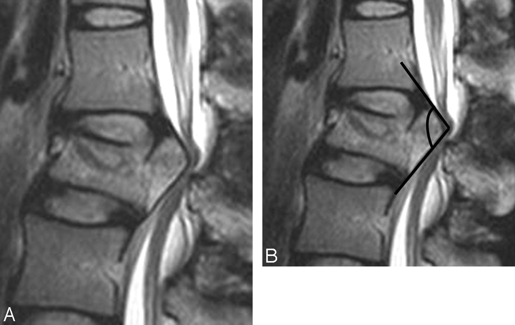
- Fig 1.
Measurement of the angle of the retropulsed segment in the spinal burst fracture. A, This was assessed in the portion having the most acute angle on the sagittal plane. B, We obtained an angle formed by crossing 2 lines drawn according to the cortical line of the retropulsed segment.
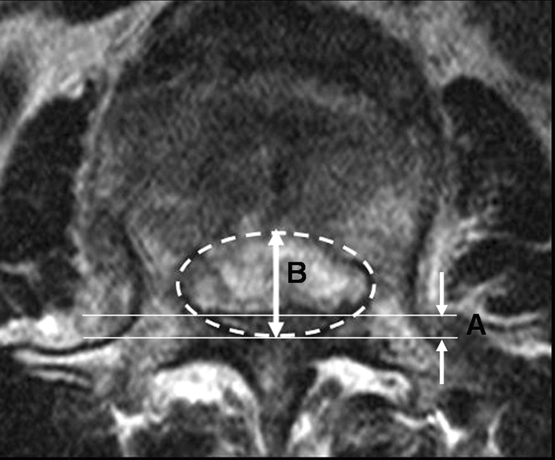
- Fig 2.
The ratio of central canal diameter. The central canal diameter was measured with each AP diameter of the normal central canal (B) and the narrowest portion of central canal (A) drawn by an imaginary line at the burst fracture level. The ratio of central canal diameter reduction was calculated dividing B by A as seen on the axial image.
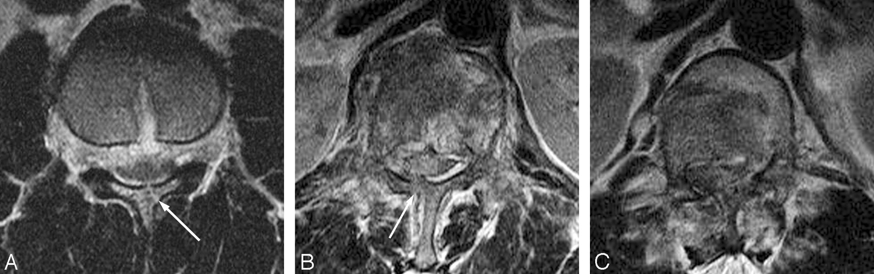
- Fig 3.
The grades of laminar fractures. The degree is classified according to the following: 0, no fracture; 1, fracture without gap (A); 2, fracture with gap (B); and 3, displaced fracture (C).
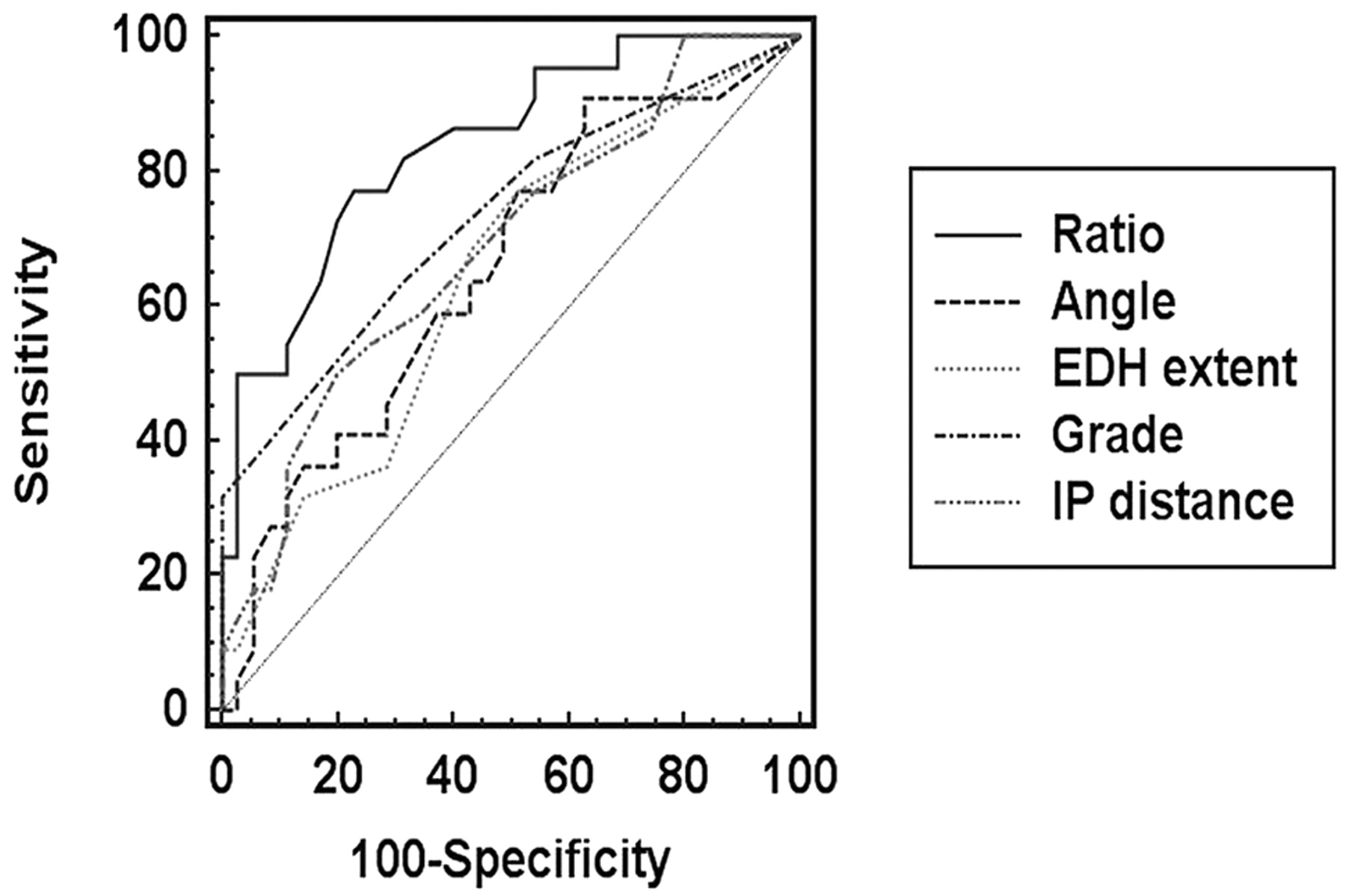
- Fig 4.
The ROC curve in each parameter. This graph shows that the ratio is the most significant among the 5 parameters used to predict a dural tear. Ratio indicates the ratio of the central canal diameter; Angle, the angle of the retropulsed segment; EDH extent, the number of vertebral bodies involved in the epidural hemorrhage; Grade, the grade of laminar fracture; IP distance, the distance between pedicles.


Tables
- Table 1:
The mean values of each parameter and the statistical significance in the study and control groups
Study Group Control Group Pvalue IP distance 28.7 (24–39) mm 26 (18–34) mm .02 Angle 112° (64–180°) 128° (66–180°) .05 Ratio of central canal 0.37 (0.13–0.65) 0.58 (0.21–0.86) .008 Lamina fracture 1.77 (0–3) 0.86 (0–2) .003 Extent of EDH 2.4 (0–6) 1.4 (0–4) .11 Note:—EDH indicates the number of vertebral bodies involved in the epidural hemorrhage.


