
Article Figures & Data
Figures
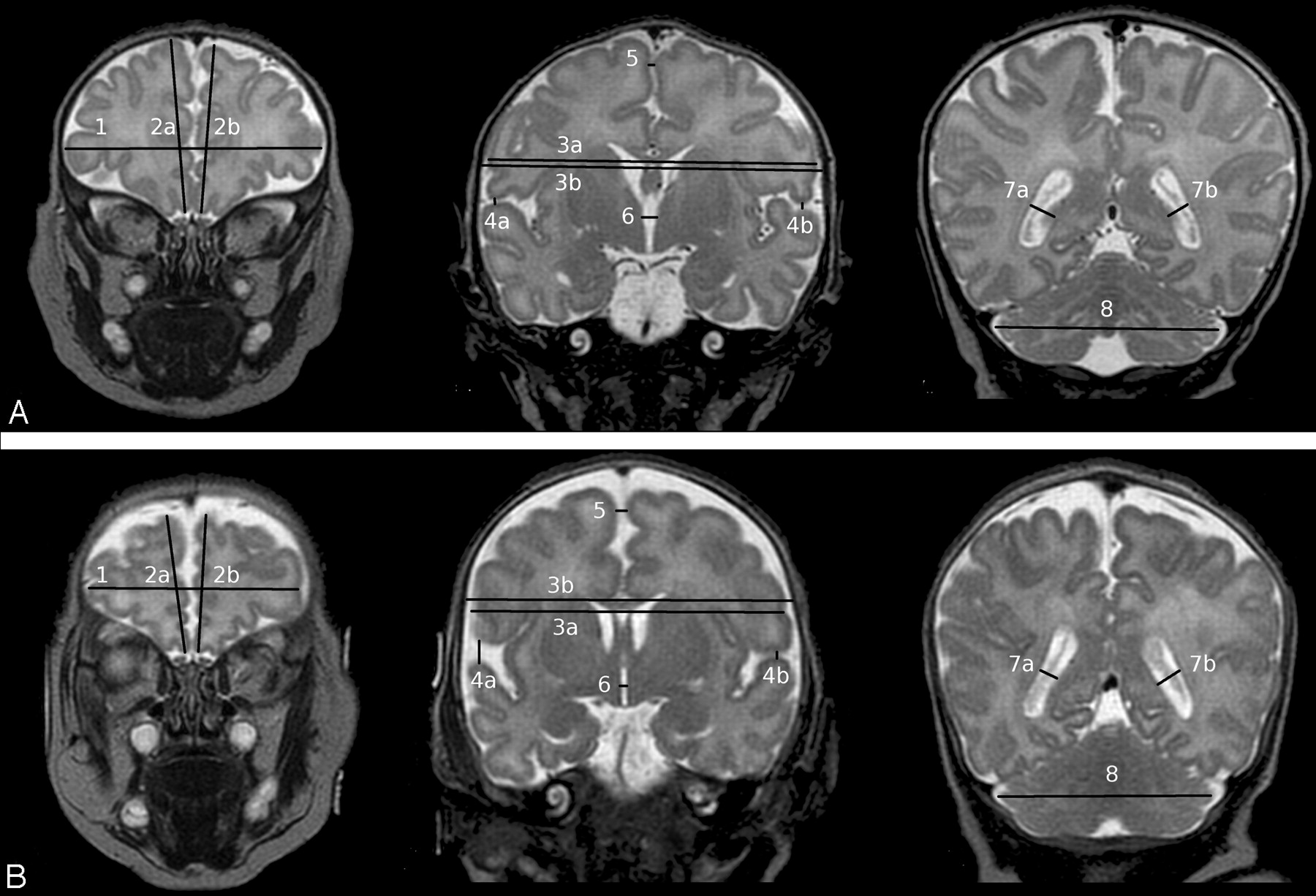
- Fig 1.
A and B, Selected sections and landmarks (see On-line Table 1) used to calculate the coronal brain metrics with examples of a full-term (A) and a preterm infant (B)
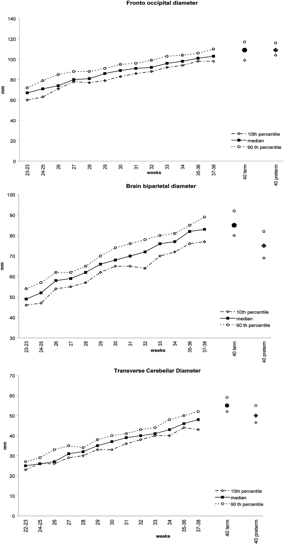
- Fig 2.
Comparison (median, 10th–90th percentiles) of the fronto-occipital, biparietal, and transverse cerebellar diameters by weeks of gestational age measured in term infants (circle) and in the preterm infants (diamond), with the results obtained during pregnancy by measuring fetal brains (square) with the same methodology. Adapted with permission from Garel.9
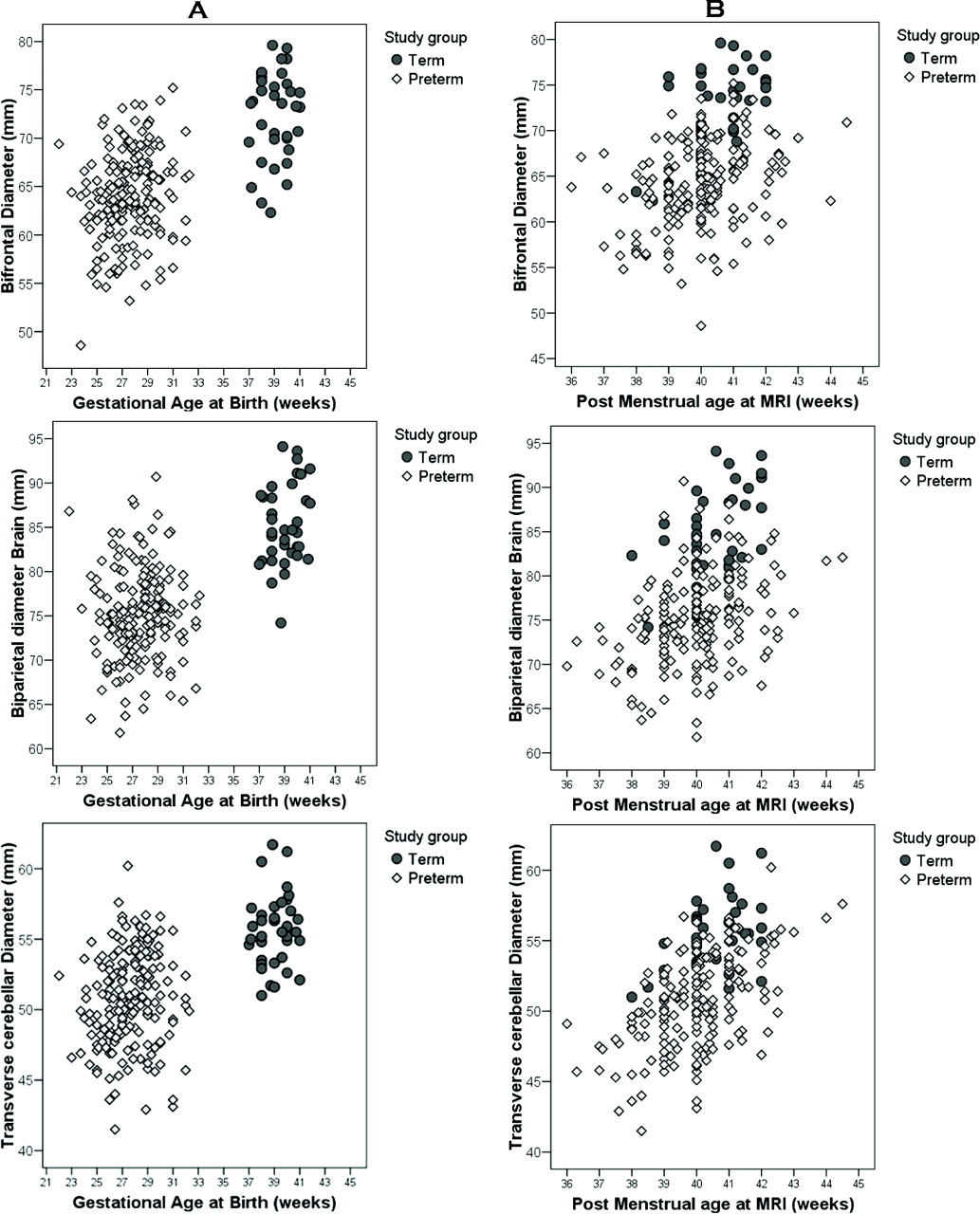
- Fig 3.
A and B, Comparison of bifrontal, biparietal, and transverse cerebellar diameters in preterm infants (diamond) and full-term infants (circle) by their gestational age at birth (A) and by their postmenstrual age at the time of MR imaging (B), demonstrating the lower values obtained in the preterm cohort.


