
Article Figures & Data
Figures
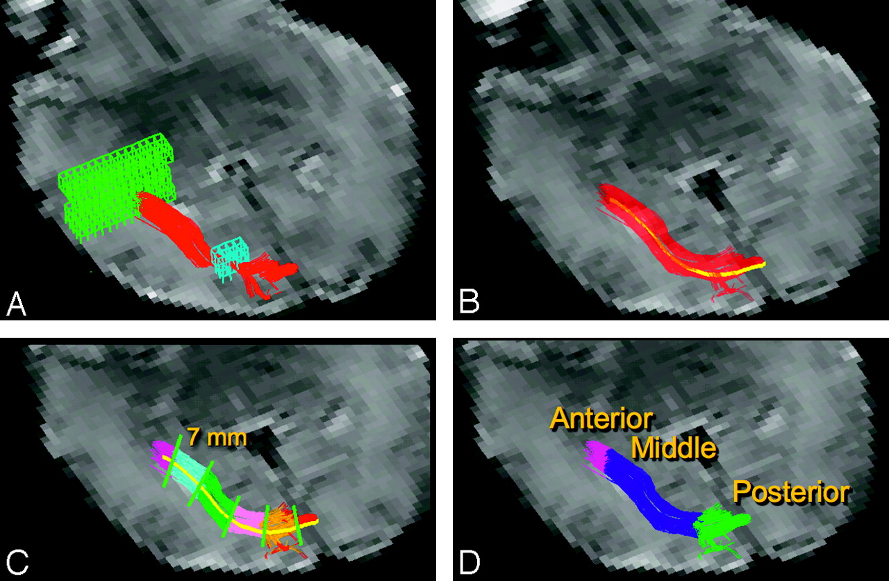
- Fig 1.
DTI fiber tracking and segmentation. A, DTI fiber tracks (red) were launched from a starting region of interest (green mesh) in a plane adjacent to the trigone of the lateral ventricle. Fiber tracks were filtered with a second region of interest (blue mesh) in a plane posterior to the lateral ventricle. B, The average fiber track (yellow) was constructed by averaging coordinates from each of the delineated optic radiation fiber tracks (red). C, The average fiber track was divided into 7-mm segments. Each voxel within the delineated optic radiation is assigned to the closest segment. Different segments are shown with various colors. D, Each discrete 7-mm segment is assigned to either the anterior, middle, or posterior anatomic segment of the optic radiation. The background image in each panel is an echo-planar image without diffusion weighting.
- Fig 2.
FA along optic radiation. Left and right optic radiations are shown in a 35-week GA premature infant. The fiber tracks originate in the thalamus and course posteriorly and adjacent to the ventricle toward the primary visual cortex. The fiber tracks are color coded by the underlying FA. The anterior portion of the tract is observed to have the highest FA.
- Fig 3.
Tract-specific diffusion metrics. Plots show diffusion metrics (FA, Dav, λ1, and λ⊥) within the anterior (red), middle (blue), and posterior (green) segments of the optic radiation.
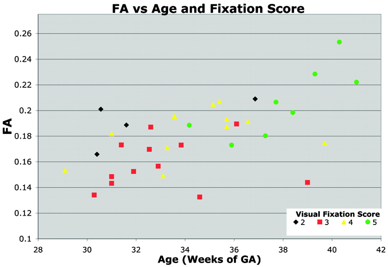
- Fig 4.
Relationship between visual fixation score, FA, and age. FA within the optic radiation increases with GA. The symbols are colored to indicate each neonate's visual fixation score on a scale from 1 to 5. Neonates with higher visual fixation scores demonstrated better visual fixation and tracking behavior. The visual fixation score correlated significantly with FA, independent of age. This relationship between visual fixation score and FA is evident within the set of neonates older than 39 weeks’ GA. Two neonates older than 39 weeks’ GA with abnormally poor visual examination performance also displayed abnormally low FA.


In this issue


Jump to section
Related Articles
Cited By...
- Involvement of the Posterior Visual Pathway Correlates with Higher-Order Visual Impairment in Childhood Stroke Patients detected by Virtual Reality/Eye Tracking Paradigm
- Quantitative Fiber Tracking in the Corpus Callosum and Internal Capsule Reveals Microstructural Abnormalities in Preterm Infants at Term-Equivalent Age
- Directional diffusivity changes in the optic nerve and optic radiation in optic neuritis
- Quantitative Fiber Tracking of the Optic Radiation Is Correlated with Visual-Evoked Potential Amplitude in Preterm Infants