Advertisement

Main menu

  • Home
  • Content
    • Current Issue
    • Accepted Manuscripts
    • Article Preview
    • Past Issue Archive
    • Video Articles
    • AJNR Case Collection
    • Case of the Week Archive
    • Case of the Month Archive
    • Classic Case Archive
  • Special Collections
    • AJNR Awards
    • Low-Field MRI
    • Alzheimer Disease
    • ASNR Foundation Special Collection
    • Photon-Counting CT
    • View All
  • Multimedia
    • AJNR Podcasts
    • AJNR SCANtastic
    • Trainee Corner
    • MRI Safety Corner
    • Imaging Protocols
  • For Authors
    • Submit a Manuscript
    • Submit a Video Article
    • Submit an eLetter to the Editor/Response
    • Manuscript Submission Guidelines
    • Statistical Tips
    • Fast Publishing of Accepted Manuscripts
    • Graphical Abstract Preparation
    • Imaging Protocol Submission
    • Author Policies
  • About Us
    • About AJNR
    • Editorial Board
    • Editorial Board Alumni
  • More
    • Become a Reviewer/Academy of Reviewers
    • Subscribers
    • Permissions
    • Alerts
    • Feedback
    • Advertisers
    • ASNR Home

User menu

  • Alerts
  • Log in

Search

  • Advanced search
American Journal of Neuroradiology

American Journal of Neuroradiology

ASHNR American Society of Functional Neuroradiology ASHNR American Society of Pediatric Neuroradiology ASSR
  • Alerts
  • Log in

Advanced Search

  • Home
  • Content
    • Current Issue
    • Accepted Manuscripts
    • Article Preview
    • Past Issue Archive
    • Video Articles
    • AJNR Case Collection
    • Case of the Week Archive
    • Case of the Month Archive
    • Classic Case Archive
  • Special Collections
    • AJNR Awards
    • Low-Field MRI
    • Alzheimer Disease
    • ASNR Foundation Special Collection
    • Photon-Counting CT
    • View All
  • Multimedia
    • AJNR Podcasts
    • AJNR SCANtastic
    • Trainee Corner
    • MRI Safety Corner
    • Imaging Protocols
  • For Authors
    • Submit a Manuscript
    • Submit a Video Article
    • Submit an eLetter to the Editor/Response
    • Manuscript Submission Guidelines
    • Statistical Tips
    • Fast Publishing of Accepted Manuscripts
    • Graphical Abstract Preparation
    • Imaging Protocol Submission
    • Author Policies
  • About Us
    • About AJNR
    • Editorial Board
    • Editorial Board Alumni
  • More
    • Become a Reviewer/Academy of Reviewers
    • Subscribers
    • Permissions
    • Alerts
    • Feedback
    • Advertisers
    • ASNR Home
  • Follow AJNR on Twitter
  • Visit AJNR on Facebook
  • Follow AJNR on Instagram
  • Join AJNR on LinkedIn
  • RSS Feeds

AJNR Awards, New Junior Editors, and more. Read the latest AJNR updates

OtherHead and Neck Imaging

A Case of Varicella Zoster Virus Polyneuropathy: Involvement of the Glossopharyngeal and Vagus Nerves Mimicking a Tumor

M. Adachi
American Journal of Neuroradiology October 2008, 29 (9) 1743-1745; DOI: https://doi.org/10.3174/ajnr.A1141
M. Adachi
  • Find this author on Google Scholar
  • Find this author on PubMed
  • Search for this author on this site
  • Article
  • Figures & Data
  • Info & Metrics
  • Responses
  • References
  • PDF
Loading

Abstract

SUMMARY: A 36-year-old woman presented with glossopharyngeal and vagus nerve palsy, which proved to be herpes zoster based on the high titers of Varicella zoster virus antibody in her serum. Thin-section T1-weighted images with contrast media demonstrated swelling and distinct contrast enhancement of the glossopharyngeal and vagus complex, mimicking a tumor. Following MR imaging, the size of the nerve complex returned to normal; however, the contrast enhancement remained longer than the symptoms.

Varicella zoster virus has a high affinity for the ganglion and causes various neuropathies including Ramsay Hunt syndrome. I was able to detect and follow abnormalities in the glossopharyngeal and vagus nerve (cranial nerves IX and X) complex in a patient with herpes zoster by using thin-section MR imaging with contrast media. Although many reports have documented the MR imaging findings of Ramsay Hunt syndrome,1–7 only a few have described abnormalities of cranial nerves IX and X. I present a series of MR images of abnormalities in the cranial nerve IX and X complex and discuss the association between these findings and the patient's symptoms.

Case Report

A 36-year-old woman had a sore throat on December 17, 2006. She consulted an otorhinologist and was prescribed drugs (600-mg ibuprofen and 1000-mg ampicillin daily). By December 19, her sore throat had worsened, and she noticed swelling of the neck lymph nodes. The following day she had trouble swallowing, and on the day after that, she had not only a slight fever and sore throat but also hoarseness. On December 25, a few blisters on the left aryepiglottic fold and paresis of the left soft palate were noticed. She was prescribed an antiherpetic drug (3000-mg valacyclovir hydrochloride daily) for 7 days (from December 25 to 31). On December 30, the fever, sore throat, and blisters on the aryepiglottic fold disappeared, and the paresis of the left soft palate and left vocal cord improved. By January 19, 2007, the swallowing disturbance and hoarseness had improved. By February 16, the paresis of the left vocal cord, hoarseness, and swallowing disturbance had disappeared completely.

Biochemical examination of blood obtained on December 19, 2006, revealed the following values: white blood cell count, 5700/μL; red blood cell count, 4,730,000/μL; lactate dehydrogenase, 208 IU/L; and C-reactive protein, <0.1 mg/dL. In serum from blood obtained on December 25, 2006, and January 12, 2007, the titers of antibodies to Varicella zoster virus, immunoglobulin G, and immunoglobulin M were significantly elevated.

To examine abnormalities of cranial nerves IX and X, I obtained MR images including T1-weighted images (TR, 520 ms; TE, 15 ms; FOV, 24 × 24 cm; section thickness, 7 mm; section gap, 2 mm; matrix, 512 × 256; NEX, 2); T2-weighted images (TR, 4000 ms; TE, 102 ms; FOV, 24 × 24 cm; section thickness, 7 mm; section gap, 2 mm; matrix, 512 × 256; NEX, 2; echo-train length, 12); routine T1-weighted images with contrast media (intravenous administration of 0.1 mmol/Kg of gadopentetate dimeglumine, Magnevist; Bayer-Schering Pharma, Osaka, Japan); and thin-section T1-weighted images (1-mm-thick) with contrast media reformed by using 3D Fourier transformation spoiled gradient-recalled-echo (SPGR) images (TR, 30 ms; TE, 4.3 ms; flip angle, 35°; FOV, 18 × 18 cm; slab thickness, 64 mm; matrix, 256 × 256 × 80; NEX, 1) on December 26, 2006 and January 19, February 16, and March 13, 2007, respectively. The MR imaging study was performed by using a 1.5T superconducting system (Signa Excite; GE Healthcare, Milwaukee, Wis) with an 8-channel neurovascular phased-array coil.

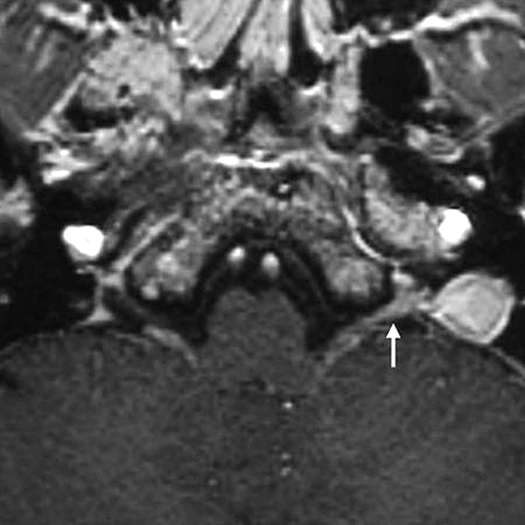
Although no abnormalities in the brain were detected on routine T1-weighted images, T2-weighted or T1-weighted images with contrast media, thin-section T1-weighted images with contrast media revealed abnormalities of cranial nerves IX and X. The nerve complex from the cisternal to the ganglionic region had thickened and had a distinct contrast enhancement resembling a tumor, such as a schwannoma (Fig 1). As time passed, the mass effect diminished and the contrast enhancement faded out; however, the enhancement remained longer than the symptoms (Figs 2–⇓4). Abnormal contrast enhancement of the facial nerve or acoustic nerve was not seen in any MR imaging examination.

Fig 1.
  • Download figure
  • Open in new tab
  • Download powerpoint
Fig 1.

On a thin-section T1-weighted image (SPGR image) with contrast media obtained on December 26, 2006, the left glossopharyngeal and vagus nerve complex from the cisternal to ganglionic region is very thick and distinctly enhanced (arrow).

Fig 2.
  • Download figure
  • Open in new tab
  • Download powerpoint
Fig 2.

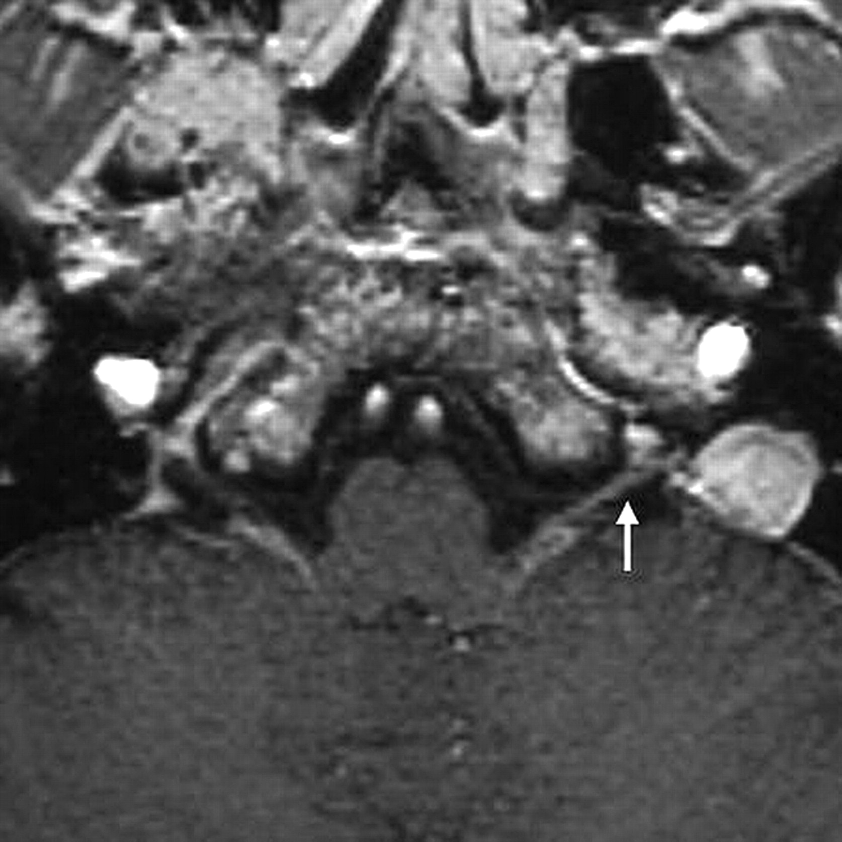
On a T1-weighted image obtained on January 19, 2007, the swelling of the nerve complex has diminished compared with the previous MR imaging study (arrow). The contrast enhancement continues.

Fig 3.
  • Download figure
  • Open in new tab
  • Download powerpoint
Fig 3.

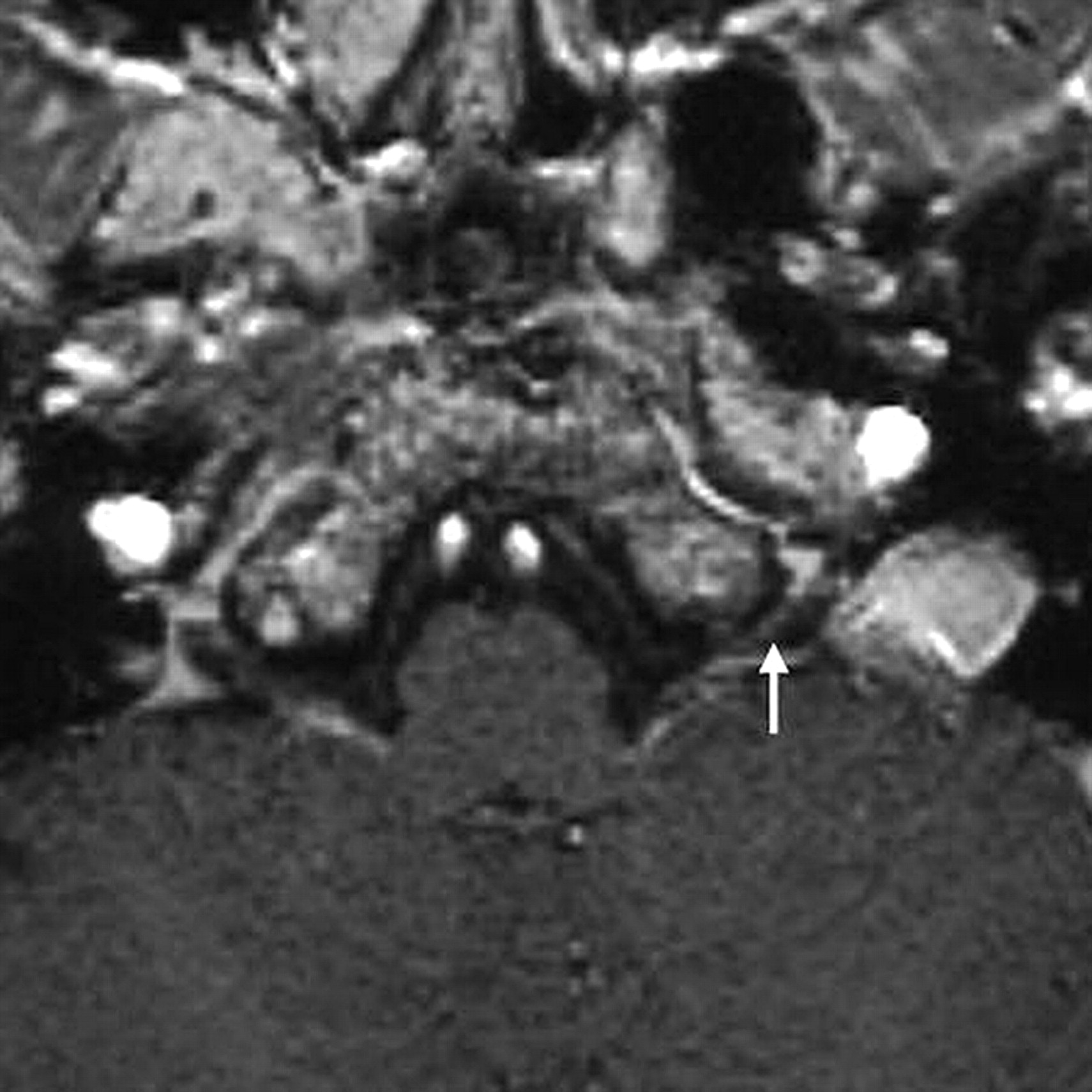
On a T1-weighted image obtained on February 16, 2007, the nerve complex has returned to normal size; however it is still distinctly enhanced (arrow).

Fig 4.
  • Download figure
  • Open in new tab
  • Download powerpoint
Fig 4.

On a T1-weighted image obtained on March 13, 2007, the contrast enhancement of the nerve complex has almost disappeared but still remains (arrow).

Discussion

Facial nerve palsy is well known as Ramsay Hunt syndrome and is caused by reactivation of the Varicella zoster virus. Patients with Ramsay Hunt syndrome typically present with a peripheral facial nerve palsy associated with sensorineural hearing loss, tinnitus, vertigo with nystagmus, and painful vesicular eruption. However, numerous articles have recently reported various cranial neuropathies, other than those of the facial and acoustic nerves.1,8–15 In particular, there have been many case reports of patients with glossopharyngeal and vagus nerve palsy caused by Varicella zoster virus infection, as in the present case.1,9–12,14,15 However, only a few have described the MR imaging findings of glossopharyngeal and vagus nerve palsy.1,15 Fortunately, I had the chance to depict and follow the abnormalities in the cranial nerve IX and X complex by using thin-section MR imaging with contrast media in a patient with Varicella zoster virus infection.

Reports of Ramsay Hunt syndrome have noted that the facial nerve under contrast enhancement might swell and resemble a small tumor.4–7 In the present case, when the symptoms were acute, the nerve complex also showed severe swelling. Previous histologic examination4 has revealed inflammatory infiltrates of lymphocytes and plasma cells in the geniculate ganglion of the facial nerve. In addition, intraneural edema in the neuronal nerve sheaths caused by breakdown of the blood–peripheral nerve barrier and by venous congestion in the epineural and perineural venous plexus was presumed.6,7 These inflammatory cells and edema most likely caused the swelling of the infected cranial nerves in the present patient.

In normal cranial nerves, contrast enhancement is not seen because of the blood-nerve barrier; however, in the first MR imaging study, cranial nerves IX and X had thickened, resembling a tumor (eg, a schwannoma) and had distinct contrast enhancement. As the mass effect of the nerve complex regressed, the symptoms improved; this change suggested some relationship between the swelling of the nerves and the symptoms. However, the contrast enhancement remained on the MR images obtained on February 16 and March 13, when the symptoms had already disappeared. A similar phenomenon of prolongation of contrast enhancement has previously been documented in cases of Ramsay Hunt syndrome and Bell palsy.7 The authors of that article suggested that no relationship could be established between the intensity of the enhancement and the prognosis for the facial palsy and that the long-lasting contrast enhancement was associated with a 2-phase breakdown in the blood-brain-nerve barrier consisting of breakdown of the endoneural barrier followed by breakdown of the perineurium.7 Therefore, swelling of the cranial nerves, not contrast enhancement, may be an indicator of the prognosis in neuropathies due to herpes zoster.

To the best of my knowledge, 2 case reports demonstrated the abnormalities of cranial nerves IX and X in patients with herpes zoster with MR imaging using contrast media.1,15 One depicted the liner and small nodular enhancement in cranial nerves IX and X and the ganglionic region,1 and the other report revealed a nodular lesion with contrast enhancement at the medial side of the left jugular foramen.15 These findings were somewhat similar to those of the present case when the symptoms were intense; however, unfortunately they did not offer a series of MR images following the abnormalities. The present case is very important for investigating the relationship between the MR imaging findings and symptoms.

Conclusions

I obtained a series of thin-section (1-mm-thick) T1-weighted images with contrast media following abnormalities in the glossopharyngeal and vagus nerve complex from infection by Varicella zoster virus. It may be that the natural course of reactivation of Varicella zoster virus is cranial nerve swelling when the symptoms are intense and prolonged contrast enhancement lasts longer than the symptoms.

References

  1. ↵
    Kikuchi H, Yoshimura T, Hara H, et al. A case of multiple cranial neuropathy due to varicella-zoster virus infection: detection on involvement of cranial ganglia with MRI [in Japanese]. Rinsho Shinkeigaku 1995;35:814–16
    PubMed
  2. Naganawa S, Sugiura M, Kawamura M, et al. Prompt contrast enhancement of cerebrospinal fluid space in the fundus of the internal auditory canal: observations in patients with meningeal diseases on 3D-FLAIR images at 3 Tesla. Magn Reson Med Sci 2006;5:151–55
    CrossRefPubMed
  3. Suzuki F, Furuta Y, Ohtani F, et al. Herpes virus reactivation and gadolinium-enhanced magnetic resonance imaging in patients with facial palsy. Otol Neurotol 2001;22:549–53
    CrossRefPubMed
  4. ↵
    Sartoretti-Schefer S, Kollias S, Valvanis A. Ramsay-Hunt syndrome associated with brain stem enhancement. AJNR Am J Neuroradiol 1999;20:278–80
    Abstract/FREE Full Text
  5. Han MH, Jabour BA, Andrews JC, et al. Nonneoplastic enhancing lesions mimicking intracanalicular acoustic neuroma on gadolinium-enhanced MR images. Radiology 1991;179:795–96
    PubMed
  6. ↵
    Sartoretti-Schefer S, Wichmann W, Valavanis A. Gadolinium-enhanced MR in patients with idiopathic, herpetic, and HIV-associated facial nerve palsies: abnormal MR enhancement patterns. AJNR Am J Neuroradiol 1994;15:479–85
    Abstract/FREE Full Text
  7. ↵
    Sartoretti-Schefer S, Brändle P, Wichmann W, et al. Intensity of MR contrast enhancement does not correspond to clinical and electroneurographic findings in acute inflammatory facial nerve palsy. AJNR Am J Neuroradiol 1996;17:1229–36
    Abstract
  8. ↵
    Nagano K, Yoshimura K, Yamasaki M. A case of Ramsay Hunt syndrome associated with local meningitis, multiple cranial neuropathy, and the second cervical nerve involvement [in Japanese]. Rinsho Shinkeigaku 1999;39:475–77
    PubMed
  9. ↵
    Miyazaki Y, Tajima Y, Sudo K, et al. A case of Ramsay Hunt syndrome initiated with hoarseness and dysphagia: consideration on spreading mechanisms of cranial neuropathy [in Japanese]. Rinsho Shinkeigaku 2002;42:855–58
    PubMed
  10. Nishioka K, Fujishima K, Kobayashi H, et al. An extremely unusual presentation of varicella zoster viral infection of cranial nerves mimicking Garcin sydrome. Clin Neural Neuro Surg 2006;108:772–74. Epub 2005 Oct 13
  11. Maeda A, Shoji T, Tsuchiya K, et al. A case of multiple cranial nerve palsy with severe dysphagia due to herpes zoster infection [in Japanese]. Rinsho Shinkeigaku 1992;32:524–26
    PubMed
  12. ↵
    Yoshioka A, Kitagawa Y, Kawada J, et al. A case of unilateral VIIth, IXth and Xth cranial nerve involvement with herpes zoster [in Japanese]. Rinsho Shinkeigaku 1990;30:413–15
    PubMed
  13. Kobayashi Y, Riku S, Ieda T, et al. A case of herpes zoster associated with multiple cranial nerve involvement [in Japanese]. Rinsho Shinkeigaku 1992;32:314–16
    PubMed
  14. ↵
    Ohashi T, Fujimoto M, Shimizu H, et al. A case of isolated vagus nerve palsy with herpes zoster [in Japanese]. Rinsho Shinkeigaku 1994;34:928–29
    PubMed
  15. ↵
    Doi H, Segawa F, Koyano S, et al. A case of Vernet's syndrome due to varicella-zoster virus infection [in Japanese]. Rinsho Shikeigaku 2001;41:695–97
  • Received February 15, 2008.
  • Accepted after revision March 3, 2008.
  • Copyright © American Society of Neuroradiology
View Abstract
PreviousNext
Back to top

In this issue

American Journal of Neuroradiology: 29 (9)
American Journal of Neuroradiology
Vol. 29, Issue 9
October 2008
  • Table of Contents
  • Index by author
Advertisement
Print
Download PDF
Email Article

Thank you for your interest in spreading the word on American Journal of Neuroradiology.

NOTE: We only request your email address so that the person you are recommending the page to knows that you wanted them to see it, and that it is not junk mail. We do not capture any email address.

Enter multiple addresses on separate lines or separate them with commas.
A Case of Varicella Zoster Virus Polyneuropathy: Involvement of the Glossopharyngeal and Vagus Nerves Mimicking a Tumor
(Your Name) has sent you a message from American Journal of Neuroradiology
(Your Name) thought you would like to see the American Journal of Neuroradiology web site.
CAPTCHA
This question is for testing whether or not you are a human visitor and to prevent automated spam submissions.
Cite this article
M. Adachi
A Case of Varicella Zoster Virus Polyneuropathy: Involvement of the Glossopharyngeal and Vagus Nerves Mimicking a Tumor
American Journal of Neuroradiology Oct 2008, 29 (9) 1743-1745; DOI: 10.3174/ajnr.A1141

Citation Manager Formats

  • BibTeX
  • Bookends
  • EasyBib
  • EndNote (tagged)
  • EndNote 8 (xml)
  • Medlars
  • Mendeley
  • Papers
  • RefWorks Tagged
  • Ref Manager
  • RIS
  • Zotero
0 Responses
Respond to this article
Share
Bookmark this article
A Case of Varicella Zoster Virus Polyneuropathy: Involvement of the Glossopharyngeal and Vagus Nerves Mimicking a Tumor
M. Adachi
American Journal of Neuroradiology Oct 2008, 29 (9) 1743-1745; DOI: 10.3174/ajnr.A1141
del.icio.us logo Twitter logo Facebook logo Mendeley logo
  • Tweet Widget
  • Facebook Like
  • Google Plus One
Purchase

Jump to section

  • Article
    • Abstract
    • Case Report
    • Discussion
    • Conclusions
    • References
  • Figures & Data
  • Info & Metrics
  • Responses
  • References
  • PDF

Related Articles

  • No related articles found.
  • PubMed
  • Google Scholar

Cited By...

  • Uncommon presentation and complications of herpes zoster infection involving the cervical, vagus and accessory nerves which caused a delay in diagnosis and treatment
  • Acute dysphagia
  • Crossref (27)
  • Google Scholar

This article has been cited by the following articles in journals that are participating in Crossref Cited-by Linking.

  • Pharyngolaryngeal Involvement by Varicella-Zoster Virus
    Lluís Nisa, Basile Nicolas Landis, Roland Giger, Igor Leuchter
    Journal of Voice 2013 27 5
  • Varicella zoster virus infection of the pharynx and larynx with multiple cranial neuropathies
    Yuan‐Yung Lin, Chuan‐Hsiang Kao, Chih‐Hung Wang
    The Laryngoscope 2011 121 8
  • The lower cranial nerves: IX, X, XI, XII
    J.-L. Sarrazin, F. Toulgoat, F. Benoudiba
    Diagnostic and Interventional Imaging 2013 94 10
  • The glossopharyngeal, vagus and spinal accessory nerves
    Cheng Kang Ong, Vincent Fook Hin Chong
    European Journal of Radiology 2010 74 2
  • Imaging of Jugular Foramen
    Cheng Kang Ong, Vincent Fook-Hin Chong
    Neuroimaging Clinics of North America 2009 19 3
  • Intracranial Infections: Key Neuroimaging Findings
    Jitender Saini, Rakesh K. Gupta, Krishan K. Jain
    Seminars in Roentgenology 2014 49 1
  • Magnetic Resonance Imaging Evidence of Varicella Zoster Virus Polyneuropathy
    Hediye Pinar Gunbey, Gokhan Kutlar, Kerim Aslan, Asli Tanrivermis Sayit, Lutfi Incesu
    Journal of Craniofacial Surgery 2016 27 3
  • Successful treatment with acyclovir and a corticosteroid for lower cranial polyneuropathy in zoster sine herpete: a case report
    Daisuke Taniguchi, Toshiki Nakahara Ph.D., Sho Nakajima, Tomoko Nakazato, Michitaka Mikasa, Yoshiaki Furukawa Ph.D.
    Rinsho Shinkeigaku 2015 55 12
  • Acute worsening of CADASIL in a patient with COVID-19 infection: illustrative case
    Jared S. Rosenblum, Jessa M. Tunacao, Matthew A. Nazari, Halle Ronk, Danielle D. Dang, Chad Downing, Zhengping Zhuang, John D. Heiss, James G. Smirniotopoulos, Avraham Bluestone, James Badia, Joseph White
    Journal of Neurosurgery: Case Lessons 2022 4 26
  • Idiopathic trigeminal neuropathy with trigeminal mass lesion on MRI: Neoplasm or not?
    Keita Sakurai, Masaki Hara, Kenji Okita, Shoji Kawashima, Takemori Yamawaki, Yuta Shibamoto
    Cephalalgia 2010 30 8

More in this TOC Section

  • ASL Sensitivity for Head and Neck Paraganglioma
  • Post SRS Peritumoral Hyperintense Signal of VSs
  • Contrast Enhanced Pituitary CISS/FIESTA
Show more Head and Neck Imaging

Similar Articles

Advertisement

Indexed Content

  • Current Issue
  • Accepted Manuscripts
  • Article Preview
  • Past Issues
  • Editorials
  • Editor's Choice
  • Fellows' Journal Club
  • Letters to the Editor
  • Video Articles

Cases

  • Case Collection
  • Archive - Case of the Week
  • Archive - Case of the Month
  • Archive - Classic Case

More from AJNR

  • Trainee Corner
  • Imaging Protocols
  • MRI Safety Corner
  • Book Reviews

Multimedia

  • AJNR Podcasts
  • AJNR Scantastics

Resources

  • Turnaround Time
  • Submit a Manuscript
  • Submit a Video Article
  • Submit an eLetter to the Editor/Response
  • Manuscript Submission Guidelines
  • Statistical Tips
  • Fast Publishing of Accepted Manuscripts
  • Graphical Abstract Preparation
  • Imaging Protocol Submission
  • Evidence-Based Medicine Level Guide
  • Publishing Checklists
  • Author Policies
  • Become a Reviewer/Academy of Reviewers
  • News and Updates

About Us

  • About AJNR
  • Editorial Board
  • Editorial Board Alumni
  • Alerts
  • Permissions
  • Not an AJNR Subscriber? Join Now
  • Advertise with Us
  • Librarian Resources
  • Feedback
  • Terms and Conditions
  • AJNR Editorial Board Alumni

American Society of Neuroradiology

  • Not an ASNR Member? Join Now

© 2025 by the American Society of Neuroradiology All rights, including for text and data mining, AI training, and similar technologies, are reserved.
Print ISSN: 0195-6108 Online ISSN: 1936-959X

Powered by HighWire