Advertisement

Main menu

  • Home
  • Content
    • Current Issue
    • Accepted Manuscripts
    • Article Preview
    • Past Issue Archive
    • Video Articles
    • AJNR Case Collection
    • Case of the Week Archive
    • Case of the Month Archive
    • Classic Case Archive
  • Special Collections
    • Advancing NeuroMRI with High-Relaxivity Contrast Agents
    • AJNR Awards
    • Low-Field MRI
    • Alzheimer Disease
    • ASNR Foundation Special Collection
    • Photon-Counting CT
    • View All
  • Multimedia
    • AJNR Podcasts
    • AJNR SCANtastic
    • Trainee Corner
    • MRI Safety Corner
    • Imaging Protocols
  • For Authors
    • Submit a Manuscript
    • Submit a Video Article
    • Submit an eLetter to the Editor/Response
    • Manuscript Submission Guidelines
    • Statistical Tips
    • Fast Publishing of Accepted Manuscripts
    • Graphical Abstract Preparation
    • Imaging Protocol Submission
    • Author Policies
  • About Us
    • About AJNR
    • Editorial Board
    • Editorial Board Alumni
  • More
    • Become a Reviewer/Academy of Reviewers
    • Subscribers
    • Permissions
    • Alerts
    • Feedback
    • Advertisers
    • ASNR Home

User menu

  • Alerts
  • Log in

Search

  • Advanced search
American Journal of Neuroradiology

American Journal of Neuroradiology

ASHNR American Society of Functional Neuroradiology ASHNR American Society of Pediatric Neuroradiology ASSR
  • Alerts
  • Log in

Advanced Search

  • Home
  • Content
    • Current Issue
    • Accepted Manuscripts
    • Article Preview
    • Past Issue Archive
    • Video Articles
    • AJNR Case Collection
    • Case of the Week Archive
    • Case of the Month Archive
    • Classic Case Archive
  • Special Collections
    • Advancing NeuroMRI with High-Relaxivity Contrast Agents
    • AJNR Awards
    • Low-Field MRI
    • Alzheimer Disease
    • ASNR Foundation Special Collection
    • Photon-Counting CT
    • View All
  • Multimedia
    • AJNR Podcasts
    • AJNR SCANtastic
    • Trainee Corner
    • MRI Safety Corner
    • Imaging Protocols
  • For Authors
    • Submit a Manuscript
    • Submit a Video Article
    • Submit an eLetter to the Editor/Response
    • Manuscript Submission Guidelines
    • Statistical Tips
    • Fast Publishing of Accepted Manuscripts
    • Graphical Abstract Preparation
    • Imaging Protocol Submission
    • Author Policies
  • About Us
    • About AJNR
    • Editorial Board
    • Editorial Board Alumni
  • More
    • Become a Reviewer/Academy of Reviewers
    • Subscribers
    • Permissions
    • Alerts
    • Feedback
    • Advertisers
    • ASNR Home
  • Follow AJNR on Twitter
  • Visit AJNR on Facebook
  • Follow AJNR on Instagram
  • Join AJNR on LinkedIn
  • RSS Feeds

AJNR Awards, New Junior Editors, and more. Read the latest AJNR updates


Improved Turnaround Times | Median time to first decision: 12 days

Research ArticleBrain

Histogram Analysis of MR Imaging–Derived Cerebral Blood Volume Maps: Combined Glioma Grading and Identification of Low-Grade Oligodendroglial Subtypes

K.E. Emblem, D. Scheie, P. Due-Tonnessen, B. Nedregaard, T. Nome, J.K. Hald, K. Beiske, T.R. Meling and A. Bjornerud
American Journal of Neuroradiology October 2008, 29 (9) 1664-1670; DOI: https://doi.org/10.3174/ajnr.A1182
K.E. Emblem
  • Find this author on Google Scholar
  • Find this author on PubMed
  • Search for this author on this site
D. Scheie
  • Find this author on Google Scholar
  • Find this author on PubMed
  • Search for this author on this site
P. Due-Tonnessen
  • Find this author on Google Scholar
  • Find this author on PubMed
  • Search for this author on this site
B. Nedregaard
  • Find this author on Google Scholar
  • Find this author on PubMed
  • Search for this author on this site
T. Nome
  • Find this author on Google Scholar
  • Find this author on PubMed
  • Search for this author on this site
J.K. Hald
  • Find this author on Google Scholar
  • Find this author on PubMed
  • Search for this author on this site
K. Beiske
  • Find this author on Google Scholar
  • Find this author on PubMed
  • Search for this author on this site
T.R. Meling
  • Find this author on Google Scholar
  • Find this author on PubMed
  • Search for this author on this site
A. Bjornerud
  • Find this author on Google Scholar
  • Find this author on PubMed
  • Search for this author on this site
  • Article
  • Figures & Data
  • Info & Metrics
  • Responses
  • References
  • PDF
Loading

Article Figures & Data

Figures

  • Tables
  • Fig 1.
    • Download figure
    • Open in new tab
    • Download powerpoint
    Fig 1.

    A, Axial T2-weighted fast spin-echo (FSE) image (TR/TE, 4000/104 ms) of a patient (subject 3) with a low-grade oligoastrocytoma without LOH on 1p/19q. B, Axial postcontrast T1-weighted spin-echo (SE) image (TR/TE, 500/7.7 ms) of subject 3. C, Coregistered rCBV map of subject 3. D, Axial T2-weighted FSE image (TR/TE, 4000/104 ms) of a patient (subject 7) with a low-grade oligoastrocytoma with LOH on 1p/19q. E, Axial postcontrast T1-weighted SE image (TR/TE, 500/7.7 ms) of subject 7. F, Coregistered rCBV map of subject 7. Note the low rCBV values in the tumor area in image C compared with the tumor area in image F, typical of low-grade oligodendroglial tumors without LOH on 1p/19q. The corresponding normalized histogram signatures are shown in Fig 2.

  • Fig 2.
    • Download figure
    • Open in new tab
    • Download powerpoint
    Fig 2.

    Resulting normalized histogram plots of the total distribution of rCBV values from the patients shown in Fig 1. The higher peak height of the low-grade oligoastrocytoma without LOH on p/19q, shown in a dotted line (subject 3, Fig 1A–C), indicates a more homogeneous rCBV distribution than the rCBV distribution of a low-grade oligoastrocytoma with LOH on 1p/19q, shown in a solid line (subject 7, Fig 1D–F).

  • Fig 3.
    • Download figure
    • Open in new tab
    • Download powerpoint
    Fig 3.

    Mean histogram peak heights with SEs of the mean for the different glioma types investigated. The values are as follows: HGG = 0.052 (0.004), LGG = 0.094 (0.004), all high-grade oligodendroglial tumors (HGO) = 0.068 (0.010), low-grade oligodendroglial tumor with LOH on 1p/19q (LGO[−]) = 0.096 (0.004), low-grade oligodendroglial tumor without LOH on 1p/19q (LGO[+]) = 0.113 (0.002), and low-grade diffuse astrocytoma (LGA) = 0.086 (0.012). The oligodendroglial tumors include both oligodendrogliomas and oligoastrocytomas.

  • Fig 4.
    • Download figure
    • Open in new tab
    • Download powerpoint
    Fig 4.

    A scatterplot showing histogram peak heights for the 52 patients included in our study. The histogram method is able to significantly differentiate between HGGs (▴) and LGGs (+) (P < .001), with an ICC of 0.902.
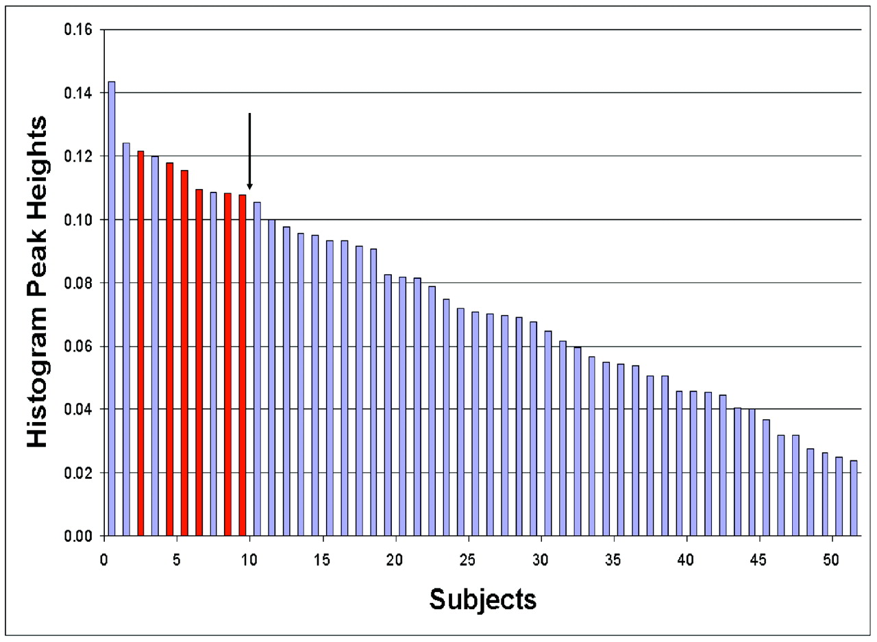
  • Fig 5.
    • Download figure
    • Open in new tab
    • Download powerpoint
    Fig 5.

    Histogram peak heights for the 52 patients investigated in our study (23 LGGs and 29 HGGs). Each histogram peak height is a mean value across the 4 observers. With a cutoff value of 0.107, the sensitivity and specificity when distinguishing the 6 patients with low-grade oligodendroglial tumors without LOH on 1p/19q (red bars) from the other 46 patients (blue bars) are 100% (6/6) and 91% (42/46), respectively.
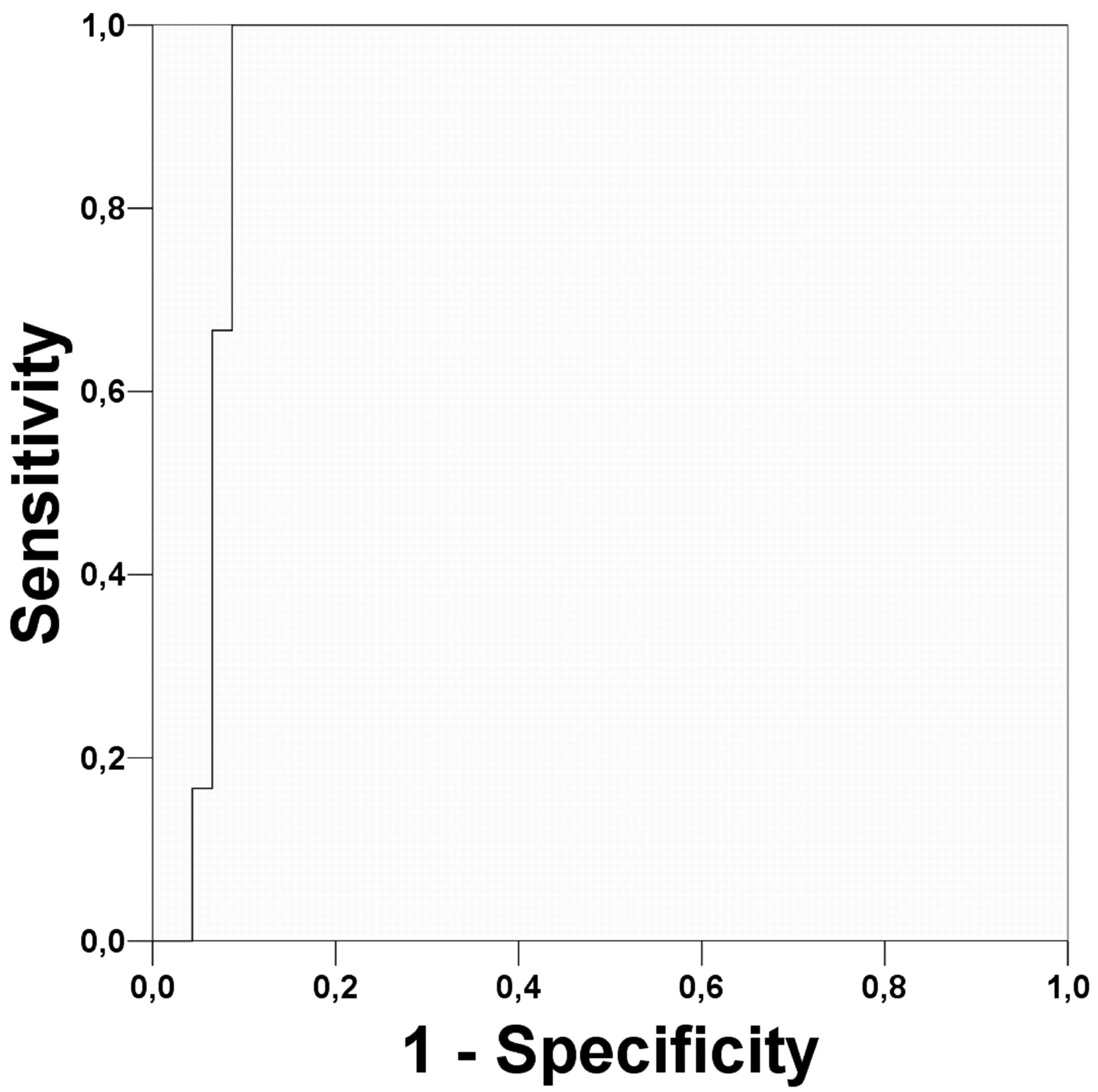
  • Fig 6.
    • Download figure
    • Open in new tab
    • Download powerpoint
    Fig 6.

    ROC curve for the histogram method when distinguishing patients with low-grade oligodendroglial tumors without LOH on 1p/19q (n = 6) from the other gliomas (n = 46). The area (±SE) under the ROC curve is Az = 0.931 ± .036.

Tables

  • Figures
  • Patient demographics, histologic features, surgical procedure, and MR imaging findings for patients diagnosed with oligodendroglial tumors

    SubjectAge (yr)SexHDWHO GradeLOH on 1p/19qClassic HistologySurgical ProcedureContrast EnhancementHistogram Peak Heights*
    162FODIIYesYesResectionNone0.120
    236FOAIIYesNoResectionNone0.095
    328MOAIINoNoBiopsyNone0.118
    463MAODIIIYesYesResectionModerate0.075
    554MAODIIIYesYesResectionModerate0.024
    654MODIIYesYesBiopsyNone0.091
    744FOAIIYesNoResectionNone0.093
    844MOAIINoNoResectionNone0.109
    940FOAIINoNoResectionExtensive0.108
    1037FODIIYesYesBiopsyNone0.070
    1137FOAIINoNoResectionNone0.108
    1230FOAIIYesYesResectionNone0.095
    1359MAODIIINoNoBiopsyExtensive0.109
    1431FODIIYesYesResectionNone0.100
    1551MAOAIIINoNoResectionNone0.081
    1629FAOAIIINoNoResectionModerate0.045
    1771MAOAIIINoNoResectionModerate0.069
    1863MAOAIIINoNoResectionModerate0.070
    1947MOAIIYesNoBiopsyModerate0.093
    2043FOAIINoNoResectionNone0.116
    2164MODIIYesYesBiopsyNone0.105
    2238MOAIINoNoResectionNone0.122
    • Note:—HD indicates histopathologic diagnosis; OA, oligoastrocytoma; OD, oligodendroglioma; AOD, anaplastic oligodendroglioma; AOA. anaplastic oligoastrocytoma; LOH, loss of heterozygosity.

    • * The histogram peak height is in units of relative frequency between 0–1.

PreviousNext
Back to top

In this issue

American Journal of Neuroradiology: 29 (9)
American Journal of Neuroradiology
Vol. 29, Issue 9
October 2008
  • Table of Contents
  • Index by author
Advertisement
Print
Download PDF
Email Article

Thank you for your interest in spreading the word on American Journal of Neuroradiology.

NOTE: We only request your email address so that the person you are recommending the page to knows that you wanted them to see it, and that it is not junk mail. We do not capture any email address.

Enter multiple addresses on separate lines or separate them with commas.
Histogram Analysis of MR Imaging–Derived Cerebral Blood Volume Maps: Combined Glioma Grading and Identification of Low-Grade Oligodendroglial Subtypes
(Your Name) has sent you a message from American Journal of Neuroradiology
(Your Name) thought you would like to see the American Journal of Neuroradiology web site.
CAPTCHA
This question is for testing whether or not you are a human visitor and to prevent automated spam submissions.
Cite this article
K.E. Emblem, D. Scheie, P. Due-Tonnessen, B. Nedregaard, T. Nome, J.K. Hald, K. Beiske, T.R. Meling, A. Bjornerud
Histogram Analysis of MR Imaging–Derived Cerebral Blood Volume Maps: Combined Glioma Grading and Identification of Low-Grade Oligodendroglial Subtypes
American Journal of Neuroradiology Oct 2008, 29 (9) 1664-1670; DOI: 10.3174/ajnr.A1182

Citation Manager Formats

  • BibTeX
  • Bookends
  • EasyBib
  • EndNote (tagged)
  • EndNote 8 (xml)
  • Medlars
  • Mendeley
  • Papers
  • RefWorks Tagged
  • Ref Manager
  • RIS
  • Zotero
0 Responses
Respond to this article
Share
Bookmark this article
Histogram Analysis of MR Imaging–Derived Cerebral Blood Volume Maps: Combined Glioma Grading and Identification of Low-Grade Oligodendroglial Subtypes
K.E. Emblem, D. Scheie, P. Due-Tonnessen, B. Nedregaard, T. Nome, J.K. Hald, K. Beiske, T.R. Meling, A. Bjornerud
American Journal of Neuroradiology Oct 2008, 29 (9) 1664-1670; DOI: 10.3174/ajnr.A1182
del.icio.us logo Twitter logo Facebook logo Mendeley logo
  • Tweet Widget
  • Facebook Like
  • Google Plus One
Purchase

Jump to section

  • Article
    • Abstract
    • Materials and Methods
    • Results
    • Discussion
    • Conclusion
    • Acknowledgments
    • Footnotes
    • References
  • Figures & Data
  • Info & Metrics
  • Responses
  • References
  • PDF

Related Articles

  • No related articles found.
  • PubMed
  • Google Scholar

Cited By...

  • Glioma grade map: a machine-learning based imaging biomarker for tumor characterization
  • Discrimination between Glioma Grades II and III Using Dynamic Susceptibility Perfusion MRI: A Meta-Analysis
  • Impact of Software Modeling on the Accuracy of Perfusion MRI in Glioma
  • Comparison of 18F-FET PET and Perfusion-Weighted MR Imaging: A PET/MR Imaging Hybrid Study in Patients with Brain Tumors
  • CT Imaging Correlates of Genomic Expression for Oral Cavity Squamous Cell Carcinoma
  • Semi-automated and automated glioma grading using dynamic susceptibility-weighted contrast-enhanced perfusion MRI relative cerebral blood volume measurements
  • The Role of Preload and Leakage Correction in Gadolinium-Based Cerebral Blood Volume Estimation Determined by Comparison with MION as a Criterion Standard
  • Biology, genetics and imaging of glial cell tumours
  • Imaging biomarkers of angiogenesis and the microvascular environment in cerebral tumours
  • Crossref
  • Google Scholar

This article has not yet been cited by articles in journals that are participating in Crossref Cited-by Linking.

More in this TOC Section

  • Multimodal CT Provides Improved Performance for Lacunar Infarct Detection
  • Optimal MRI Sequence for Identifying Occlusion Location in Acute Stroke: Which Value of Time-Resolved Contrast-Enhanced MRA?
  • Evaluating the Effects of White Matter Multiple Sclerosis Lesions on the Volume Estimation of 6 Brain Tissue Segmentation Methods
Show more Brain

Similar Articles

Advertisement

Indexed Content

  • Current Issue
  • Accepted Manuscripts
  • Article Preview
  • Past Issues
  • Editorials
  • Editor's Choice
  • Fellows' Journal Club
  • Letters to the Editor
  • Video Articles

Cases

  • Case Collection
  • Archive - Case of the Week
  • Archive - Case of the Month
  • Archive - Classic Case

More from AJNR

  • Trainee Corner
  • Imaging Protocols
  • MRI Safety Corner
  • Book Reviews

Multimedia

  • AJNR Podcasts
  • AJNR Scantastics

Resources

  • Turnaround Time
  • Submit a Manuscript
  • Submit a Video Article
  • Submit an eLetter to the Editor/Response
  • Manuscript Submission Guidelines
  • Statistical Tips
  • Fast Publishing of Accepted Manuscripts
  • Graphical Abstract Preparation
  • Imaging Protocol Submission
  • Evidence-Based Medicine Level Guide
  • Publishing Checklists
  • Author Policies
  • Become a Reviewer/Academy of Reviewers
  • News and Updates

About Us

  • About AJNR
  • Editorial Board
  • Editorial Board Alumni
  • Alerts
  • Permissions
  • Not an AJNR Subscriber? Join Now
  • Advertise with Us
  • Librarian Resources
  • Feedback
  • Terms and Conditions
  • AJNR Editorial Board Alumni

American Society of Neuroradiology

  • Not an ASNR Member? Join Now

© 2025 by the American Society of Neuroradiology All rights, including for text and data mining, AI training, and similar technologies, are reserved.
Print ISSN: 0195-6108 Online ISSN: 1936-959X

Powered by HighWire