
Article Figures & Data
Figures
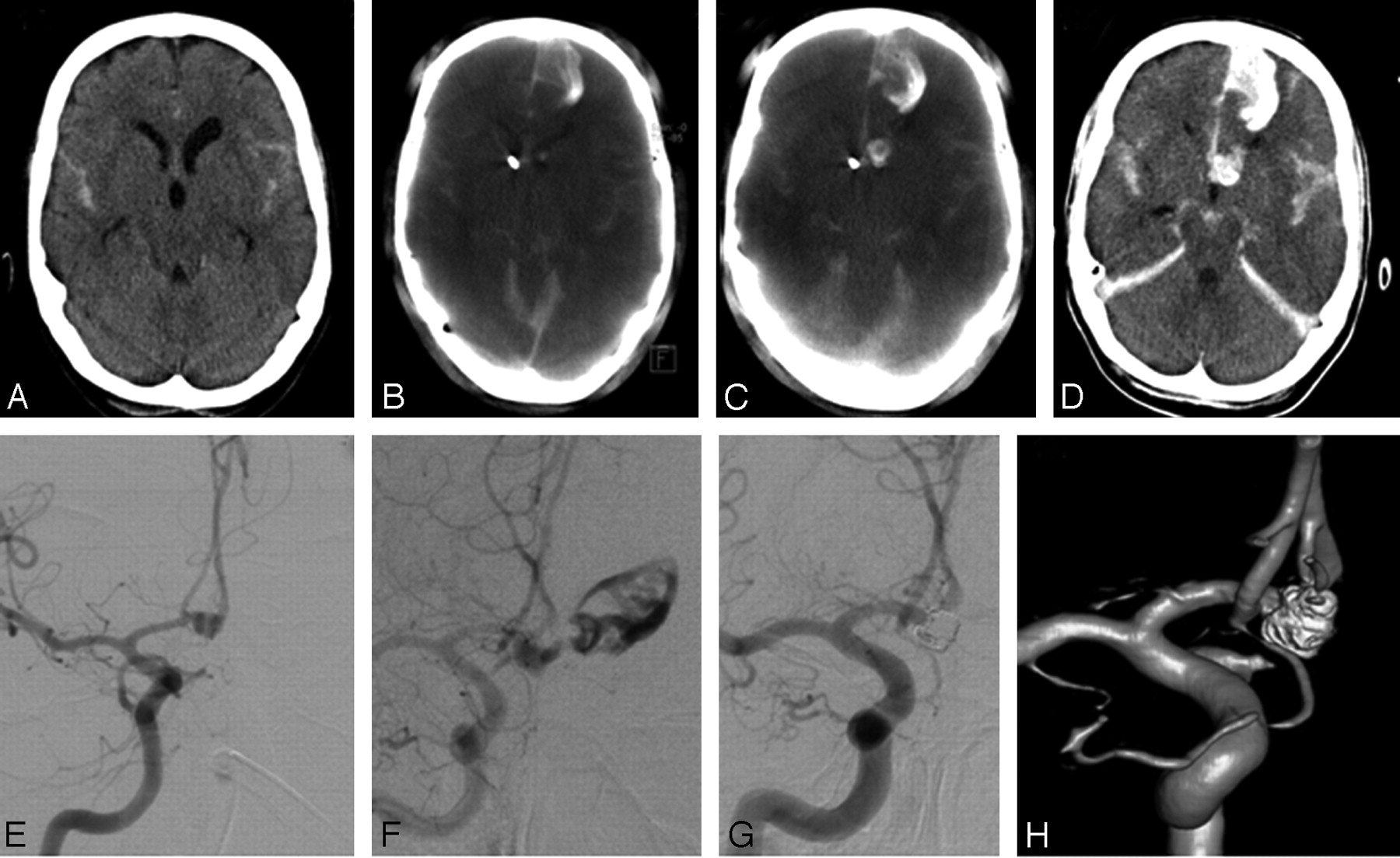
- Fig 1.
Patient 1. A, Initial axial CT scan before coil embolization shows only slight subarachnoid hemorrhage without intraparenchymal bleeding. B, Axial ACT scan reveals rebleeding mainly in the anterior interhemispheric gap and in the left frontal lobe before the onset of diagnostic angiography. C, Axial ACT scan after aneurysmal rupture and installation of the first 2 coils demonstrates no significant space-occupying rebleeding compared with initial ACT. D, Axial CT scan after coil embolization shows only subtle additional subarachnoid hemorrhage compared with intraprocedural ACT (C). E, Right internal carotid artery (ICA) arteriogram (anterior-posterior [AP] projection) reveals an anterior communicating artery aneurysm. F, Right ICA arteriogram (AP projection) demonstrates massive extravasation of contrast media before onset of endovascular therapy. G and H, Right ICA arteriogram (AP projection) and rotational 3D angiography show complete embolization after installation of 8 Guglielmi detachable coils.
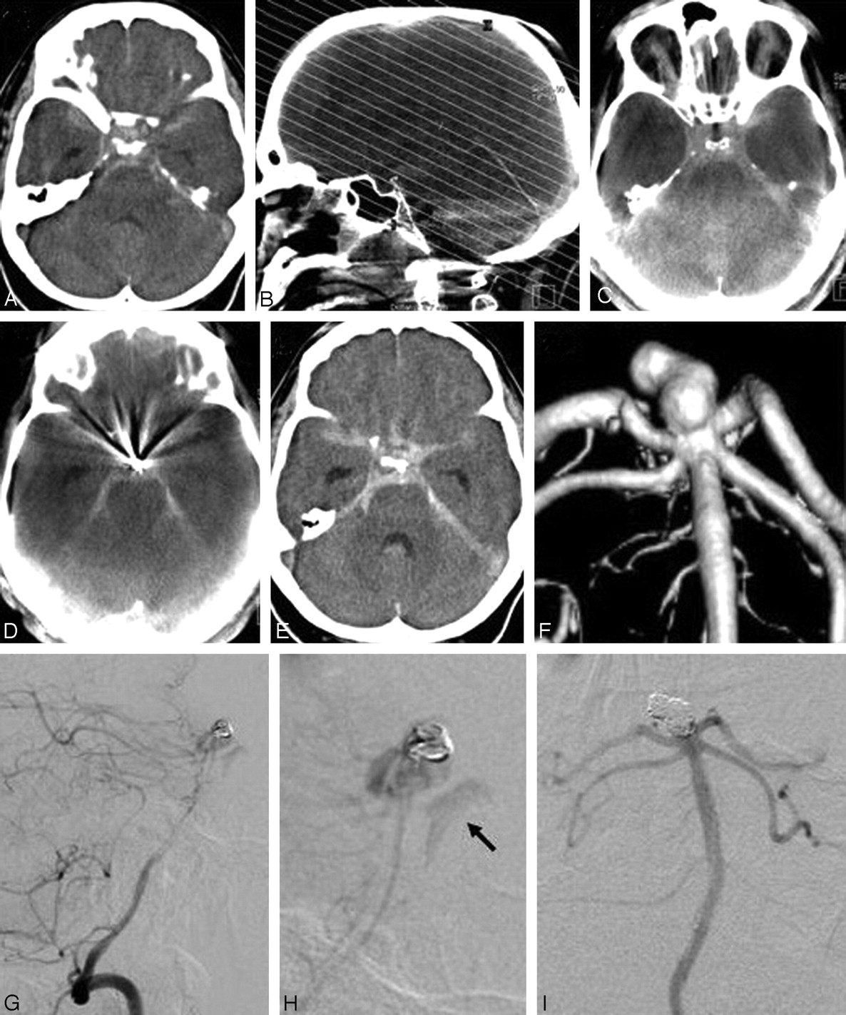
- Fig 2.
Patient 2. A, Axial CT scan before onset of endovascular treatment. B, After postprocessing, MPRs in axial orientation parallel to the skull base identical to conventional CT were calculated. C, Axial ACT scan (preprocedural) reveals no rebleeding in the time between conventional CT and onset of angiography. D, Axial ACT scan (intraprocedural) after aneurysmal rupture and installation of 2 coils shows no significant increase in subarachnoid hemorrhage compared with preprocedural ACT. E, Axial CT scan after endovascular treatment shows no rebleeding after installation of the first 2 coils. F, Rotational 3D angiography shows an irregular, lobulated basilar tip aneurysm. G and H, Basilar artery arteriogram (lateral projection) demonstrates slight extravasation of contrast media (arrow) after installation of the first coil. I, Basilar artery arteriogram (AP projection) shows complete embolization after installation of 7 coils.


In this issue


Jump to section
Related Articles
Cited By...
- Clinical evaluation of volume of interest imaging combined with metal artifact reduction reconstruction techniques in coiling and stent assisted coiling during neurointerventional procedures
- Three-dimensional image fusion of CTA and angiography for real-time guidance during neurointerventional procedures
- Intravenous C-Arm Conebeam CT Angiography following Long-Term Flow-Diverter Implantation: Technologic Evaluation and Preliminary Results
- Technology developments in endovascular treatment of intracranial aneurysms
- A prospective, multicenter pilot study investigating the utility of flat detector derived parenchymal blood volume maps to estimate cerebral blood volume in stroke patients
- Predictive value of flat-panel CT for haemorrhagic transformations in patients with acute stroke treated with thrombectomy
- Angiographic CT for Intraprocedural Monitoring of Complex Neuroendovascular Procedures
- Subarachnoid Hyperattenuation on Flat Panel Detector-Based Conebeam CT Immediately after Uneventful Coil Embolization of Unruptured Intracranial Aneurysms
- Feasibility of Cerebral Blood Volume Mapping by Flat Panel Detector CT in the Angiography Suite: First Experience in Patients with Acute Middle Cerebral Artery Occlusions
- Applicability of Tableside Flat Panel Detector CT Parenchymal Cerebral Blood Volume Measurement in Neurovascular Interventions: Preliminary Clinical Experience
- Use of Angiographic CT Imaging in the Cardiac Catheterization Laboratory for Congenital Heart Disease
- Flat Detector CT in the Evaluation of Brain Parenchyma, Intracranial Vasculature, and Cerebral Blood Volume: A Pilot Study in Patients with Acute Symptoms of Cerebral Ischemia
- Visualisation of Tumour Regression after Local Chemotherapy with Magnetic Nanoparticles - A Pilot Study
- Metal Artifact Reduction for Clipping and Coiling in Interventional C-Arm CT
- Advances in Interventional Neuroradiology