Advertisement

Main menu

  • Home
  • Content
    • Current Issue
    • Accepted Manuscripts
    • Article Preview
    • Past Issue Archive
    • Video Articles
    • AJNR Case Collection
    • Case of the Week Archive
    • Case of the Month Archive
    • Classic Case Archive
  • Special Collections
    • Advancing NeuroMRI with High-Relaxivity Contrast Agents
    • AJNR Awards
    • Low-Field MRI
    • Alzheimer Disease
    • ASNR Foundation Special Collection
    • Photon-Counting CT
    • View All
  • Multimedia
    • AJNR Podcasts
    • AJNR SCANtastic
    • Trainee Corner
    • MRI Safety Corner
    • Imaging Protocols
  • For Authors
    • Submit a Manuscript
    • Submit a Video Article
    • Submit an eLetter to the Editor/Response
    • Manuscript Submission Guidelines
    • Statistical Tips
    • Fast Publishing of Accepted Manuscripts
    • Graphical Abstract Preparation
    • Imaging Protocol Submission
    • Author Policies
  • About Us
    • About AJNR
    • Editorial Board
    • Editorial Board Alumni
  • More
    • Become a Reviewer/Academy of Reviewers
    • Subscribers
    • Permissions
    • Alerts
    • Feedback
    • Advertisers
    • ASNR Home

User menu

  • Alerts
  • Log in

Search

  • Advanced search
American Journal of Neuroradiology

American Journal of Neuroradiology

ASHNR American Society of Functional Neuroradiology ASHNR American Society of Pediatric Neuroradiology ASSR
  • Alerts
  • Log in

Advanced Search

  • Home
  • Content
    • Current Issue
    • Accepted Manuscripts
    • Article Preview
    • Past Issue Archive
    • Video Articles
    • AJNR Case Collection
    • Case of the Week Archive
    • Case of the Month Archive
    • Classic Case Archive
  • Special Collections
    • Advancing NeuroMRI with High-Relaxivity Contrast Agents
    • AJNR Awards
    • Low-Field MRI
    • Alzheimer Disease
    • ASNR Foundation Special Collection
    • Photon-Counting CT
    • View All
  • Multimedia
    • AJNR Podcasts
    • AJNR SCANtastic
    • Trainee Corner
    • MRI Safety Corner
    • Imaging Protocols
  • For Authors
    • Submit a Manuscript
    • Submit a Video Article
    • Submit an eLetter to the Editor/Response
    • Manuscript Submission Guidelines
    • Statistical Tips
    • Fast Publishing of Accepted Manuscripts
    • Graphical Abstract Preparation
    • Imaging Protocol Submission
    • Author Policies
  • About Us
    • About AJNR
    • Editorial Board
    • Editorial Board Alumni
  • More
    • Become a Reviewer/Academy of Reviewers
    • Subscribers
    • Permissions
    • Alerts
    • Feedback
    • Advertisers
    • ASNR Home
  • Follow AJNR on Twitter
  • Visit AJNR on Facebook
  • Follow AJNR on Instagram
  • Join AJNR on LinkedIn
  • RSS Feeds

AJNR Awards, New Junior Editors, and more. Read the latest AJNR updates


Improved Turnaround Times | Median time to first decision: 12 days

Research ArticleHead and Neck Imaging

Sinonasal Inverted Papilloma: Value of Convoluted Cerebriform Pattern on MR Imaging

T.Y. Jeon, H.-J. Kim, S.-K. Chung, H.-J. Dhong, H.Y. Kim, Y.J. Yim, S.T. Kim, P. Jeon and K.H. Kim
American Journal of Neuroradiology September 2008, 29 (8) 1556-1560; DOI: https://doi.org/10.3174/ajnr.A1128
T.Y. Jeon
  • Find this author on Google Scholar
  • Find this author on PubMed
  • Search for this author on this site
H.-J. Kim
  • Find this author on Google Scholar
  • Find this author on PubMed
  • Search for this author on this site
S.-K. Chung
  • Find this author on Google Scholar
  • Find this author on PubMed
  • Search for this author on this site
H.-J. Dhong
  • Find this author on Google Scholar
  • Find this author on PubMed
  • Search for this author on this site
H.Y. Kim
  • Find this author on Google Scholar
  • Find this author on PubMed
  • Search for this author on this site
Y.J. Yim
  • Find this author on Google Scholar
  • Find this author on PubMed
  • Search for this author on this site
S.T. Kim
  • Find this author on Google Scholar
  • Find this author on PubMed
  • Search for this author on this site
P. Jeon
  • Find this author on Google Scholar
  • Find this author on PubMed
  • Search for this author on this site
K.H. Kim
  • Find this author on Google Scholar
  • Find this author on PubMed
  • Search for this author on this site
  • Article
  • Figures & Data
  • Info & Metrics
  • Responses
  • References
  • PDF
Loading

Article Figures & Data

Figures

  • Tables
  • Fig 1.
    • Download figure
    • Open in new tab
    • Download powerpoint
    Fig 1.

    Characteristic MR imaging appearance of a CCP in IP. Coronal T2-weighted (A) and contrast-enhanced fat-suppressed T1-weighted (B) MR images show alternating hypointense and hyperintense striations throughout the tumor involving the left maxillary sinus and nasal cavity.
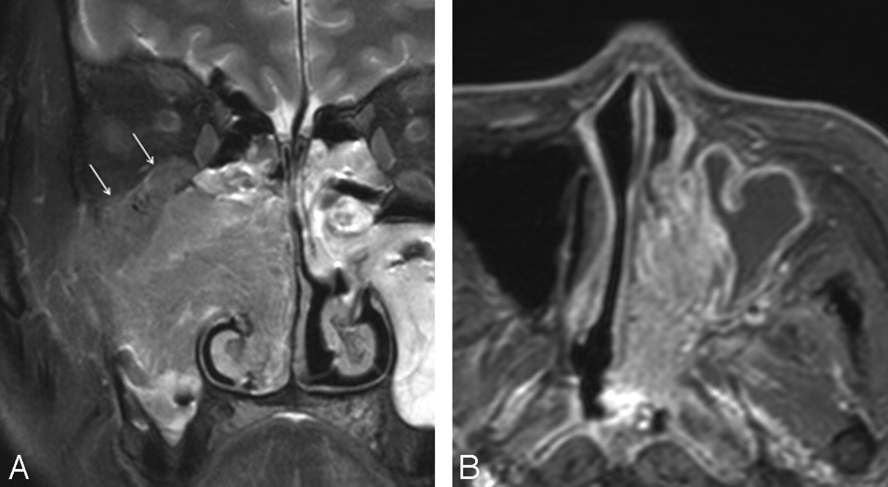
  • Fig 2.
    • Download figure
    • Open in new tab
    • Download powerpoint
    Fig 2.

    MR images of the IPs concomitant with SCC in 2 different patients. A, Coronal contrast-enhanced, fat-suppressed, T1-weighted image shows a large irregular mass in the right nasal cavity and maxillary sinus. Although the nasal mass displays the characteristic CCP, it is lost in most of the mass of the maxillary sinus. Note the bone destruction at the superior and lateral walls of the maxillary sinus with associated orbital invasion (arrows). B, Axial contrast-enhanced, fat-suppressed, T1-weighted image shows an expansile mass in the left nasal cavity, which displays a diffuse CCP throughout the lesion, indistinguishable from the IP without associated carcinoma. The histology revealed multiple foci of microscopic carcinoma scattered within the tumor.

  • Fig 3.
    • Download figure
    • Open in new tab
    • Download powerpoint
    Fig 3.

    Contrast-enhanced fat-suppressed MR images of 4 different malignant sinonasal tumors displaying a CCP either diffusely (A–C) or partially (D). A, SCC. B, Primitive neuroectodermal tumor. C, Mucoepidermoid carcinoma. D, Esthesioneuroblastoma. In the case of esthesioneuroblastoma, the MR image shows a large mass in the left nasal cavity, extending to the orbit and cranial cavity. Note that, whereas the nasal component of the mass displays a typical appearance of CCP, it is not seen in the rather homogeneously enhancing orbital and intracranial components (asterisks in D).

Tables

  • Figures
  • Comparison of visualization of a CCP on MR imaging between inverted papilloma and various malignant sinonasal tumors

    TumorNo. of Total SubjectsNo. of Subjects Showing CCP on MR Imaging
    DiffusePartialT2WICE-T1WI
    Inverted papilloma302642830
        Without SCC222202022
        With SCC84488
    Malignant tumors1286111317
        SCC492668
        Lymphoma390000
        Adenoid cystic carcinoma101112
        Malignant melanoma80101
        Adenocarcinoma51011
        Rhabdomyosarcoma40000
        Esthesioneuroblastoma30222
        Plasmacytoma30000
        Primitive neuroectodermal tumor21011
        Ameloblastic carcinoma10000
        Chordoma10000
        Mucoepidermoid carcinoma11011
        Undifferentiated carcinoma10111
        Non-keratinizing carcinoma10000
    • Note:—CCP indicates convoluted cerebriform pattern; T2WI, T2-weighted image; CE-T1WI, contrast-enhanced T1-weighted image; SCC, squamous cell carcinoma.

PreviousNext
Back to top

In this issue

American Journal of Neuroradiology: 29 (8)
American Journal of Neuroradiology
Vol. 29, Issue 8
September 2008
  • Table of Contents
  • Index by author
Advertisement
Print
Download PDF
Email Article

Thank you for your interest in spreading the word on American Journal of Neuroradiology.

NOTE: We only request your email address so that the person you are recommending the page to knows that you wanted them to see it, and that it is not junk mail. We do not capture any email address.

Enter multiple addresses on separate lines or separate them with commas.
Sinonasal Inverted Papilloma: Value of Convoluted Cerebriform Pattern on MR Imaging
(Your Name) has sent you a message from American Journal of Neuroradiology
(Your Name) thought you would like to see the American Journal of Neuroradiology web site.
CAPTCHA
This question is for testing whether or not you are a human visitor and to prevent automated spam submissions.
Cite this article
T.Y. Jeon, H.-J. Kim, S.-K. Chung, H.-J. Dhong, H.Y. Kim, Y.J. Yim, S.T. Kim, P. Jeon, K.H. Kim
Sinonasal Inverted Papilloma: Value of Convoluted Cerebriform Pattern on MR Imaging
American Journal of Neuroradiology Sep 2008, 29 (8) 1556-1560; DOI: 10.3174/ajnr.A1128

Citation Manager Formats

  • BibTeX
  • Bookends
  • EasyBib
  • EndNote (tagged)
  • EndNote 8 (xml)
  • Medlars
  • Mendeley
  • Papers
  • RefWorks Tagged
  • Ref Manager
  • RIS
  • Zotero
0 Responses
Respond to this article
Share
Bookmark this article
Sinonasal Inverted Papilloma: Value of Convoluted Cerebriform Pattern on MR Imaging
T.Y. Jeon, H.-J. Kim, S.-K. Chung, H.-J. Dhong, H.Y. Kim, Y.J. Yim, S.T. Kim, P. Jeon, K.H. Kim
American Journal of Neuroradiology Sep 2008, 29 (8) 1556-1560; DOI: 10.3174/ajnr.A1128
del.icio.us logo Twitter logo Facebook logo Mendeley logo
  • Tweet Widget
  • Facebook Like
  • Google Plus One
Purchase

Jump to section

  • Article
    • Abstract
    • Materials and Methods
    • Results
    • Discussion
    • Conclusion
    • References
  • Figures & Data
  • Info & Metrics
  • Responses
  • References
  • PDF

Related Articles

  • No related articles found.
  • PubMed
  • Google Scholar

Cited By...

  • Melanoma of the Sinonasal Tract: Value of a Septate Pattern on Precontrast T1-Weighted MR Imaging
  • MRI-Based Texture Analysis to Differentiate Sinonasal Squamous Cell Carcinoma from Inverted Papilloma
  • Routine and Dynamic MR Imaging Study of Lobular Capillary Hemangioma of the Nasal Cavity with Comparison to Inverting Papilloma
  • Sinonasal imaging
  • Apparent Diffusion Coefficient Mapping for Sinonasal Diseases: Differentiation of Benign and Malignant Lesions
  • Crossref
  • Google Scholar

This article has not yet been cited by articles in journals that are participating in Crossref Cited-by Linking.

More in this TOC Section

  • Diagnostic Performance of US in Neck Node NIRADS 2
  • Hydrops Herniation into the Semicircular Canals
  • ASL Sensitivity for Head and Neck Paraganglioma
Show more HEAD AND NECK IMAGING

Similar Articles

Advertisement

Indexed Content

  • Current Issue
  • Accepted Manuscripts
  • Article Preview
  • Past Issues
  • Editorials
  • Editor's Choice
  • Fellows' Journal Club
  • Letters to the Editor
  • Video Articles

Cases

  • Case Collection
  • Archive - Case of the Week
  • Archive - Case of the Month
  • Archive - Classic Case

More from AJNR

  • Trainee Corner
  • Imaging Protocols
  • MRI Safety Corner
  • Book Reviews

Multimedia

  • AJNR Podcasts
  • AJNR Scantastics

Resources

  • Turnaround Time
  • Submit a Manuscript
  • Submit a Video Article
  • Submit an eLetter to the Editor/Response
  • Manuscript Submission Guidelines
  • Statistical Tips
  • Fast Publishing of Accepted Manuscripts
  • Graphical Abstract Preparation
  • Imaging Protocol Submission
  • Evidence-Based Medicine Level Guide
  • Publishing Checklists
  • Author Policies
  • Become a Reviewer/Academy of Reviewers
  • News and Updates

About Us

  • About AJNR
  • Editorial Board
  • Editorial Board Alumni
  • Alerts
  • Permissions
  • Not an AJNR Subscriber? Join Now
  • Advertise with Us
  • Librarian Resources
  • Feedback
  • Terms and Conditions
  • AJNR Editorial Board Alumni

American Society of Neuroradiology

  • Not an ASNR Member? Join Now

© 2025 by the American Society of Neuroradiology All rights, including for text and data mining, AI training, and similar technologies, are reserved.
Print ISSN: 0195-6108 Online ISSN: 1936-959X

Powered by HighWire