
Article Figures & Data
Figures
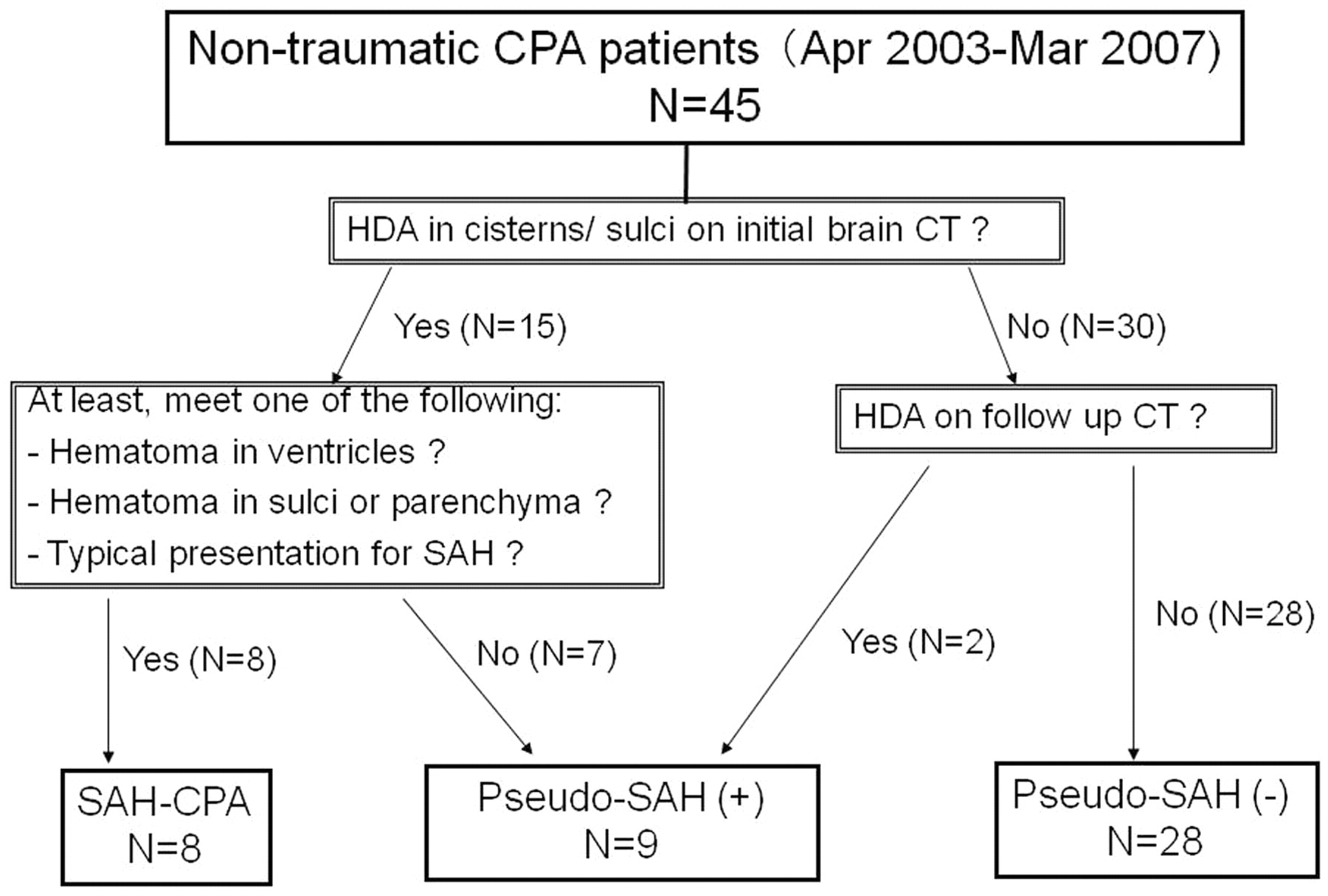
- Fig 1.
Classification of nontraumatic CPA groups.
- Fig 2.
Definition of regions of interest. A, An oval region of interest is defined on the HDA of the Sylvian vallecula, providing the CT number. B, A round or oval region of interest is defined in the brain parenchyma just ventral to the Sylvian vallecula.
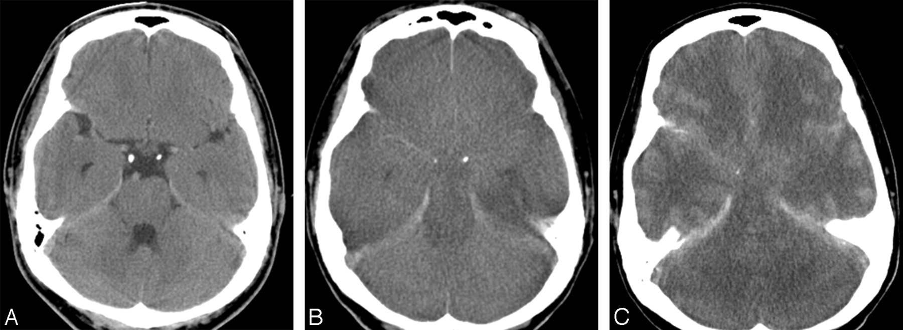
- Fig 3.
A 34-year-old man of the pseudo-SAH(+) group. He had CPA due to suffocation. He was resuscitated immediately but did not recover from coma. A, On the first day, no abnormal finding is seen. B, On the eighth day, the brain shows diffuse low attenuation with obliteration of cisterns and cerebral sulci and narrowed ventricles. HDAs mimicking SAH are noted along the bilateral Sylvian valleculae and tentorium cerebelli. CT values of the HDA of the Sylvian vallecula and the brain parenchyma are 36 and 23.5 HU, respectively. C, On the 129th day, brain edema becomes more severe. The SAH-like HDAs become more prominent.
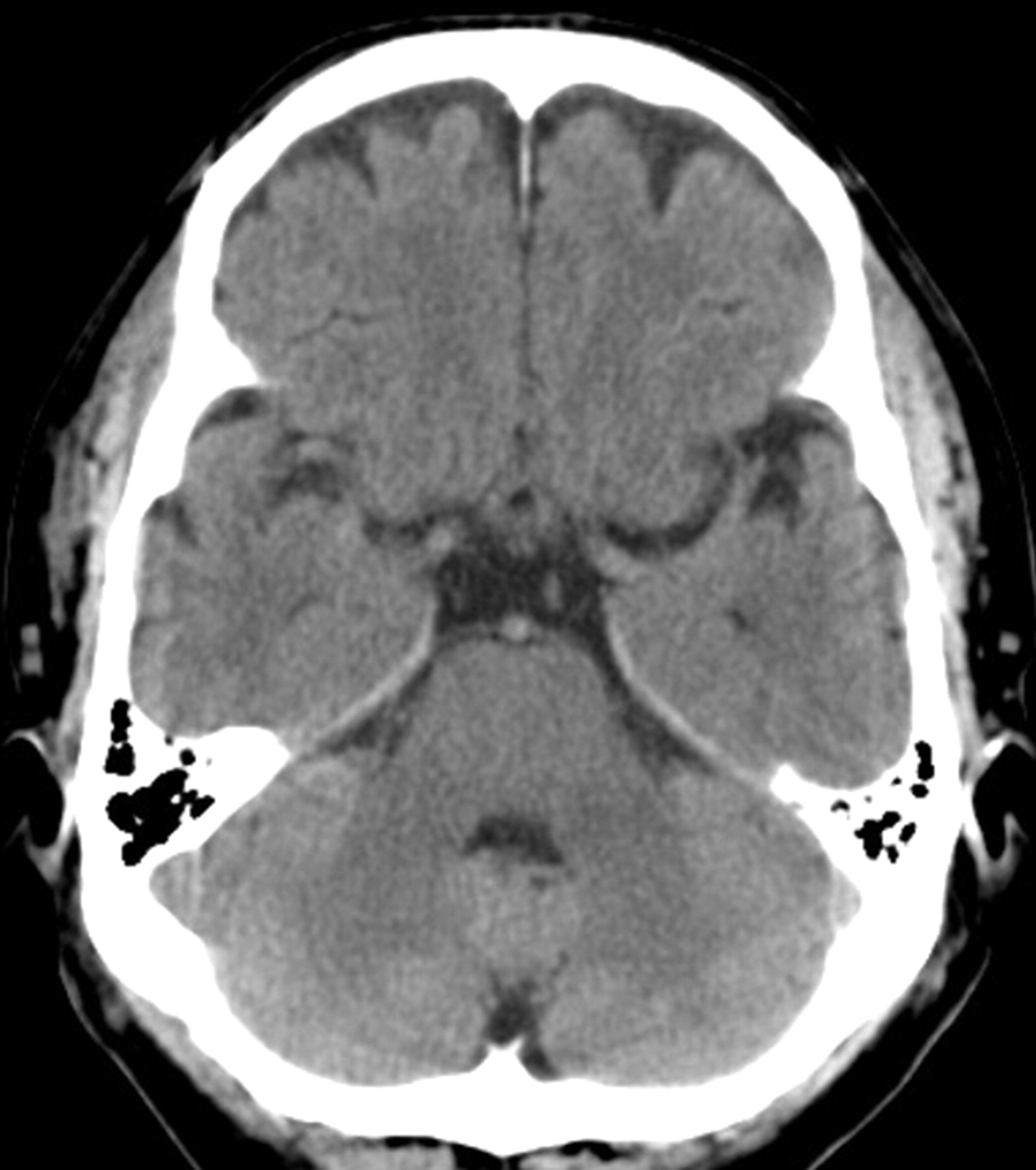
- Fig 4.
A 60-year-old man of the pseudo-SAH(−) group. He had CPA due to ventricular fibrillation followed by immediate resuscitation and resultant full recovery. On the thirteenth day, CT shows no demonstrable abnormality.
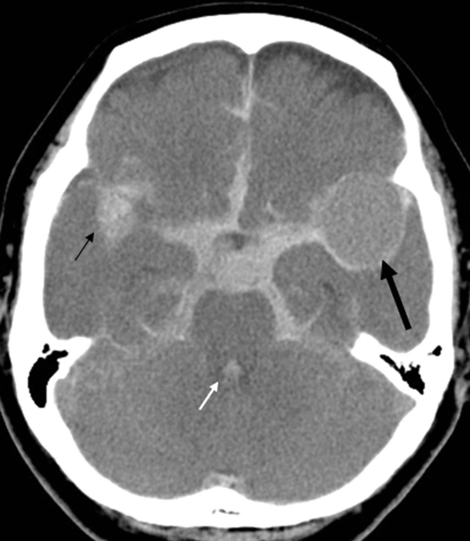
- Fig 5.
A 72-year-old woman of the SAH-CPA group. She suddenly fell unconscious and experienced CPA. The first-day CT shows diffuse HDAs in the basal cisterns, Sylvian valleculae/fissures, and cerebral sulci. Note hematoma in the right Sylvian vallecula (small arrow) and in the fourth ventricle (white arrow). The brain shows diffuse subtle low attenuation with obliteration of the cerebral sulci. The CT values of the HDA and the brain are 49 and 29 HU, respectively. In the left Sylvian vallecula, there is a round filling defect in the hematoma that is thought to represent an aneurysm (large arrow).
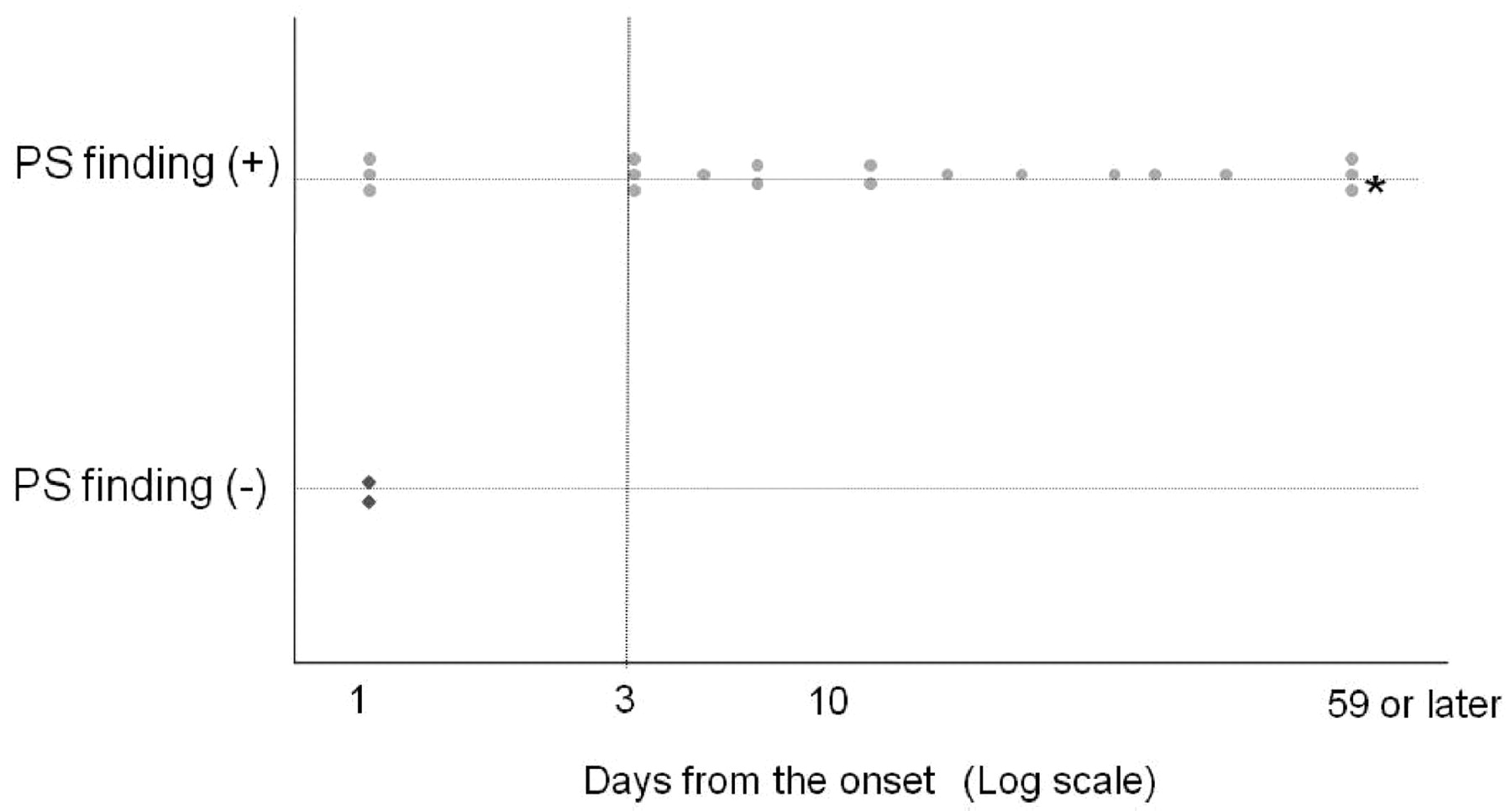
- Fig 6.
The relationship between the days from the onset and the pseudo-SAH finding in patients eventually falling into the pseudo-SAH (+) group. Every dot indicates 1 occasion of CT examination. The asterisk indicates that these 3 examinations were performed on the 59th, 129th, and 268th days. PS indicates pseudo-SAH.
- Fig 7.
Survival curves comparing the pseudo-SAH(+) and the pseudo-SAH(−) groups.


Tables
Pseudo-SAH Finding Degree of Cerebral Edema None Mild Severe Total Positive 0 0 19 19 Negative 35 22 4 61 Total 35 22 23 80 * P < .0001 (χ2 test).
† Numeric values in the cells represent the number of CT examinations.
Groups CT Values (HU) No. Mean SD Range (Min-Max) Pseudo-SAH(+) 18 37.6† 3.3 30.0–42.0 CPA-SAH 14 56.6 7.8 41.0–67.0 SAH 23 53.7 5.1 42.0–60.0 Note:—No. indicates number of hemispheres; Min, minimum; Max, maximum.
* ANOVA, P < .0001.
† P < .0001, as compared with the other 2 groups (Bonferroni post hoc test).
Groups CT Values (HU) No. Mean SD Range (Min-Max) Pseudo-SAH(+) 18 26.8†‡ 3.1 19.0–31.0 Pseudo-SAH(−)§ 54 29.8‡ 1.9 24.0–34.0 SAH-CPA 16 30.0‡ 2.3 27.0–36.0 SAH 26 31.2 1.9 28.0–35.0 Healthy 40 32.5 1.7 30.0–35.0 Note:—No. indicates number of hemispheres, Min, minimum; Max, maximum.
* ANOVA, P < .0001.
† P < .0001, as compared with the other 4 groups (Bonferroni post hoc test).
‡ P < .0001, as compared with healthy groups (Bonferroni post hoc test).
§ One case of Pseudo-SAH(−) group was excluded because of contrast media injection.


