Advertisement

Main menu

  • Home
  • Content
    • Current Issue
    • Accepted Manuscripts
    • Article Preview
    • Past Issue Archive
    • Video Articles
    • AJNR Case Collection
    • Case of the Week Archive
    • Case of the Month Archive
    • Classic Case Archive
  • Special Collections
    • AJNR Awards
    • Low-Field MRI
    • Alzheimer Disease
    • ASNR Foundation Special Collection
    • Photon-Counting CT
    • View All
  • Multimedia
    • AJNR Podcasts
    • AJNR SCANtastic
    • Trainee Corner
    • MRI Safety Corner
    • Imaging Protocols
  • For Authors
    • Submit a Manuscript
    • Submit a Video Article
    • Submit an eLetter to the Editor/Response
    • Manuscript Submission Guidelines
    • Statistical Tips
    • Fast Publishing of Accepted Manuscripts
    • Graphical Abstract Preparation
    • Imaging Protocol Submission
    • Author Policies
  • About Us
    • About AJNR
    • Editorial Board
    • Editorial Board Alumni
  • More
    • Become a Reviewer/Academy of Reviewers
    • Subscribers
    • Permissions
    • Alerts
    • Feedback
    • Advertisers
    • ASNR Home

User menu

  • Alerts
  • Log in

Search

  • Advanced search
American Journal of Neuroradiology

American Journal of Neuroradiology

ASHNR American Society of Functional Neuroradiology ASHNR American Society of Pediatric Neuroradiology ASSR
  • Alerts
  • Log in

Advanced Search

  • Home
  • Content
    • Current Issue
    • Accepted Manuscripts
    • Article Preview
    • Past Issue Archive
    • Video Articles
    • AJNR Case Collection
    • Case of the Week Archive
    • Case of the Month Archive
    • Classic Case Archive
  • Special Collections
    • AJNR Awards
    • Low-Field MRI
    • Alzheimer Disease
    • ASNR Foundation Special Collection
    • Photon-Counting CT
    • View All
  • Multimedia
    • AJNR Podcasts
    • AJNR SCANtastic
    • Trainee Corner
    • MRI Safety Corner
    • Imaging Protocols
  • For Authors
    • Submit a Manuscript
    • Submit a Video Article
    • Submit an eLetter to the Editor/Response
    • Manuscript Submission Guidelines
    • Statistical Tips
    • Fast Publishing of Accepted Manuscripts
    • Graphical Abstract Preparation
    • Imaging Protocol Submission
    • Author Policies
  • About Us
    • About AJNR
    • Editorial Board
    • Editorial Board Alumni
  • More
    • Become a Reviewer/Academy of Reviewers
    • Subscribers
    • Permissions
    • Alerts
    • Feedback
    • Advertisers
    • ASNR Home
  • Follow AJNR on Twitter
  • Visit AJNR on Facebook
  • Follow AJNR on Instagram
  • Join AJNR on LinkedIn
  • RSS Feeds

AJNR Awards, New Junior Editors, and more. Read the latest AJNR updates

LetterLetter

A Developmental Venous Anomaly Presenting Atypical Findings on Susceptibility-Weighted Imaging

Y. Fushimi, Y. Miki, K. Togashi, K. Kikuta, N. Hashimoto and H. Fukuyama
American Journal of Neuroradiology August 2008, 29 (7) e56; DOI: https://doi.org/10.3174/ajnr.A1074
Y. Fushimi
  • Find this author on Google Scholar
  • Find this author on PubMed
  • Search for this author on this site
Y. Miki
  • Find this author on Google Scholar
  • Find this author on PubMed
  • Search for this author on this site
K. Togashi
  • Find this author on Google Scholar
  • Find this author on PubMed
  • Search for this author on this site
K. Kikuta
  • Find this author on Google Scholar
  • Find this author on PubMed
  • Search for this author on this site
N. Hashimoto
  • Find this author on Google Scholar
  • Find this author on PubMed
  • Search for this author on this site
H. Fukuyama
  • Find this author on Google Scholar
  • Find this author on PubMed
  • Search for this author on this site
  • Article
  • Figures & Data
  • Info & Metrics
  • Responses
  • References
  • PDF
Loading

Developmental venous anomaly (DVA) is the most common type of vascular malformation found at autopsy, with a prevalence of 2%.1 DVA is composed of radially arranged venous complexes converging to a centrally located venous trunk, which drains the normal brain parenchyma.2 Susceptibility-weighted imaging (SWI) shows low signal intensity for DVA due to the blood oxygen level–dependent (BOLD) effect in the abnormal medullary and draining veins.3 SWI has been successfully proved to have better detectability of venous structures than conventional T2*-weighted imaging.4 Our letter will present a DVA that showed isointensity compared with the surrounding cerebral parenchyma on SWI.

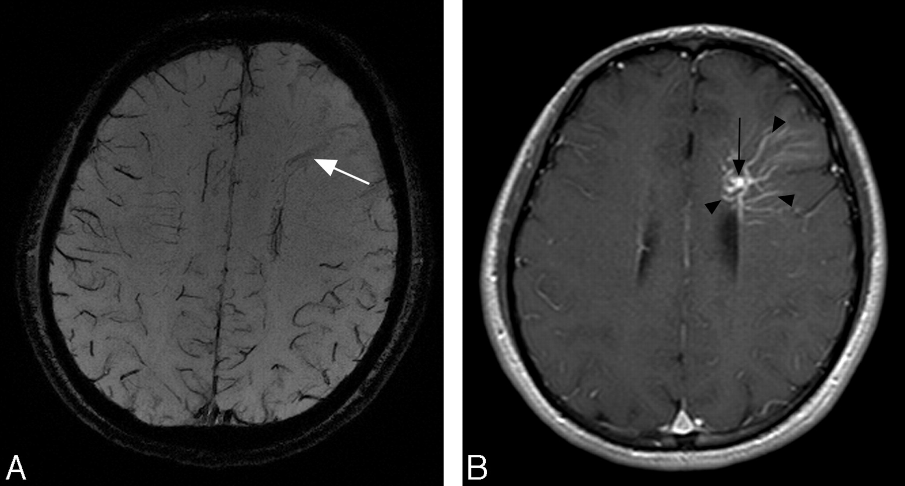
A 26-year-old man was referred to our institution due to an incidentally found DVA with no symptoms attributable to it. He underwent MR imaging examination, including SWI and MR angiography, in a 3T MR imaging scanner (Magnetom Trio; Siemens, Erlangen, Germany). MR imaging parameters of SWI were as follows: TR, 27 ms; TE, 20 ms; flip angle, 15°; FOV, 230 × 186 mm; matrix, 512 × 416; section thickness, 1.2 mm; 80 imaging sections; and scanning time, 6 minutes 50 seconds. Minimum intensity projection (MIP) images over 6 sections were created from phase and magnitude images. An MIP image of SWI showed a moderately low-signal-intensity structure corresponding to the draining vein; however, no low-signal-intensity structures corresponding to medullary veins (Fig 1A) were seen. Low-signal-intensity structures corresponding to the cortical veins of the other lobes were clearly shown; however, no apparent signals of cortical veins in the left frontal lobe were identified. Postcontrast T1-weighted images at 1.5T obtained 2 days after precontrast images revealed enhanced medullary veins and a draining vein (Fig 1B).

Signals on SWI are not compromised by low-velocity venous flow; therefore, SWI has successfully demonstrated low-flow vascular formations such as DVA.4 SWI at high-field MR imaging such as 3T allowed high-spatial-resolution images and better delineation of venous structures because the BOLD effect is related to the strength of the main magnetic field.3 In our case, SWI at 3T revealed no medullary veins of DVA, which implies that a relatively higher flow in the DVA reduced the amount of deoxyhemoglobin inside the DVA.

Our letter will add some information for interpretation of imaging findings on SWI, especially in the case of DVA with relatively higher flow. Neuroradiologists should keep such exceptional findings in mind in the interpretation of SWI.

This work was supported in part by a Health and Labour Sciences Research grant of Japan.

  Fig 1.
  • Download figure
  • Open in new tab
  • Download powerpoint
Fig 1.

A 26-year-old man with a DVA. A, MIP image of SWI shows a moderate low-signal-intensity structure corresponding to the draining vein (arrow); however, no low-signal-intensity structures corresponding to medullary veins are seen. Low-signal-intensity structures corresponding to the cortical veins of the other lobes are clearly shown; however, no apparent signals of cortical veins in the left frontal lobe are identified. B, Postcontrast T1-weighted images at 1.5T obtained 2 days after precontrast images reveal the enhanced draining vein (arrow) and medullary veins (arrowheads).

References

  1. ↵
    Garner TB, Del Curling O Jr, Kelly DL Jr, et al. The natural history of intracranial venous angiomas. J Neurosurg 1991;75:715–22
    PubMed
  2. ↵
    Abe T, Singer RJ, Marks MP, et al. Coexistence of occult vascular malformations and developmental venous anomalies in the central nervous system: MR evaluation. AJNR Am J Neuroradiol 1998;19:51–57
    Abstract
  3. ↵
    Reichenbach JR, Barth M, Haacke EM, et al. High-resolution MR venography at 3.0 Tesla. J Comput Assist Tomogr 2000;24:949–57
    CrossRefPubMed
  4. ↵
    Lee BC, Vo KD, Kido DK, et al. MR high-resolution blood oxygenation level-dependent venography of occult (low-flow) vascular lesions. AJNR Am J Neuroradiol 1999;20:1239–42
    Abstract/FREE Full Text
  • Copyright © American Society of Neuroradiology
PreviousNext
Back to top

In this issue

American Journal of Neuroradiology: 29 (7)
American Journal of Neuroradiology
Vol. 29, Issue 7
August 2008
  • Table of Contents
  • Index by author
Advertisement
Print
Download PDF
Email Article

Thank you for your interest in spreading the word on American Journal of Neuroradiology.

NOTE: We only request your email address so that the person you are recommending the page to knows that you wanted them to see it, and that it is not junk mail. We do not capture any email address.

Enter multiple addresses on separate lines or separate them with commas.
A Developmental Venous Anomaly Presenting Atypical Findings on Susceptibility-Weighted Imaging
(Your Name) has sent you a message from American Journal of Neuroradiology
(Your Name) thought you would like to see the American Journal of Neuroradiology web site.
CAPTCHA
This question is for testing whether or not you are a human visitor and to prevent automated spam submissions.
Cite this article
Y. Fushimi, Y. Miki, K. Togashi, K. Kikuta, N. Hashimoto, H. Fukuyama
A Developmental Venous Anomaly Presenting Atypical Findings on Susceptibility-Weighted Imaging
American Journal of Neuroradiology Aug 2008, 29 (7) e56; DOI: 10.3174/ajnr.A1074

Citation Manager Formats

  • BibTeX
  • Bookends
  • EasyBib
  • EndNote (tagged)
  • EndNote 8 (xml)
  • Medlars
  • Mendeley
  • Papers
  • RefWorks Tagged
  • Ref Manager
  • RIS
  • Zotero
0 Responses
Respond to this article
Share
Bookmark this article
A Developmental Venous Anomaly Presenting Atypical Findings on Susceptibility-Weighted Imaging
Y. Fushimi, Y. Miki, K. Togashi, K. Kikuta, N. Hashimoto, H. Fukuyama
American Journal of Neuroradiology Aug 2008, 29 (7) e56; DOI: 10.3174/ajnr.A1074
del.icio.us logo Twitter logo Facebook logo Mendeley logo
  • Tweet Widget
  • Facebook Like
  • Google Plus One
Purchase

Jump to section

  • Article
    • References
  • Figures & Data
  • Info & Metrics
  • Responses
  • References
  • PDF

Related Articles

  • No related articles found.
  • PubMed
  • Google Scholar

Cited By...

  • No citing articles found.
  • Crossref (23)
  • Google Scholar

This article has been cited by the following articles in journals that are participating in Crossref Cited-by Linking.

  • Susceptibility-Weighted Imaging: Technical Aspects and Clinical Applications, Part 1
    E.M. Haacke, S. Mittal, Z. Wu, J. Neelavalli, Y.-C.N. Cheng
    American Journal of Neuroradiology 2009 30 1
  • Susceptibility-weighted imaging for differential diagnosis of cerebral vascular pathology: A pictorial review
    Yu-Kun Tsui, Fong Y. Tsai, Anton N. Hasso, Fred Greensite, Binh V. Nguyen
    Journal of the Neurological Sciences 2009 287 1-2
  • The association between cerebral developmental venous anomaly and concomitant cavernous malformation: an observational study using magnetic resonance imaging
    Guolu Meng, Chuanfeng Bai, Tengfei Yu, Zhen Wu, Xing Liu, Junting Zhang, Jizong zhao
    BMC Neurology 2014 14 1
  • Susceptibility weighted magnetic resonance imaging of cerebral cavernous malformations: prospects, drawbacks, and first experience at ultra–high field strength (7-Tesla) magnetic resonance imaging
    Philipp Dammann, Markus Barth, Yuan Zhu, Stefan Maderwald, Marc Schlamann, Mark E. Ladd, Ulrich Sure
    Neurosurgical Focus 2010 29 3
  • Susceptibility-weighted imaging in pediatric neuroimaging
    Thangamadhan Bosemani, Andrea Poretti, Thierry A.G.M. Huisman
    Journal of Magnetic Resonance Imaging 2014 40 3
  • Developmental venous anomalies of the brain in children — imaging spectrum and update
    Luke L. Linscott, James L. Leach, Blaise V. Jones, Todd A. Abruzzo
    Pediatric Radiology 2016 46 3
  • Clinical characteristics of multiple primary carcinomas of the oral cavity
    Yumi Mochizuki, Hiroyuki Harada, Minoru Ikuta, Hiroaki Shimamoto, Hirofumi Tomioka, Kae Tanaka, Hideaki Hirai, Ken Omura
    Oral Oncology 2015 51 2
  • Investigating the oxygenation of brain arteriovenous malformations using quantitative susceptibility mapping
    Emma Biondetti, Alvaro Rojas-Villabona, Magdalena Sokolska, Francesca B. Pizzini, Hans Rolf Jäger, David L. Thomas, Karin Shmueli
    NeuroImage 2019 199
  • The relation between angioarchitectural factors of developmental venous anomaly and concomitant sporadic cavernous malformation
    Tengfei Yu, Xing Liu, Xiangjiang Lin, Chuanfeng Bai, Jizong Zhao, Junting Zhang, Liwei Zhang, Zhen Wu, Shuo Wang, Yuanli Zhao, Guolu Meng
    BMC Neurology 2016 16 1
  • Developmental venous anomaly coexisting with a true arteriovenous malformation: a rare clinical entity
    Eren Erdem, Adewumi O Amole, Mehmet S Akdol, Rohan S Samant, Gazi M Yaşargil
    Journal of NeuroInterventional Surgery 2012 4 4

More in this TOC Section

  • Letter to the Editor regarding “Automated Volumetric Software in Dementia: Help or Hindrance to the Neuroradiologist?”
  • Reply:
  • Brain AVM’s Nidus: What if We Hadn’t Understood Anything?
Show more LETTERS

Similar Articles

Advertisement

Indexed Content

  • Current Issue
  • Accepted Manuscripts
  • Article Preview
  • Past Issues
  • Editorials
  • Editor's Choice
  • Fellows' Journal Club
  • Letters to the Editor
  • Video Articles

Cases

  • Case Collection
  • Archive - Case of the Week
  • Archive - Case of the Month
  • Archive - Classic Case

More from AJNR

  • Trainee Corner
  • Imaging Protocols
  • MRI Safety Corner

Multimedia

  • AJNR Podcasts
  • AJNR Scantastics

Resources

  • Turnaround Time
  • Submit a Manuscript
  • Submit a Video Article
  • Submit an eLetter to the Editor/Response
  • Manuscript Submission Guidelines
  • Statistical Tips
  • Fast Publishing of Accepted Manuscripts
  • Graphical Abstract Preparation
  • Imaging Protocol Submission
  • Evidence-Based Medicine Level Guide
  • Publishing Checklists
  • Author Policies
  • Become a Reviewer/Academy of Reviewers
  • News and Updates

About Us

  • About AJNR
  • Editorial Board
  • Editorial Board Alumni
  • Alerts
  • Permissions
  • Not an AJNR Subscriber? Join Now
  • Advertise with Us
  • Librarian Resources
  • Feedback
  • Terms and Conditions
  • AJNR Editorial Board Alumni

American Society of Neuroradiology

  • Not an ASNR Member? Join Now

© 2025 by the American Society of Neuroradiology All rights, including for text and data mining, AI training, and similar technologies, are reserved.
Print ISSN: 0195-6108 Online ISSN: 1936-959X

Powered by HighWire