
Article Figures & Data
Figures
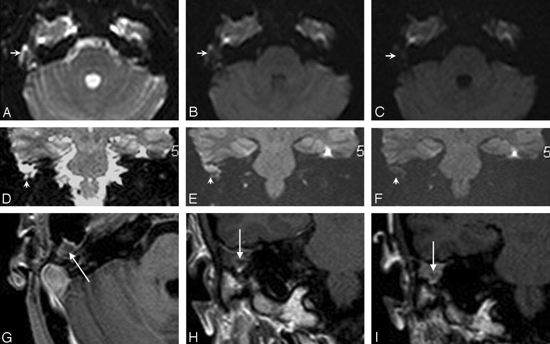
- Fig 1.
EPI DWI and DPI for residual cholesteatoma detection. DWI axial b = 0 (A), b = 500 (B), b = 1000 (C) and coronal b = 0 (D), b = 500 (E), b = 1000 (F) EPI sequences. Early (G and I) and delayed (H and J) postconstrast T1-weighted axial (G and H) and coronal (I and J) sequences. With DWI, an increasing signal intensity (arrow) between b = 0 (A and D), b = 500 (B and E), and b = 1000 (C and F) sequences was observed in the left hypotympanum. A hypointense space-occupying lesion (arrows) was also observed in the left hypotympanum with early enhanced T1-weighted sequences (G and I). This lesion was more conspicuous on delayed imaging (H and J, large arrows), whereas scar tissue was intensely enhanced (small arrows). Thus, surgery confirmed a residual cholesteatoma of the left hypotympanum.
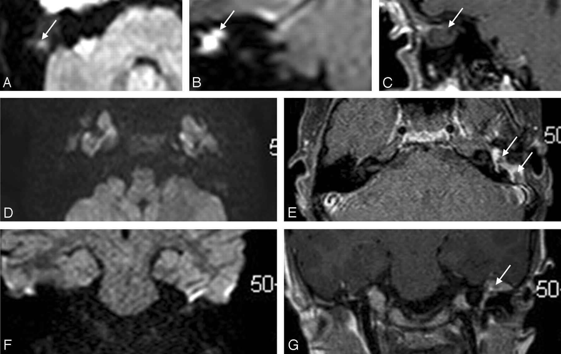
- Fig 2.
Mismatch between DWI and DPI, with a small lesion. EPI DWI axial b = 0 (A), b = 500 (B), b = 1000 (C) and coronal b = 0 (D), b = 500 (E), b = 1000 (F) sequences. Early coronal (H) and delayed postcontrast T1-weighted axial (G) and coronal (I) sequences. Progressive signal-intensity decay was observed in the right mastoid with DWI (A–F, small arrows). DWI was considered negative for residual cholesteatoma. Conversely, a hypointense lesion was observed on early-enhanced T1-weighted MR images (H, large arrow), with a ring enhancement on DPI (G and I, large arrows). At surgery, a 3-mm large pearl-like residual cholesteatoma was found.
- Fig 3.
FP examinations with DWI and DPI in 2 different patients. Axial (A) and coronal (B) EPI DWI b = 1000 and coronal DPI (C) sequences. Hyperintense foci were observed in the right mastoid with axial and coronal DWI (A and B, arrows). Lack of enhancement on coronal delayed T1-weighted MR image (C, arrow) led us to suspect a residual cholesteatoma. At surgery, only a silastic sheet filling the right mastoid was found. Axial (D) and coronal (F) EPI DWI b = 1000 and axial (E) and coronal (G) DPIs in another patient. DWI was negative for residual cholesteatoma (D and F). Note nonenhancing nodular masses in the left mesotympanum and mastoid on axial and coronal delayed T1-weighted MR images (E and G, arrows). Small-sized residual cholesteatomas were suspected. At surgery, only scar tissue was found.


Tables
Patient DWI Cholesteatoma DPI Cholesteatoma Surgery Tissue Surgery Size (mm)/Type 1 N N Scar 2 Y Y Cholesteatoma 3/pearl 3 N N Inflammatory 4 Y Y Cholesteatoma 5/infiltrating 5 Y Y Scar* 6 Y Y Cholesteatoma 4/pearl 7 N Y Cholesteatoma 3/pearl 8 Y Y Scar* 9 Y Y Cholesteatoma 3/pearl 10 Y Y Cholesteatoma 10/infiltrating 11 Y Y Inflammatory 12 N Y Cholesteatoma 2/pearl 13 N Y Scar* 14 N N Cholesteatoma 3/pearl 15 Y Y Cholesteatoma 10/infiltrating 16 Y Y Cholesteatoma 8/infiltrating 17 N N Scar 18 N Y Cholesteatoma 3/infiltrating 19 N Y Cholesteatoma 2/pearl 20 Y Y Cholesteatoma 3/pearl 21 N N Inflammatory 22 N N Cholesteatoma 3/pearl 23 Y Y Cholesteatoma 6/infiltrating 24 N Y Cholesteatoma 4/infiltrating 25 N N Scar 26 Y Y Cholesteatoma 10/infiltrating 27 Y Y Cholesteatoma 10/infiltrating 28 N N Scar 29 N Y Cholesteatoma 5/pearl 30 N N Scar 31 N Y Scar* Note:—Y indicates yes; N, no; pearl, pearl-like cholesteatoma; infiltrating, infiltrating form of cholesteatoma.
* Presence of a silastic sheet.
- Table 2:
Summary of the diagnostic values of DWI and DPI sequences according to residual cholesteatoma size
DWI DPI Total Series Tumor >3 mm Tumor >5 mm Total Series Tumor >3 mm Tumor >5 mm Se 60.00 75.00 100.00 90.00 100.00 100.00 Spe 72.73 84.21 88.00 54.55 75.00 80.00 PPV 80.00 75.00 66.67 78.26 68.75 54.55 NPV 50.00 84.21 100.00 75.00 100.00 100.00 Note:—Se indicates sensitivity; Spe, specificity; PPV, positive predictive value; NPV, negative predictive value.
- Table 3:
Summary of the diagnostic values of DWI and/or DPI sequences according to residual cholesteatoma size*
DWI or DPI DWI and DPI Total Series Tumor >3 mm Tumor >5 mm Total Series Tumor >3 mm Tumor >5 mm Se 90.00 100.00 100.00 60.00 75.00 100.00 Spe 54.55 75.00 80.00 72.73 84.21 88.00 PPV 78.26 68.75 54.55 80.00 75.00 66.67 NPV 75.00 100.00 100.00 50.00 84.21 100.00 Note:—Se indicates sensitivity; Spe, specificity; PPV, positive predictive value; NPV, negative predictive value.
* DWI or DPI diagnosis of cholesteatoma with any of these 2 examinations is sufficient to suggest a residual lesion. DWI and DPI diagnosis of cholesteatoma with both examinations is necessary to suggest a residual lesion.
In this issue


Jump to section
Related Articles
Cited By...
- Optimal Duration of MRI Follow-up to Safely Identify Middle Ear Residual Cholesteatoma
- MRI Findings of a Middle Ear Cholesteatoma in a Dog
- Detection of Middle Ear Cholesteatoma by Diffusion-Weighted MR Imaging: Multishot Echo-Planar Imaging Compared with Single-Shot Echo-Planar Imaging
- The Utility of Diffusion-Weighted Imaging for Cholesteatoma Evaluation
- Neuroradiology of Cholesteatomas