
Article Figures & Data
Figures
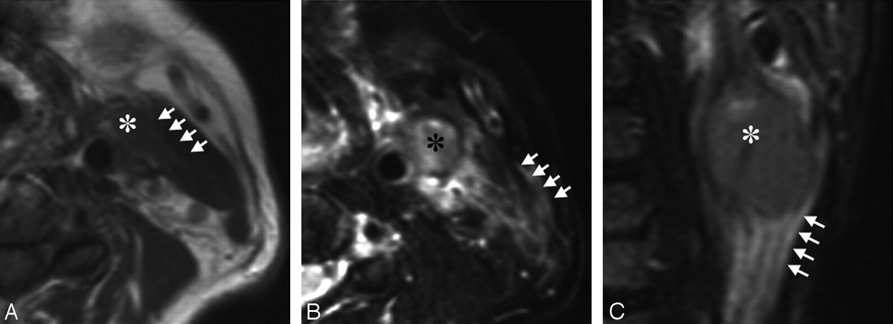
- Fig 1.
A 63-year-old man with tongue carcinoma. A, Axial T1-weighted (TR/TE = 500/15) image shows an ENS-positive metastatic node (asterisk) at level II. Note the vanishing border sign (arrows) with obliteration of the fat layer between the node and the neighboring sternocleidomastoid muscle. B, Axial fat-suppressed T2-weighted (TR/TE = 4674/80 ms) image shows the flare sign that is around and extending from the same ENS-positive metastatic node (asterisk) as in A. Note that high-intensity signals are present in the interstitial tissues between the sternocleidomastoid muscle and the subcutaneous fat (arrows). C, Coronal fat-suppressed T2-weighted (TR/TE = 4674/80 ms) image shows the flare sign (arrows) caudal to the same ENS-positive metastatic node (asterisk) as in A and B.
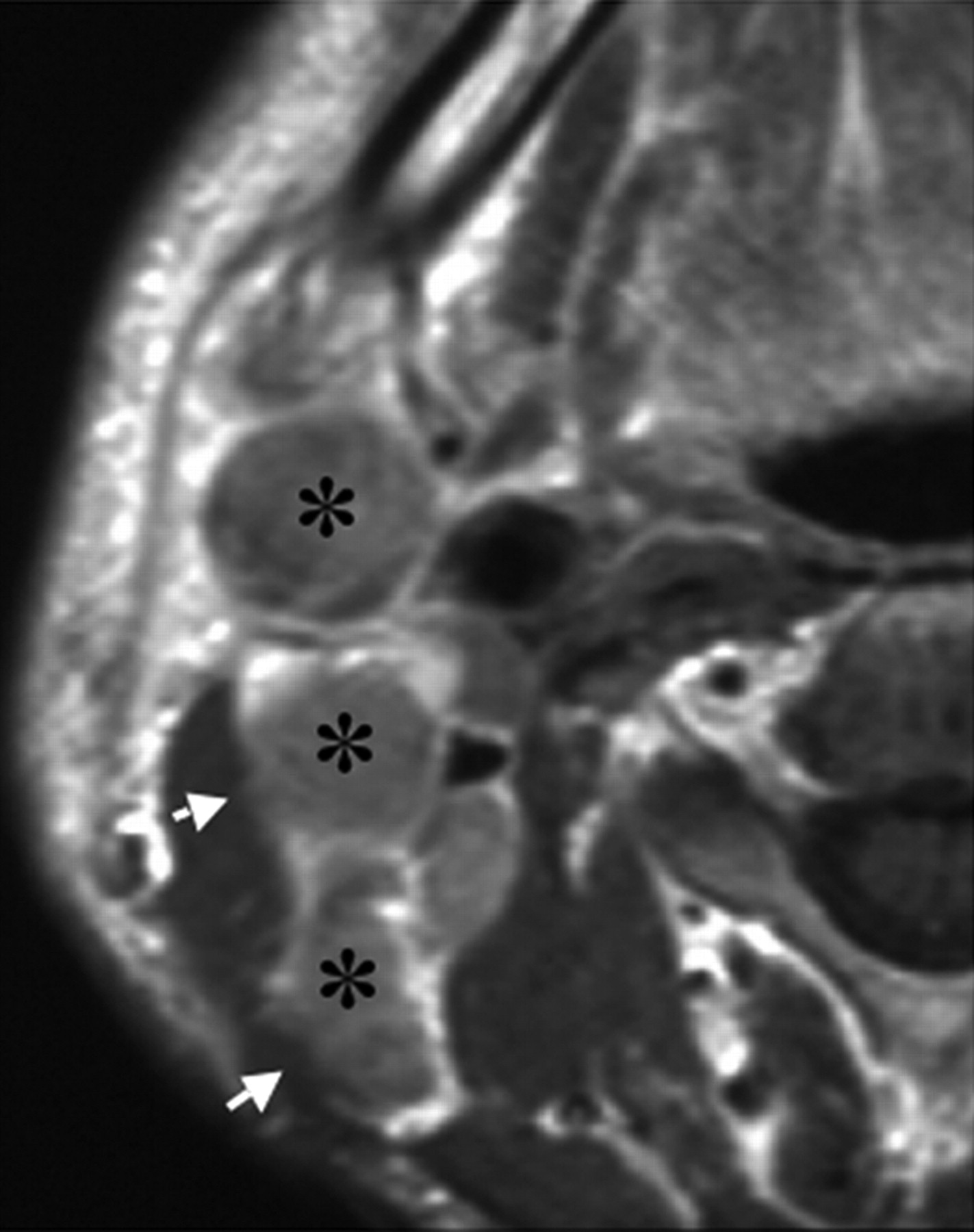
- Fig 2.
A 51-year-old man with upper gingival carcinoma. Gadolinium-enhanced axial T1-weighted (TR/TE = 500/15) image shows the irregular boundary (shaggy margin, arrows) of contrast-enhanced metastatic nodes with ENS (asterisk) at levels II and III.
- Fig 3.
Comparison of nodal sizes of ENS-positive and -negative metastatic nodes in the neck. Graph (boxplots) shows distributions of short-axis diameters of ENS-negative (−) (n = 130) and ENS-positive (+) (n = 47) metastatic nodes in the necks from 109 patients with head and neck cancers. The horizontal line is a median (50th percentile) of the measured volumes; the tops and bottoms of the boxes represent the 25th and 75th percentiles, respectively, and whiskers indicate the range from the largest to smallest observed data points within the 1.5 interquartile range presented by the box. The short-axis diameters of ENS (+) metastatic nodes are significantly greater than those of ENS (−) metastatic nodes (P value, Mann-Whitney U test).


Tables
- Table 1:
Incidence (%) of MR imaging findings in the necks of patients with head and neck cancers by consensus of 2 radiologists
Vanishing Border Sign* (T1WI) Flare Sign* (fsT2WI) Shaggy Margin* (CET1WI) Present Absent Present Absent Present Absent ENS (+) 91 6 90 7 97 0 ENS (−) 10 33 8 35 12 31 Total 101 39 98 42 109 31 Note:—T1WI indicates T1-weighted imaging; fT2WI, fat-suppressed T2-weighted imaging; CET1WI, gadolinium-enhanced T1-weighted imaging.
* The incidence of MR imaging criteria is significantly different between ENS (+) and ENS (−) necks (P < .0001, Fisher exact test).
Variable Coefficient 95% CI SE Odds Ratio P Short-axis diameter −0.029 −0.045 to −0.016 0.009 6.8 .017* Vanishing border sign 0.325 0.213 to 0.647 0.258 6.8 .350 Flare sign −0.773 −0.952 to −0.556 0.325 8.3 .005* Shaggy margin −0.334 −0.22 to −0.462 0.274 7.5 <.001* Note:—CI indicates confidence interval.
* Independent and significant MR imaging findings.
MR Imaging Findings Diagnostic Ability (%) Sensitivity Specificity Accuracy PPV NPV Flare sign (fsT2WI) 77 93 88 83 90 Shaggy margin (CET1WI) 65 99 89 86 86 Short-axis diameter 15 mm 93 66 75 61 94 16 mm 80 85 84 76 88 17 mm 78 84 81 74 87 Note:—fsT2WI indicates fat-suppressed T2-weighted imaging; CET1WI, gadolinium-enhanced T1-weighted imaging; PPV, positive predictive value; NPV, negative predictive value.
In this issue


Jump to section
Related Articles
Cited By...
- No citing articles found.