Advertisement

Main menu

  • Home
  • Content
    • Current Issue
    • Accepted Manuscripts
    • Article Preview
    • Past Issue Archive
    • Video Articles
    • AJNR Case Collection
    • Case of the Week Archive
    • Case of the Month Archive
    • Classic Case Archive
  • Special Collections
    • AJNR Awards
    • Low-Field MRI
    • Alzheimer Disease
    • ASNR Foundation Special Collection
    • Photon-Counting CT
    • View All
  • Multimedia
    • AJNR Podcasts
    • AJNR SCANtastic
    • Trainee Corner
    • MRI Safety Corner
    • Imaging Protocols
  • For Authors
    • Submit a Manuscript
    • Submit a Video Article
    • Submit an eLetter to the Editor/Response
    • Manuscript Submission Guidelines
    • Statistical Tips
    • Fast Publishing of Accepted Manuscripts
    • Graphical Abstract Preparation
    • Imaging Protocol Submission
    • Author Policies
  • About Us
    • About AJNR
    • Editorial Board
    • Editorial Board Alumni
  • More
    • Become a Reviewer/Academy of Reviewers
    • Subscribers
    • Permissions
    • Alerts
    • Feedback
    • Advertisers
    • ASNR Home

User menu

  • Alerts
  • Log in

Search

  • Advanced search
American Journal of Neuroradiology

American Journal of Neuroradiology

ASHNR American Society of Functional Neuroradiology ASHNR American Society of Pediatric Neuroradiology ASSR
  • Alerts
  • Log in

Advanced Search

  • Home
  • Content
    • Current Issue
    • Accepted Manuscripts
    • Article Preview
    • Past Issue Archive
    • Video Articles
    • AJNR Case Collection
    • Case of the Week Archive
    • Case of the Month Archive
    • Classic Case Archive
  • Special Collections
    • AJNR Awards
    • Low-Field MRI
    • Alzheimer Disease
    • ASNR Foundation Special Collection
    • Photon-Counting CT
    • View All
  • Multimedia
    • AJNR Podcasts
    • AJNR SCANtastic
    • Trainee Corner
    • MRI Safety Corner
    • Imaging Protocols
  • For Authors
    • Submit a Manuscript
    • Submit a Video Article
    • Submit an eLetter to the Editor/Response
    • Manuscript Submission Guidelines
    • Statistical Tips
    • Fast Publishing of Accepted Manuscripts
    • Graphical Abstract Preparation
    • Imaging Protocol Submission
    • Author Policies
  • About Us
    • About AJNR
    • Editorial Board
    • Editorial Board Alumni
  • More
    • Become a Reviewer/Academy of Reviewers
    • Subscribers
    • Permissions
    • Alerts
    • Feedback
    • Advertisers
    • ASNR Home
  • Follow AJNR on Twitter
  • Visit AJNR on Facebook
  • Follow AJNR on Instagram
  • Join AJNR on LinkedIn
  • RSS Feeds

AJNR Awards, New Junior Editors, and more. Read the latest AJNR updates

OtherBRAIN

MR Imaging–Based Decision in Thrombolytic Therapy for Stroke on Awakening: Report of 2 Cases

C. Iosif, C. Oppenheim, D. Trystram, V. Domigo and J.-F. Méder
American Journal of Neuroradiology August 2008, 29 (7) 1314-1316; DOI: https://doi.org/10.3174/ajnr.A1069
C. Iosif
  • Find this author on Google Scholar
  • Find this author on PubMed
  • Search for this author on this site
C. Oppenheim
  • Find this author on Google Scholar
  • Find this author on PubMed
  • Search for this author on this site
D. Trystram
  • Find this author on Google Scholar
  • Find this author on PubMed
  • Search for this author on this site
V. Domigo
  • Find this author on Google Scholar
  • Find this author on PubMed
  • Search for this author on this site
J.-F. Méder
  • Find this author on Google Scholar
  • Find this author on PubMed
  • Search for this author on this site
  • Article
  • Figures & Data
  • Info & Metrics
  • Responses
  • References
  • PDF
Loading

Abstract

SUMMARY: Patients with stroke on awakening are denied the potential benefit of thrombolysis on the grounds that the onset time is unknown. Relying on clinical and MR imaging to indicate the most appropriate treatment could be more rational. We report 2 cases of stroke with unknown onset time. In both cases, anamnesis and MR imaging indicated that we might still be within 6 hours from stroke onset, with salvageable tissue. Arterial recanalization was successfully performed in both cases.

Onset time is uncertain in more than one quarter of patients with acute ischemic stroke.1,2 Because the exact time of stroke onset cannot be determined, these patients are ineligible for thrombolysis using recombinant tissue plasminogen activator (rtPA). However, clinical and MR imaging data of some of the patients with awakening stroke are comparable with those of other patients with known stroke onset time, who are eligible for acute stroke therapy.1,2 Individuals who are admitted to the hospital rapidly after waking up with stroke may be appropriate candidates for thrombolysis, provided that they have favorable MR imaging characteristics.1,2 We report 2 such patients in whom information from clinical deficits combined with multiparametric MR imaging data was used to make the thrombolysis decision despite an unknown time of onset.

Case Reports

Case 1

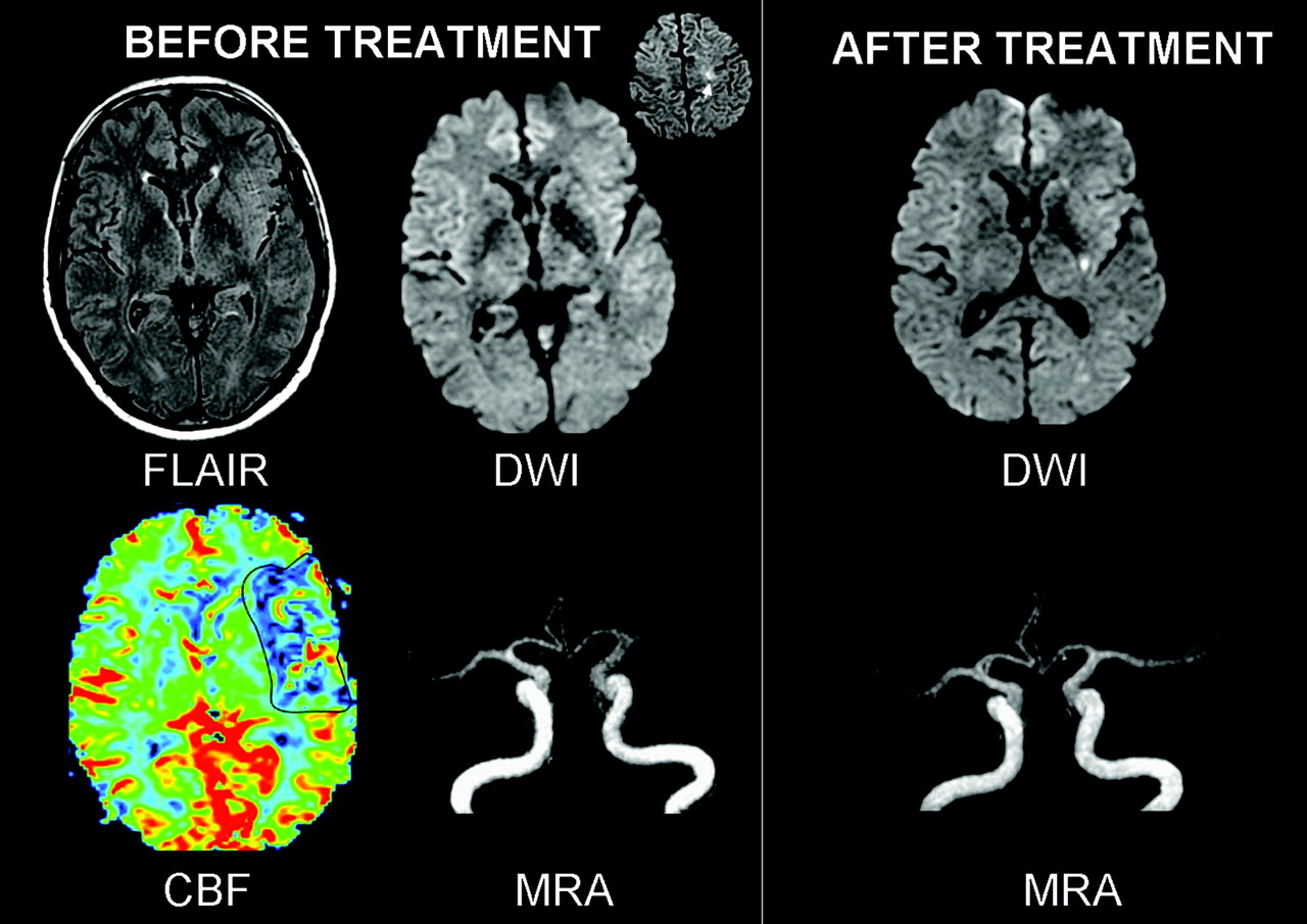
A 34-year-old man, who went to sleep at 2:00 am, woke up at 9:00 am with right hemiplegia and global aphasia, as witnessed by his wife. He reached the hospital at 11:15 am with global aphasia and right hemiplegia with a National Institutes of Health Stroke Scale (NIHSS) score of 15. No headaches or neck pain was reported, and he had no risk factors for stroke. Brain MR imaging (Signa 1.5T; GE Healthcare, Milwaukee, Wis), initiated at 11:30 am, showed a mild hyperintensity in the left deep middle cerebral artery (MCA) territory on diffusion-weighted imaging (DWI) (TR/TE = 6600/91.9 ms, b = 0\N1000 mm2/s, thickness = 6 mm, no gap, FOV = 24 × 24 cm, matrix = 128 × 128) with a decreased apparent diffusion coefficient (mean = 665 μm2/s, 75% of contralateral values). Fluid-attenuated inversion recovery (FLAIR) (parameters are unchanged except TR/TE/TI = 9800/156.9/2300 ms, matrix = 256 × 192) and T2*-weighted imaging (TR/TE = 480/13 ms, flip angle = 25°, matrix = 256 × 224) showed no parenchymal signal-intensity changes. 3D time-of-flight MR angiography (MRA) (TR/TE = 24/3.1, thickness = 1.2 mm, FOV = 20 × 17.6, matrix = 352 × 224) showed a proximal occlusion of the left MCA. First-pass gadolinium perfusion-weighted imaging (PWI) (TR/TE = 2000/60 ms, matrix = 96 × 64, 25 phases) showed prolonged mean transit time (MTT) and low cerebral blood flow (CBF) (MTT = 231%, CBF = 46% compared with contralateral hemisphere) in the superficial left MCA territory, indicating a PWI/DWI mismatch (Fig 1).

Fig. 1.
  • Download figure
  • Open in new tab
  • Download powerpoint
Fig. 1.

Case 1. Before treatment, FLAIR was normal and DWI showed a mildly hyperintense left putamen. Perfusion abnormalities (CBF decrease) existed in the superficial MCA territory, which was normal on DWI (PWI-DWI mismatch). MRA showed a left proximal MCA occlusion. After IA thrombolysis, the flow was restored in the left MCA. A deep MCA infarct with petechiae and a small cortical infarct in the left inferior frontal gyrus (not illustrated) were seen on 24-hour DWI follow-up.

This PWI/DWI mismatch, along with the lack of signal-intensity changes on FLAIR, indicated that the patient may still have been within the 6 hours of stroke onset. At 12:00 pm, the decision was made to proceed with intra-arterial thrombolysis. Mechanical and chemical thrombolysis (50 mg of rtPA) resulted in complete recanalization of the left proximal M1 occlusion at 1:30 pm, except for an anterior insular branch. The 24-hour MR imaging follow-up showed petechial hemorrhage in the deep MCA infarct and a small cortical infarct in the inferior frontal gyrus. The patient was discharged on day 9 with no motor deficit and mild language deficit, which slowly resolved (modified Rankin Scale score of 1 at 2 months).

Case 2

A 56-year-old man was suddenly heard breathing noisily during sleep by his wife. She immediately woke him up at 7:15 am and noticed a language and motor deficit. The patient was a former smoker, overweight, and dyslipidemic with type 2 diabetes. He had been hospitalized 5 days prior to stroke onset for an atrial fibrillation, which resolved after cardioversion. Oral anticoagulation had been initiated 2 days prior to the stroke. At admission (8:40 am), he had a global aphasia, right hemianopsia, and hemiplegia (NIHSS = 16). His heart rhythm was irregular with a serum glucose level of 9.6 mmol/L and an international normalized ratio of 1.31. Brain MR imaging, initiated at 8:50 am, showed punctuate DWI signal-intensity changes in the superficial MCA territory without FLAIR signal-intensity changes. PWI showed an abnormally perfused area (increased MTT, decreased CBF) in the superficial MCA territory (Fig 2). Hyperintense vessels were visible on FLAIR in the left Sylvian fissure, and a T2* hyposignal suggested the presence of a fresh thrombus in the left MCA, which was occluded on MR angiography (MRA). The large PWI/DWI mismatch along with the lack of brain signal-intensity changes indicated that the patient may still have been within 6 hours from stroke onset. The motor deficit partially regressed, but major language deficit persisted (NIHSS = 9).

Fig. 2.
  • Download figure
  • Open in new tab
  • Download powerpoint
Fig. 2.

Case 2. Before treatment, FLAIR was normal while DWI showed punctuate hyperintensities in the superficial MCA territory (inset) and a left MCA occlusion. Perfusion abnormalities existed in the superficial MCA territory, consistent with a large PWI/DWI mismatch. 24-hour MR imaging follow-up showed an additional lesion in the left putamen and MCA recanalisation after IA thrombolysis.

Despite early improvement of motor deficit, the disabling language deficit at the time of treatment decision making and the MR imaging pattern favored the decision to treat. At 11:00 am, the decision was made to proceed with intra-arterial mechanical and chemical thrombolysis (15 mg of rtPA), which resulted in complete recanalization of the left M1 embolic occlusion at 12:30 pm. The neurologic symptoms regressed rapidly (NIHSS = 4 after 2 hours, NIHSS = 2 at 24 hours). On the 24-hour follow-up, MRA and PWI findings had normalized, and a punctuate infarction was visible in the deep MCA territory. Anticoagulation was initiated and interrupted 4 days later because of a transient aphasia due to a subcortical temporal hematoma. Normal sinus heart rhythm was restored with amiodarones, and anticoagulation was reintroduced 2 weeks after thrombolysis. The patient was discharged home on day 16, free of symptoms.

Discussion

Because the exact time of onset cannot be determined, patients with stroke on awakening have been excluded from acute stroke trials and are denied the potential benefits of thrombolysis. A subset of these patients, however, may wake up soon after stroke onset and be good candidates for thrombolysis.1,2 MR imaging parameters may be valuable in the estimation of stroke-lesion age.3 However, owing to the difficulty in repeatedly imaging patients with hyperacute stroke, data on the accurate estimation of stroke age by using multiparametric MR imaging are scarce.2 Multimodal MR imaging can estimate the potential individual benefit and risk of thrombolysis. Beyond the established 3-hour time window, a substantial number of patients could be successfully treated on the basis of a penumbral pattern seen on stroke MR imaging.4 In these 2 patients, the following MR imaging pattern was similar to that seen in patients eligible for thrombolysis: MCA occlusion, small diffusion lesion, and substantial PWI/DWI mismatch. Moreover, such an MR imaging pattern has been associated with poor functional prognosis and higher risk of infarct growth, if not treated.

A few stroke centers advocate the use of imaging criteria for deciding whether to initiate thrombolysis rather than relying solely on inclusion and exclusion criteria derived from CT-based international trials.5 Neuroimaging data have been used to extend the benefit of thrombolysis to patients with seizure at stroke onset, who would otherwise have been excluded according to current rtPA guidelines.6 Recently, 2 patients with stroke on awakening have been successfully reperfused on the basis of CT perfusion findings.7 To our knowledge, this is the first report of MR imaging\Nguided decision making of thrombolysis in patients with stroke on awakening. Provided that they reach the hospital soon after awakening, a subset of patients with awakening stroke may benefit from timely multimodal brain imaging and be considered for reperfusion.

References

  1. ↵
    Serena J, Davalos A, Segura T, et al. Stroke on awakening: looking for a more rational management. Cerebrovasc Dis 2003;16:128–33
    CrossRefPubMed
  2. ↵
    Fink JN, Kumar S, Horkan C, et al. The stroke patient who woke up: clinical and radiological features, including diffusion and perfusion MRI. Stroke 2002;33:988–93
    Abstract/FREE Full Text
  3. ↵
    Lansberg MG, Thijs VN, O’Brien MW, et al. Evolution of apparent diffusion coefficient, diffusion-weighted, and T2-weighted signal intensity of acute stroke. AJNR Am J Neuroradiol 2001;22:637–44
    Abstract/FREE Full Text
  4. ↵
    Schellinger PD, Thomalla G, Fiehler J, et al. MRI-based and CT-based thrombolytic therapy in acute stroke within and beyond established time windows: an analysis of 1210 patients. Stroke 2007;38:2640–45
    Abstract/FREE Full Text
  5. ↵
    Schellinger PD, Fiebach JB, Hacke W. Imaging-based decision making in thrombolytic therapy for ischemic stroke: present status. Stroke 2003;34:575–83
    Abstract/FREE Full Text
  6. ↵
    Sylaja PN, Dzialowski I, Krol A, et al. Role of CT angiography in thrombolysis decision-making for patients with presumed seizure at stroke onset. Stroke 2006;37:915–17
    Abstract/FREE Full Text
  7. ↵
    Hellier KD, Hampton JL, Guadagno JV, et al. Perfusion CT helps decision making for thrombolysis when there is no clear time of onset. J Neurol Neurosurg Psychiatry 2006;77:417–19
    Abstract/FREE Full Text
  • Received November 29, 2007.
  • Accepted after revision February 5, 2008.
  • Copyright © American Society of Neuroradiology
View Abstract
PreviousNext
Back to top

In this issue

American Journal of Neuroradiology: 29 (7)
American Journal of Neuroradiology
Vol. 29, Issue 7
August 2008
  • Table of Contents
  • Index by author
Advertisement
Print
Download PDF
Email Article

Thank you for your interest in spreading the word on American Journal of Neuroradiology.

NOTE: We only request your email address so that the person you are recommending the page to knows that you wanted them to see it, and that it is not junk mail. We do not capture any email address.

Enter multiple addresses on separate lines or separate them with commas.
MR Imaging–Based Decision in Thrombolytic Therapy for Stroke on Awakening: Report of 2 Cases
(Your Name) has sent you a message from American Journal of Neuroradiology
(Your Name) thought you would like to see the American Journal of Neuroradiology web site.
CAPTCHA
This question is for testing whether or not you are a human visitor and to prevent automated spam submissions.
Cite this article
C. Iosif, C. Oppenheim, D. Trystram, V. Domigo, J.-F. Méder
MR Imaging–Based Decision in Thrombolytic Therapy for Stroke on Awakening: Report of 2 Cases
American Journal of Neuroradiology Aug 2008, 29 (7) 1314-1316; DOI: 10.3174/ajnr.A1069

Citation Manager Formats

  • BibTeX
  • Bookends
  • EasyBib
  • EndNote (tagged)
  • EndNote 8 (xml)
  • Medlars
  • Mendeley
  • Papers
  • RefWorks Tagged
  • Ref Manager
  • RIS
  • Zotero
0 Responses
Respond to this article
Share
Bookmark this article
MR Imaging–Based Decision in Thrombolytic Therapy for Stroke on Awakening: Report of 2 Cases
C. Iosif, C. Oppenheim, D. Trystram, V. Domigo, J.-F. Méder
American Journal of Neuroradiology Aug 2008, 29 (7) 1314-1316; DOI: 10.3174/ajnr.A1069
del.icio.us logo Twitter logo Facebook logo Mendeley logo
  • Tweet Widget
  • Facebook Like
  • Google Plus One
Purchase

Jump to section

  • Article
    • Abstract
    • Case Reports
    • Discussion
    • References
  • Figures & Data
  • Info & Metrics
  • Responses
  • References
  • PDF

Related Articles

  • No related articles found.
  • PubMed
  • Google Scholar

Cited By...

  • Reperfusion Therapies for Wake-Up Stroke: Systematic Review
  • We Are on the Clock: MRI as a Surrogate Marker of Lesion Age in Acute Ischemic Stroke
  • Crossref (21)
  • Google Scholar

This article has been cited by the following articles in journals that are participating in Crossref Cited-by Linking.

  • Negative fluid‐attenuated inversion recovery imaging identifies acute ischemic stroke at 3 hours or less
    Götz Thomalla, Philipp Rossbach, Michael Rosenkranz, Susanne Siemonsen, Anna Krützelmann, Jens Fiehler, Christian Gerloff
    Annals of Neurology 2009 65 6
  • Intravenous thrombolysis in unwitnessed stroke onset: MR WITNESS trial results
    Lee H. Schwamm, Ona Wu, Shlee S. Song, Lawrence L. Latour, Andria L. Ford, Amie W. Hsia, Alona Muzikansky, Rebecca A. Betensky, Albert J. Yoo, Michael H. Lev, Gregoire Boulouis, Arne Lauer, Pedro Cougo, William A. Copen, Gordon J. Harris, Steven Warach
    Annals of Neurology 2018 83 5
  • MR Imaging Helps Predict Time from Symptom Onset in Patients with Acute Stroke: Implications for Patients with Unknown Onset Time
    Mina Petkova, Sebastian Rodrigo, Catherine Lamy, Georges Oppenheim, Emmanuel Touzé, Jean-Louis Mas, Jean-François Méder, Catherine Oppenheim
    Radiology 2010 257 3
  • Wake-Up Stroke: Clinical Characteristics, Imaging Findings, and Treatment Option – an Update
    D. Leander Rimmele, Götz Thomalla
    Frontiers in Neurology 2014 5
  • Wake-Up Stroke and Stroke of Unknown Onset: A Critical Review
    Anke Wouters, Robin Lemmens, Patrick Dupont, Vincent Thijs
    Frontiers in Neurology 2014 5
  • Feasibility and Safety of Magnetic Resonance Imaging-Based Thrombolysis in Patients with Stroke on Awakening: Initial Single-Centre Experience
    L. Breuer, P. D. Schellinger, H. B. Huttner, R. Halwachs, T. Engelhorn, A. Doerfler, M. Köhrmann
    International Journal of Stroke 2010 5 2
  • Safety of endovascular treatment beyond the 6‐h time window in 205 patients
    S. Jung, J. Gralla, U. Fischer, M.‐L. Mono, A. Weck, R. Lüdi, M. R. Heldner, O. Findling, M. El‐Koussy, C. Brekenfeld, G. Schroth, H. P. Mattle, M. Arnold
    European Journal of Neurology 2013 20 6
  • What to do With Wake-Up Stroke
    Mark N. Rubin, Kevin M. Barrett
    The Neurohospitalist 2015 5 3
  • Alberta Stroke Program Early CT Score (ASPECTS) in Patients With Wake-Up Stroke
    Branko N. Huisa, Rema Raman, Karin Ernstrom, Gilda Tafreshi, Andrew Stemer, Brett C. Meyer, Thomas Hemmen
    Journal of Stroke and Cerebrovascular Diseases 2010 19 6
  • Prospective, open‐label safety study of intravenous recombinant tissue plasminogen activator in wake‐up stroke
    Andrew D. Barreto, Christopher V. Fanale, Andrei V. Alexandrov, Kevin C. Gaffney, Farhaan S. Vahidy, Claude B. Nguyen, Amrou Sarraj, Mohammad Rahbar, James C. Grotta, Sean I. Savitz
    Annals of Neurology 2016 80 2

More in this TOC Section

  • Enhanced Axonal Metabolism during Early Natalizumab Treatment in Relapsing-Remitting Multiple Sclerosis
  • SWI or T2*: Which MRI Sequence to Use in the Detection of Cerebral Microbleeds? The Karolinska Imaging Dementia Study
  • Progression of Microstructural Damage in Spinocerebellar Ataxia Type 2: A Longitudinal DTI Study
Show more BRAIN

Similar Articles

Advertisement

Indexed Content

  • Current Issue
  • Accepted Manuscripts
  • Article Preview
  • Past Issues
  • Editorials
  • Editor's Choice
  • Fellows' Journal Club
  • Letters to the Editor
  • Video Articles

Cases

  • Case Collection
  • Archive - Case of the Week
  • Archive - Case of the Month
  • Archive - Classic Case

More from AJNR

  • Trainee Corner
  • Imaging Protocols
  • MRI Safety Corner
  • Book Reviews

Multimedia

  • AJNR Podcasts
  • AJNR Scantastics

Resources

  • Turnaround Time
  • Submit a Manuscript
  • Submit a Video Article
  • Submit an eLetter to the Editor/Response
  • Manuscript Submission Guidelines
  • Statistical Tips
  • Fast Publishing of Accepted Manuscripts
  • Graphical Abstract Preparation
  • Imaging Protocol Submission
  • Evidence-Based Medicine Level Guide
  • Publishing Checklists
  • Author Policies
  • Become a Reviewer/Academy of Reviewers
  • News and Updates

About Us

  • About AJNR
  • Editorial Board
  • Editorial Board Alumni
  • Alerts
  • Permissions
  • Not an AJNR Subscriber? Join Now
  • Advertise with Us
  • Librarian Resources
  • Feedback
  • Terms and Conditions
  • AJNR Editorial Board Alumni

American Society of Neuroradiology

  • Not an ASNR Member? Join Now

© 2025 by the American Society of Neuroradiology All rights, including for text and data mining, AI training, and similar technologies, are reserved.
Print ISSN: 0195-6108 Online ISSN: 1936-959X

Powered by HighWire