Advertisement

Main menu

  • Home
  • Content
    • Current Issue
    • Accepted Manuscripts
    • Article Preview
    • Past Issue Archive
    • Video Articles
    • AJNR Case Collection
    • Case of the Week Archive
    • Case of the Month Archive
    • Classic Case Archive
  • Special Collections
    • AJNR Awards
    • Low-Field MRI
    • Alzheimer Disease
    • ASNR Foundation Special Collection
    • Photon-Counting CT
    • View All
  • Multimedia
    • AJNR Podcasts
    • AJNR SCANtastic
    • Trainee Corner
    • MRI Safety Corner
    • Imaging Protocols
  • For Authors
    • Submit a Manuscript
    • Submit a Video Article
    • Submit an eLetter to the Editor/Response
    • Manuscript Submission Guidelines
    • Statistical Tips
    • Fast Publishing of Accepted Manuscripts
    • Graphical Abstract Preparation
    • Imaging Protocol Submission
    • Author Policies
  • About Us
    • About AJNR
    • Editorial Board
    • Editorial Board Alumni
  • More
    • Become a Reviewer/Academy of Reviewers
    • Subscribers
    • Permissions
    • Alerts
    • Feedback
    • Advertisers
    • ASNR Home

User menu

  • Alerts
  • Log in

Search

  • Advanced search
American Journal of Neuroradiology

American Journal of Neuroradiology

ASHNR American Society of Functional Neuroradiology ASHNR American Society of Pediatric Neuroradiology ASSR
  • Alerts
  • Log in

Advanced Search

  • Home
  • Content
    • Current Issue
    • Accepted Manuscripts
    • Article Preview
    • Past Issue Archive
    • Video Articles
    • AJNR Case Collection
    • Case of the Week Archive
    • Case of the Month Archive
    • Classic Case Archive
  • Special Collections
    • AJNR Awards
    • Low-Field MRI
    • Alzheimer Disease
    • ASNR Foundation Special Collection
    • Photon-Counting CT
    • View All
  • Multimedia
    • AJNR Podcasts
    • AJNR SCANtastic
    • Trainee Corner
    • MRI Safety Corner
    • Imaging Protocols
  • For Authors
    • Submit a Manuscript
    • Submit a Video Article
    • Submit an eLetter to the Editor/Response
    • Manuscript Submission Guidelines
    • Statistical Tips
    • Fast Publishing of Accepted Manuscripts
    • Graphical Abstract Preparation
    • Imaging Protocol Submission
    • Author Policies
  • About Us
    • About AJNR
    • Editorial Board
    • Editorial Board Alumni
  • More
    • Become a Reviewer/Academy of Reviewers
    • Subscribers
    • Permissions
    • Alerts
    • Feedback
    • Advertisers
    • ASNR Home
  • Follow AJNR on Twitter
  • Visit AJNR on Facebook
  • Follow AJNR on Instagram
  • Join AJNR on LinkedIn
  • RSS Feeds

AJNR Awards, New Junior Editors, and more. Read the latest AJNR updates

OtherPediatric Neuroimaging

Poststreptococcal Dystonia with Bilateral Striatal Enlargement: MR Imaging and Spectroscopic Findings

A.T. Karagulle Kendi, C. Krenzel, F.W. Ott, J.R. Brace, S.K. Norberg and S.A. Kieffer
American Journal of Neuroradiology August 2008, 29 (7) 1276-1278; DOI: https://doi.org/10.3174/ajnr.A1065
A.T. Karagulle Kendi
  • Find this author on Google Scholar
  • Find this author on PubMed
  • Search for this author on this site
C. Krenzel
  • Find this author on Google Scholar
  • Find this author on PubMed
  • Search for this author on this site
F.W. Ott
  • Find this author on Google Scholar
  • Find this author on PubMed
  • Search for this author on this site
J.R. Brace
  • Find this author on Google Scholar
  • Find this author on PubMed
  • Search for this author on this site
S.K. Norberg
  • Find this author on Google Scholar
  • Find this author on PubMed
  • Search for this author on this site
S.A. Kieffer
  • Find this author on Google Scholar
  • Find this author on PubMed
  • Search for this author on this site
  • Article
  • Figures & Data
  • Info & Metrics
  • Responses
  • References
  • PDF
Loading

Abstract

SUMMARY: Isolated bilateral striatal necrosis is an abnormality of the basal ganglia associated with acute dystonia in children. This report describes the development of dystonic movements in a 7-year-old male patient 2 weeks after streptococcal pharyngitis.

Dystonia with basal ganglial imaging abnormalities is usually related to structural, metabolic, toxic, traumatic, or vascular disorders.1,2 Swedo et al3 coined the term “pediatric autoimmune neuropsychiatric disorders associated with streptococcal infection” (PANDAS) to describe them. MR imaging in patients with suspected PANDAS has demonstrated a lack of structural abnormality, though volumetric evaluations revealed enlargement of the basal ganglia.4

This report describes a patient presenting with dystonia and basal ganglial imaging abnormalities following streptococcal infection. Because there is no specific test to confirm this diagnosis, poststreptococcal dystonia was presumptive. To our knowledge, this is the first case report addressing the findings on diffusion-weighted MR imaging and on MR spectroscopy in a patient with poststreptococcal dystonia and striatal enlargement.

Case Report

A 7-year-old boy presented with rigidity and difficulty breathing. Diagnosis of streptococcal pharyngitis was made, and he was treated with an aminopenicillin. Two days later, he was unable to get out of bed, and persistent drooling was noted. Dexamethasone was begun, but his condition deteriorated with increasing rigidity, dystonia, drooling, and tremor of the hands. Rigidity and dystonia rapidly progressed, and he developed mutism, dysphagia, and incontinence. Laboratory testing demonstrated borderline high antistreptolysin O titer and a high level of antideoxyribonuclease antibody.

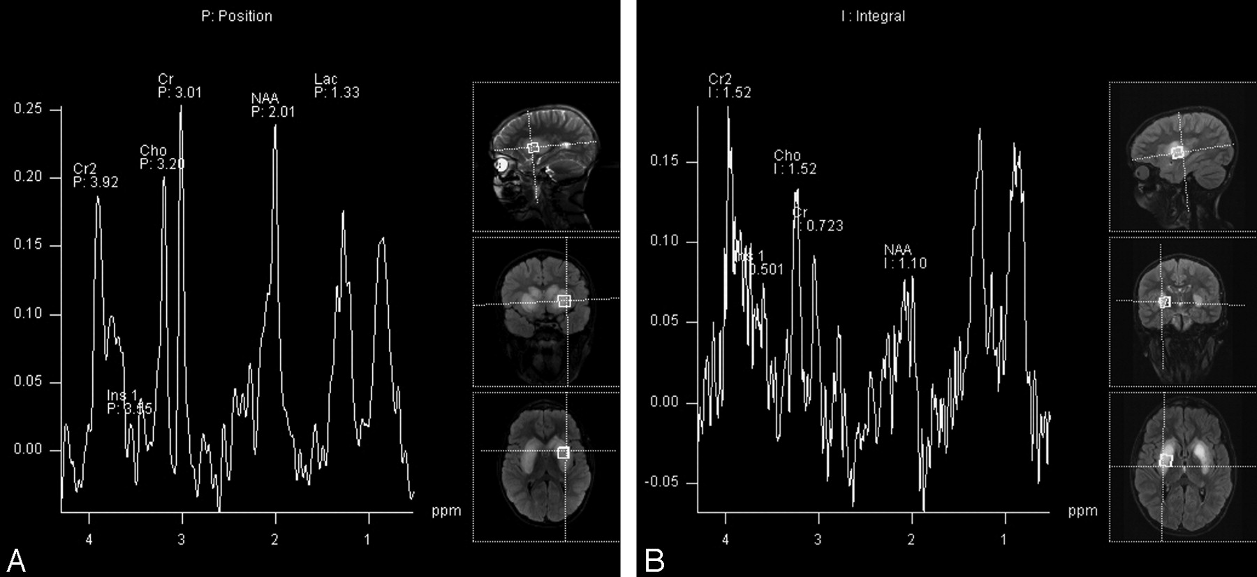
Cranial MR imaging demonstrated bilateral T2 hyperintensity in the heads of the caudate nuclei, the putamina, and the external/extreme capsules. The heads of the caudate nuclei and the putamina were enlarged (Fig 1) and demonstrated minimal restricted diffusion on diffusion-weighted images (Fig 2). Single-voxel MR spectroscopy of the left lentiform nucleus demonstrated elevation of the lipid/lactate peak and a decreased N-acetylaspartate (NAA) peak (Fig 3A).

Fig 1.
  • Download figure
  • Open in new tab
  • Download powerpoint
Fig 1.

Axial turbo fluid-attenuated inversion recovery image demonstrates bilateral T2 hyperintensity within the heads of the caudate nuclei, the putamina, and the external/extreme capsules. The heads of the caudate/putamina are enlarged.

Fig 2.
  • Download figure
  • Open in new tab
  • Download powerpoint
Fig 2.

Axial apparent diffusion coefficent map shows restricted diffusion in the heads of the caudate and the putamina bilaterally.

Fig 3.
  • Download figure
  • Open in new tab
  • Download powerpoint
Fig 3.

A, Initial single-voxel MR spectroscopy of the left putamen demonstrates an elevated lipid/lactate (Lac) peak and a decreased NAA peak. B, Follow-up MR spectroscopy of the right putamen shows further decrease in the NAA level with an increase in the choline (Cho)/creatine (Cr) ratio. Ins indicates myo-inositol.

Follow-up MR imaging 1 month later showed no significant change. MR spectroscopy demonstrated continued depression of the NAA peak with interval increase in the choline/creatine ratio compared with the initial MR spectroscopy (Fig 3B).

At the time of discharge, the patient exhibited mild improvement in neurologic functions. He participated in aggressive physical therapy, and 12-month follow-up demonstrated significant overall clinical improvement. Twelve-month follow-up MR imaging revealed significant improvement in the size and the extent of T2 hyperintensity in the heads of the caudate nuclei, the putamina, and the external/extreme capsules. There is now residual abnormal T2 hyperintensity only in the lateral aspects of both putamina. Enlargement of the heads of the caudate nuclei and putamina has completely resolved (Fig 4A), and no restricted diffusion is apparent in these structures (Fig 4B).

Fig 4.
  • Download figure
  • Open in new tab
  • Download powerpoint
Fig 4.

A, Axial fluid-attenuated inversion recovery image demonstrates only a small rim of residual T2 hyperintensity at the lateral margin of the right putamen. Slightly greater T2 hyperintensity remains in the left putamen. B, Axial apparent diffusion coefficient map demonstrates complete resolution of restricted diffusion in the heads of the caudate nuclei and putamina bilaterally.

Discussion

Although the classic poststreptococcal neuropsychiatric disorder is Sydenham chorea, recently other poststreptococcal basal ganglia phenotypes have been described in patients who do not meet the criteria for Sydenham chorea. One of these disorders is poststreptococcal dystonia.5 There is evidence suggesting that these disorders are autoimmune-mediated with antibodies attacking certain parts of the brain, specifically the basal ganglia.5

A variety of MR imaging findings have been reported in patients with Sydenham chorea, varying from normal-to-abnormal signal intensity involving the basal ganglia.6–9 Dale et al2 described 2 children with poststreptococcal dystonia. The putamina and the heads of the caudate nuclei demonstrated T2 hyperintensity, but no mention was made regarding the size of these nuclei.

MR spectroscopic studies of the basal ganglia in children with Sydenham chorea and rheumatic fever have revealed reduction in NAA, increase in the choline/creatine ratio, and abnormal peaks at 3.5–4.2 ppm6 and at 1.5 ppm.7 Because choline is involved in neuronal and glial phospholipid metabolism, an elevated choline/creatine ratio may imply disturbed cell membrane turnover, whereas a decreased NAA/creatine ratio may indicate neuronal dysfunction or loss.6 An increased choline/creatine ratio may also reflect imbalance between dopaminergic and cholinergic systems because an increased choline/creatine ratio has been found in the basal ganglia of patients with Parkinson disease and attention deficit/hyperactivity disorder.10 The peaks at 3.5–4.2 and 1.5 ppm are thought to reflect breakdown products of lipids and amino acids.6,7 Carbohydrates also resonate between 3.5–5 ppm. This supports the presence of antibodies attacking the basal ganglia because antibodies against streptolysin O are known to be mostly immunoglobulin M type, and they are composed of 15% carbohydrates.7

Single-voxel MR spectroscopy of the abnormal basal ganglia in our patient showed reduction of the NAA peak (a sign of neuronal injury) and an increased lactate peak, corresponding to conversion from aerobic energy production to anaerobic glycolysis.11 Follow-up MR spectroscopy 1 month after the initial examination demonstrated further decrease in NAA and interval increase in choline with reversal of the choline/creatine ratio, consistent with increased neuronal injury and increased cell membrane turnover. In other pathologies marked by cell membrane breakdown (eg, neurodegenerative disorders), bound moieties may be liberated into the free choline pool, contributing to an increase in choline resonance.6 Recent studies by using immunofluorescence documented serum anti-basal ganglial antibodies in the poststreptococcal basal ganglia.6 MR spectroscopy findings in our patient support the hypothesis that the pathophysiologic mechanism for poststreptococcal dystonia with striatal necrosis could be an antibody-mediated immune reaction.

Follow-up MR imaging at 1 year demonstrated interval improvement in size, T2 signal intensity, and diffusion of the basal ganglia and appears to correlate with a slowly improving clinical course. These findings suggest that the underlying pathology in isolated basal ganglial “necrosis” may not represent irreversible necrosis after all.

References

  1. ↵
    Goutieres F, Aicardi J. Acute neurological dysfunction associated with destructive lesions of the basal ganglia in children. Ann Neurol 1982;12:328–32
    CrossRefPubMed
  2. ↵
    Dale RC, Church AJ, Surtees RA, et al. Post-streptococcal autoimmune dystonia with isolated bilateral striatal necrosis. Dev Med Child Neurol 2002;44:485–89
    CrossRefPubMed
  3. ↵
    Swedo SE, Leonard HL, Garvey M, et al. Pediatric autoimmune neuropsychiatric disorders associated with streptococcal infections: clinical description of the first 50 cases. Am J Psychiatry 1998;155:264–71
    PubMed
  4. ↵
    Giedd JN, Rapoport JL, Garvey MA, et al. MRI assessment of children with obsessive compulsive disorder or tics associated with streptococcal infection. Am J Psychiatry 2000;157:281–83
    CrossRefPubMed
  5. ↵
    Dale RC, Church AJ, Surtees RAH, et al. Post-streptococcal autoimmune neuropsychiatric disease presenting as paroxysmal dystonic choreoathetosis. Mov Disord 2002;17:817–20
    CrossRefPubMed
  6. ↵
    Margari L, Ventura P, Portoghese C, et al. Brain magnetic resonance spectroscopy in Sydenham's chorea and ADHD. Pediatric Neurol 2006;34:467–73
    CrossRefPubMed
  7. ↵
    Castillo M, Kwock L, Arbelaez A. Syndenham's chorea: MRI and proton spectroscopy. Neuroradiology 1999;41:943–45
    CrossRefPubMed
  8. Emery ES, Vieco PT. Sydenham chorea: magnetic resonance imaging reveals permanent basal ganglia injury. Neurology 1997;48:531–33
    Abstract/FREE Full Text
  9. ↵
    Traill Z, Pike M, Byrne J. Sydenham's chorea: a case showing reversible striatal abnormalities on CT and MRI. Dev Med Child Neurol 1995;37:270–73
    PubMed
  10. ↵
    Jin Z, Zang YF, Zeng YW, et al. Striatal neuronal loss or dysfunction and choline rise in children with attention-deficit hyperactivity disorder: a 1H-magnetic resonance spectroscopy study. Neurosci Lett 2001;23:45–48
  11. ↵
    Michel SJ, Given CA, Robertson WC. Imaging of the brain, including diffusion-weighted imaging in methylmalonic acidemia. Pediatr Radiol 2004;34:580–82. Epub 2004 Mar 9
    PubMed
  • Received December 3, 2007.
  • Accepted after revision January 9, 2008.
  • Copyright © American Society of Neuroradiology
View Abstract
PreviousNext
Back to top

In this issue

American Journal of Neuroradiology: 29 (7)
American Journal of Neuroradiology
Vol. 29, Issue 7
August 2008
  • Table of Contents
  • Index by author
Advertisement
Print
Download PDF
Email Article

Thank you for your interest in spreading the word on American Journal of Neuroradiology.

NOTE: We only request your email address so that the person you are recommending the page to knows that you wanted them to see it, and that it is not junk mail. We do not capture any email address.

Enter multiple addresses on separate lines or separate them with commas.
Poststreptococcal Dystonia with Bilateral Striatal Enlargement: MR Imaging and Spectroscopic Findings
(Your Name) has sent you a message from American Journal of Neuroradiology
(Your Name) thought you would like to see the American Journal of Neuroradiology web site.
CAPTCHA
This question is for testing whether or not you are a human visitor and to prevent automated spam submissions.
Cite this article
A.T. Karagulle Kendi, C. Krenzel, F.W. Ott, J.R. Brace, S.K. Norberg, S.A. Kieffer
Poststreptococcal Dystonia with Bilateral Striatal Enlargement: MR Imaging and Spectroscopic Findings
American Journal of Neuroradiology Aug 2008, 29 (7) 1276-1278; DOI: 10.3174/ajnr.A1065

Citation Manager Formats

  • BibTeX
  • Bookends
  • EasyBib
  • EndNote (tagged)
  • EndNote 8 (xml)
  • Medlars
  • Mendeley
  • Papers
  • RefWorks Tagged
  • Ref Manager
  • RIS
  • Zotero
0 Responses
Respond to this article
Share
Bookmark this article
Poststreptococcal Dystonia with Bilateral Striatal Enlargement: MR Imaging and Spectroscopic Findings
A.T. Karagulle Kendi, C. Krenzel, F.W. Ott, J.R. Brace, S.K. Norberg, S.A. Kieffer
American Journal of Neuroradiology Aug 2008, 29 (7) 1276-1278; DOI: 10.3174/ajnr.A1065
del.icio.us logo Twitter logo Facebook logo Mendeley logo
  • Tweet Widget
  • Facebook Like
  • Google Plus One
Purchase

Jump to section

  • Article
    • Abstract
    • Case Report
    • Discussion
    • References
  • Figures & Data
  • Info & Metrics
  • Responses
  • References
  • PDF

Related Articles

  • No related articles found.
  • PubMed
  • Google Scholar

Cited By...

  • No citing articles found.
  • Crossref (11)
  • Google Scholar

This article has been cited by the following articles in journals that are participating in Crossref Cited-by Linking.

  • Striatal structure and function in mood disorders: a comprehensive review
    William R Marchand, Deborah Yurgelun‐Todd
    Bipolar Disorders 2010 12 8
  • Neurological Disorders Associated with Striatal Lesions: Classification and Diagnostic Approach
    Davide Tonduti, Luisa Chiapparini, Isabella Moroni, Anna Ardissone, Giovanna Zorzi, Federica Zibordi, Sergio Raspante, Celeste Panteghini, Barbara Garavaglia, Nardo Nardocci
    Current Neurology and Neuroscience Reports 2016 16 6
  • MR differential diagnosis of acute deep grey matter pathology in paediatric patients
    Carlo Cosimo Quattrocchi, Daniela Longo, Luciana Nogueira Delfino, Yuri Errante, Costanza Aiello, Giuseppe Fariello, Bruno Bernardi
    Pediatric Radiology 2013 43 6
  • Acute Movement Disorders in Childhood
    Giacomo Garone, Federica Graziola, Melissa Grasso, Alessandro Capuano
    Journal of Clinical Medicine 2021 10 12
  • Streptococcus pneumoniae Meningoencephalitis With Bilateral Basal Ganglia Necrosis
    Jessy Magnus, Paul M. Parizel, Berten Ceulemans, Patrick Cras, Marloes Luijks, Philippe G. Jorens
    Journal of Child Neurology 2011 26 11
  • Post-streptococcal chorea in an adult with bilateral striatal encephalitis
    Koichi Hagiwara, Kayo Tominaga, Yoko Okada, Miwako Kido, Ryo Yamasaki, Norihiko Shida, Yoriaki Yamashita
    Journal of Clinical Neuroscience 2011 18 5
  • Infection-associated dystonia
    Rahul Yadav, Vijay Shankar, Soaham Desai
    Annals of Movement Disorders 2021 4 3
  • Transient bilateral striatal lesion associated with varicella infection
    Roberta Dantas Azevedo, Fabiano Reis
    Radiologia Brasileira 2017 50 5
  • Neuropsychiatric disorder with basal ganglia lesions
    Prashant Bhatele, Arunmozhimaran Elavarasi, Ajay Garg
    BMJ Case Reports 2022 15 1
  • Movement Disorder Emergencies
    Roser Pons
    2022

More in this TOC Section

  • fetal brain development of 10 weeks gestation
  • CHARGE fetal MRI clival cleft
  • Neuroimaging Delineation and Progression of SLSMD
Show more Pediatric Neuroimaging

Similar Articles

Advertisement

Indexed Content

  • Current Issue
  • Accepted Manuscripts
  • Article Preview
  • Past Issues
  • Editorials
  • Editor's Choice
  • Fellows' Journal Club
  • Letters to the Editor
  • Video Articles

Cases

  • Case Collection
  • Archive - Case of the Week
  • Archive - Case of the Month
  • Archive - Classic Case

More from AJNR

  • Trainee Corner
  • Imaging Protocols
  • MRI Safety Corner

Multimedia

  • AJNR Podcasts
  • AJNR Scantastics

Resources

  • Turnaround Time
  • Submit a Manuscript
  • Submit a Video Article
  • Submit an eLetter to the Editor/Response
  • Manuscript Submission Guidelines
  • Statistical Tips
  • Fast Publishing of Accepted Manuscripts
  • Graphical Abstract Preparation
  • Imaging Protocol Submission
  • Evidence-Based Medicine Level Guide
  • Publishing Checklists
  • Author Policies
  • Become a Reviewer/Academy of Reviewers
  • News and Updates

About Us

  • About AJNR
  • Editorial Board
  • Editorial Board Alumni
  • Alerts
  • Permissions
  • Not an AJNR Subscriber? Join Now
  • Advertise with Us
  • Librarian Resources
  • Feedback
  • Terms and Conditions
  • AJNR Editorial Board Alumni

American Society of Neuroradiology

  • Not an ASNR Member? Join Now

© 2025 by the American Society of Neuroradiology All rights, including for text and data mining, AI training, and similar technologies, are reserved.
Print ISSN: 0195-6108 Online ISSN: 1936-959X

Powered by HighWire