Advertisement

Main menu

  • Home
  • Content
    • Current Issue
    • Accepted Manuscripts
    • Article Preview
    • Past Issue Archive
    • Video Articles
    • AJNR Case Collection
    • Case of the Week Archive
    • Case of the Month Archive
    • Classic Case Archive
  • Special Collections
    • AJNR Awards
    • Low-Field MRI
    • Alzheimer Disease
    • ASNR Foundation Special Collection
    • Photon-Counting CT
    • View All
  • Multimedia
    • AJNR Podcasts
    • AJNR SCANtastic
    • Trainee Corner
    • MRI Safety Corner
    • Imaging Protocols
  • For Authors
    • Submit a Manuscript
    • Submit a Video Article
    • Submit an eLetter to the Editor/Response
    • Manuscript Submission Guidelines
    • Statistical Tips
    • Fast Publishing of Accepted Manuscripts
    • Graphical Abstract Preparation
    • Imaging Protocol Submission
    • Author Policies
  • About Us
    • About AJNR
    • Editorial Board
    • Editorial Board Alumni
  • More
    • Become a Reviewer/Academy of Reviewers
    • Subscribers
    • Permissions
    • Alerts
    • Feedback
    • Advertisers
    • ASNR Home

User menu

  • Alerts
  • Log in

Search

  • Advanced search
American Journal of Neuroradiology

American Journal of Neuroradiology

ASHNR American Society of Functional Neuroradiology ASHNR American Society of Pediatric Neuroradiology ASSR
  • Alerts
  • Log in

Advanced Search

  • Home
  • Content
    • Current Issue
    • Accepted Manuscripts
    • Article Preview
    • Past Issue Archive
    • Video Articles
    • AJNR Case Collection
    • Case of the Week Archive
    • Case of the Month Archive
    • Classic Case Archive
  • Special Collections
    • AJNR Awards
    • Low-Field MRI
    • Alzheimer Disease
    • ASNR Foundation Special Collection
    • Photon-Counting CT
    • View All
  • Multimedia
    • AJNR Podcasts
    • AJNR SCANtastic
    • Trainee Corner
    • MRI Safety Corner
    • Imaging Protocols
  • For Authors
    • Submit a Manuscript
    • Submit a Video Article
    • Submit an eLetter to the Editor/Response
    • Manuscript Submission Guidelines
    • Statistical Tips
    • Fast Publishing of Accepted Manuscripts
    • Graphical Abstract Preparation
    • Imaging Protocol Submission
    • Author Policies
  • About Us
    • About AJNR
    • Editorial Board
    • Editorial Board Alumni
  • More
    • Become a Reviewer/Academy of Reviewers
    • Subscribers
    • Permissions
    • Alerts
    • Feedback
    • Advertisers
    • ASNR Home
  • Follow AJNR on Twitter
  • Visit AJNR on Facebook
  • Follow AJNR on Instagram
  • Join AJNR on LinkedIn
  • RSS Feeds

AJNR Awards, New Junior Editors, and more. Read the latest AJNR updates


Improved Turnaround Times | Median time to first decision: 12 days

OtherReview Articles

Arterial Spin-Labeling in Routine Clinical Practice, Part 2: Hypoperfusion Patterns

A.R. Deibler, J.M. Pollock, R.A. Kraft, H. Tan, J.H. Burdette and J.A. Maldjian
American Journal of Neuroradiology August 2008, 29 (7) 1235-1241; DOI: https://doi.org/10.3174/ajnr.A1033
A.R. Deibler
  • Find this author on Google Scholar
  • Find this author on PubMed
  • Search for this author on this site
J.M. Pollock
  • Find this author on Google Scholar
  • Find this author on PubMed
  • Search for this author on this site
R.A. Kraft
  • Find this author on Google Scholar
  • Find this author on PubMed
  • Search for this author on this site
H. Tan
  • Find this author on Google Scholar
  • Find this author on PubMed
  • Search for this author on this site
J.H. Burdette
  • Find this author on Google Scholar
  • Find this author on PubMed
  • Search for this author on this site
J.A. Maldjian
  • Find this author on Google Scholar
  • Find this author on PubMed
  • Search for this author on this site
  • Article
  • Figures & Data
  • Info & Metrics
  • Responses
  • References
  • PDF
Loading

Article Figures & Data

Figures

  • Tables
  • Fig 1.
    • Download figure
    • Open in new tab
    • Download powerpoint
    Fig 1.

    Ischemic penumbra. DWI reveals multiple borderzone infarcts in the left cerebral hemisphere (arrow). MR angiogram (center) demonstrates a left internal carotid artery occlusion. ASL map shows the left ischemic penumbra, corresponding to the perfusion > DWI mismatch.

  • Fig 2.
    • Download figure
    • Open in new tab
    • Download powerpoint
    Fig 2.

    Carotid occlusion and tissue at risk. Axial contrast-enhanced spoiled gradient-recalled-echo image (left) shows chronic occlusion of the left internal carotid artery (arrow). FLAIR (center) shows no evidence of ischemia. Flow asymmetry on the ASL CBF map (ellipse) represents tissue at risk in the left cerebral hemisphere.
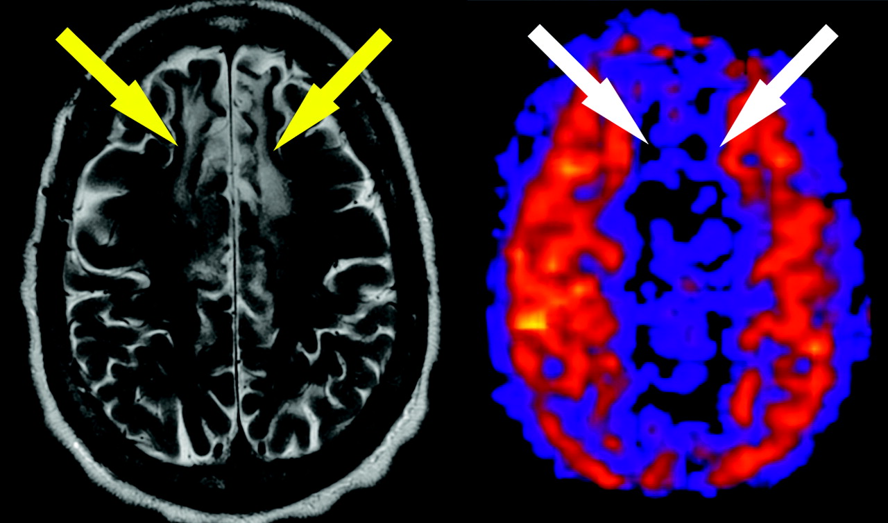
  • Fig 3.
    • Download figure
    • Open in new tab
    • Download powerpoint
    Fig 3.

    Encephalomalacia. Axial T2-weighted image shows high signal intensity in the frontal lobes representing the remote bilateral anterior cerebral artery (ACA) territory infarcts (yellow arrows). ASL CBF map reveals corresponding focal decrease in flow in the ACA territories (white arrows).
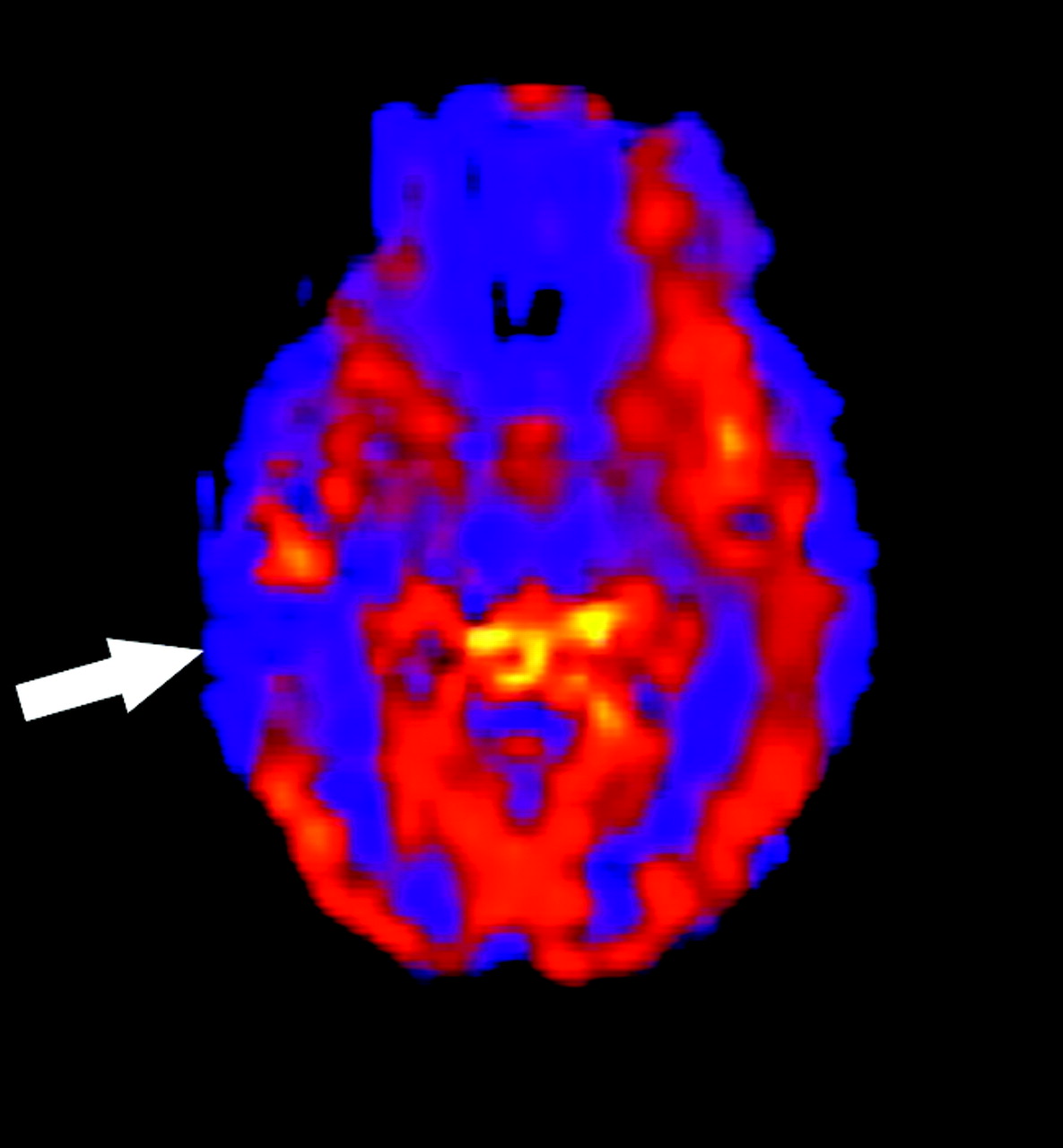
  • Fig 4.
    • Download figure
    • Open in new tab
    • Download powerpoint
    Fig 4.

    Localized hypoperfusion in temporal lobe epilepsy. Interictal ASL CBF map in a 37-year-old woman demonstrates low signal intensity in the right lateral temporal lobe (arrow) and right hippocampus. Electroencephalography confirmed a right temporal seizure focus.
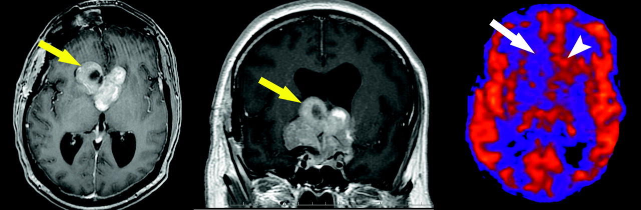
  • Fig 5.
    • Download figure
    • Open in new tab
    • Download powerpoint
    Fig 5.

    Hypoperfused tumor on ASL. Axial (left) and coronal (center) gadolinium-enhanced T1-weighted images demonstrate a heterogeneously enhancing mass invading the third and lateral ventricles (yellow arrows). ASL (right) shows regional hypoperfusion within the more cystic portion of the tumor (white arrow). The solid portions show hyperperfusion on ASL (arrowhead). The tumor proved to be a pituitary macroadenoma at surgery.

  • Fig 6.
    • Download figure
    • Open in new tab
    • Download powerpoint
    Fig 6.

    Paradoxically low signal intensity on ASL. T2*-weighted gradient-recall image reveals a calcified mass arising from the left sphenoid wing. The mass showed avid contrast enhancement (not shown) consistent with meningioma. Despite the enhancement, ASL CBF map shows no increased flow within the tumor (arrow). Underlying hyperperfusion may be masked by competing susceptibility effects.

  • Fig 7.
    • Download figure
    • Open in new tab
    • Download powerpoint
    Fig 7.

    Hypoperfused glioma on ASL. Coronal FLAIR (upper left) and axial T2 (upper right) images demonstrate a high-signal-intensity mass in the left thalamus (white arrows). Minimal enhancement is seen after contrast (lower left), with corresponding low perfusion on the CBF map (white arrow, bottom right). The rim of normal perfusion (arrowheads) corresponds to the residual normal tissue in the adjacent thalamus.
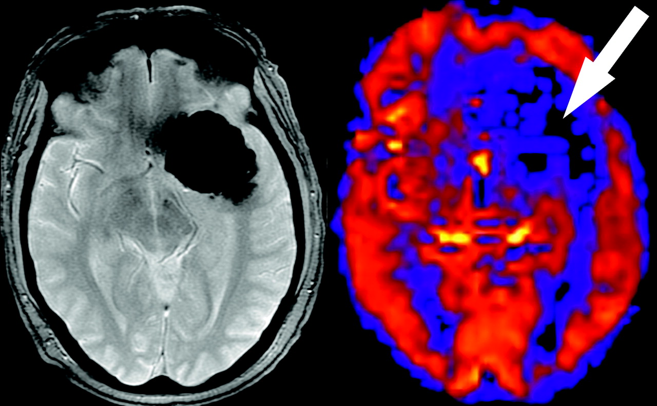
  • Fig 8.
    • Download figure
    • Open in new tab
    • Download powerpoint
    Fig 8.

    Blood products on ASL. Axial unenhanced CT scan reveals acute parenchymal hemorrhage in the right frontal lobe (yellow arrow). Focal loss of signal intensity on the ASL CBF map (white arrow) corresponds to the area of hemorrhage. Note flow asymmetry with decreased signal intensity throughout the right hemisphere.
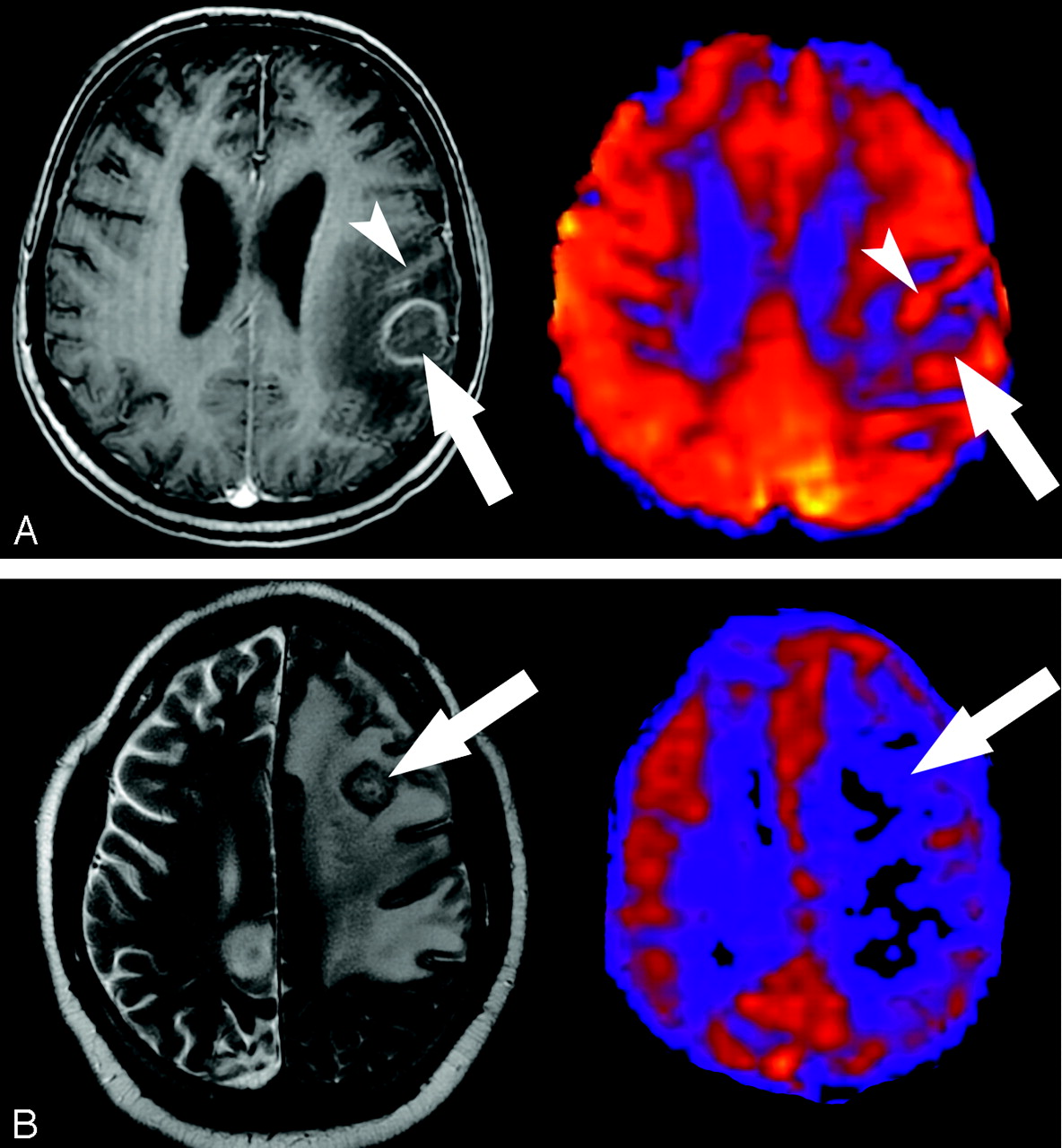
  • Fig 9.
    • Download figure
    • Open in new tab
    • Download powerpoint
    Fig 9.

    A, Toxoplasmosis on ASL. Decreased perfusion to a toxoplasmosis abscess. Ring-enhancing lesion is demonstrated on corresponding postgadolinium T1-weighted image (arrow). The edema surrounding the abscess decreases the perfusion of the white matter, accentuating the normal perfusion of the adjacent gyri. The area of high signal intensity on ASL surrounding the abscess (arrowhead) represents the normal adjacent gray matter. B, Toxoplasmosis on ASL. Axial T2-weighted image (left) shows a mass with vasogenic edema in the left frontal lobe (arrow). Corresponding ASL image (right) shows diffusely decreased perfusion in the lesion and adjacent white matter (arrow).

  • Fig 10.
    • Download figure
    • Open in new tab
    • Download powerpoint
    Fig 10.

    Steal phenomenon associated with AVM. ASL CBF map shows characteristic hyperperfusion corresponding to the AVM (arrow). Also seen is a regional zone of hypoperfusion representing the associated steal phenomenon (arrowhead).
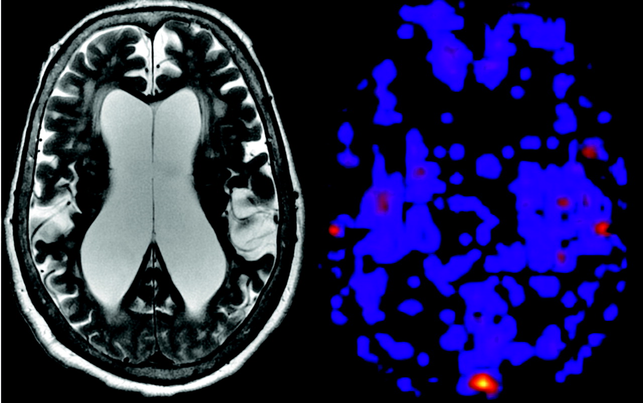
  • Fig 11.
    • Download figure
    • Open in new tab
    • Download powerpoint
    Fig 11.

    Cerebral atrophy as a cause of globally decreased ASL signal intensity. T2-weighted image (left) reveals advanced cerebral volume loss, ex vacuo ventricular enlargement, and white matter hyperintensities. ASL map demonstrates poor perfusion signal intensity throughout.
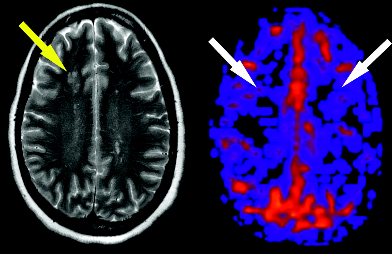
  • Fig 12.
    • Download figure
    • Open in new tab
    • Download powerpoint
    Fig 12.

    Bilateral hypoperfusion in CNS vasculitis. Multiple white matter lesions are shown on the T2-weighted image (yellow arrow). ASL CBF map demonstrates symmetrically decreased signal intensity in the bilateral cerebral hemispheres (white arrows) in a 38-year-old woman with CNS vasculitis.

Tables

  • Figures
  • Patterns of hypoperfusion encountered at clinical spin-tag perfusion imaging

    Hypoperfusion PatternCauses
    FocalIschemic core
    Ischemic penumbra
    At-risk tissue
    Leukoaraiosis
    Remote insult
    Seizure activity (interictal)
    Ventriculomegaly
    GlobalPoor cardiac output
    Vasospasm
    Brain death
    Cerebral atrophy
    Exogenous drugs
PreviousNext
Back to top

In this issue

American Journal of Neuroradiology: 29 (7)
American Journal of Neuroradiology
Vol. 29, Issue 7
August 2008
  • Table of Contents
  • Index by author
Advertisement
Print
Download PDF
Email Article

Thank you for your interest in spreading the word on American Journal of Neuroradiology.

NOTE: We only request your email address so that the person you are recommending the page to knows that you wanted them to see it, and that it is not junk mail. We do not capture any email address.

Enter multiple addresses on separate lines or separate them with commas.
Arterial Spin-Labeling in Routine Clinical Practice, Part 2: Hypoperfusion Patterns
(Your Name) has sent you a message from American Journal of Neuroradiology
(Your Name) thought you would like to see the American Journal of Neuroradiology web site.
CAPTCHA
This question is for testing whether or not you are a human visitor and to prevent automated spam submissions.
Cite this article
A.R. Deibler, J.M. Pollock, R.A. Kraft, H. Tan, J.H. Burdette, J.A. Maldjian
Arterial Spin-Labeling in Routine Clinical Practice, Part 2: Hypoperfusion Patterns
American Journal of Neuroradiology Aug 2008, 29 (7) 1235-1241; DOI: 10.3174/ajnr.A1033

Citation Manager Formats

  • BibTeX
  • Bookends
  • EasyBib
  • EndNote (tagged)
  • EndNote 8 (xml)
  • Medlars
  • Mendeley
  • Papers
  • RefWorks Tagged
  • Ref Manager
  • RIS
  • Zotero
0 Responses
Respond to this article
Share
Bookmark this article
Arterial Spin-Labeling in Routine Clinical Practice, Part 2: Hypoperfusion Patterns
A.R. Deibler, J.M. Pollock, R.A. Kraft, H. Tan, J.H. Burdette, J.A. Maldjian
American Journal of Neuroradiology Aug 2008, 29 (7) 1235-1241; DOI: 10.3174/ajnr.A1033
del.icio.us logo Twitter logo Facebook logo Mendeley logo
  • Tweet Widget
  • Facebook Like
  • Google Plus One
Purchase

Jump to section

  • Article
    • Abstract
    • Discussion
    • Conclusion
    • Acknowledgments
    • Footnotes
    • References
  • Figures & Data
  • Info & Metrics
  • Responses
  • References
  • PDF

Related Articles

  • No related articles found.
  • PubMed
  • Google Scholar

Cited By...

  • Anoxic Brain Injury Detection with the Normalized Diffusion to ASL Perfusion Ratio: Implications for Blood-Brain Barrier Injury and Permeability
  • Cerebral Blood Flow and Marrow Diffusion Alterations in Children with Sickle Cell Anemia after Bone Marrow Transplantation and Transfusion
  • Clinical Value of Hybrid TOF-PET/MR Imaging-Based Multiparametric Imaging in Localizing Seizure Focus in Patients with MRI-Negative Temporal Lobe Epilepsy
  • Comparison of Arterial Spin Labeling and Bolus Perfusion-Weighted Imaging for Detecting Mismatch in Acute Stroke
  • Quantitative Blood Flow Measurements in Gliomas Using Arterial Spin-Labeling at 3T: Intermodality Agreement and Inter- and Intraobserver Reproducibility Study
  • Arterial spin labeling and altered cerebral blood flow patterns in the minimally conscious state
  • Arterial Spin-Labeling MRI Can Identify the Presence and Intensity of Collateral Perfusion in Patients With Moyamoya Disease
  • Initial Experience in Using Continuous Arterial Spin-Labeled MR Imaging for Early Detection of Alzheimer Disease
  • Noninvasive MR imaging of cerebral perfusion in patients with a carotid artery stenosis
  • The Acetazolamide Challenge: Techniques and Applications in the Evaluation of Chronic Cerebral Ischemia
  • Hypercapnia-Induced Cerebral Hyperperfusion: An Underrecognized Clinical Entity
  • Crossref
  • Google Scholar

This article has not yet been cited by articles in journals that are participating in Crossref Cited-by Linking.

More in this TOC Section

  • An Atlas of Neonatal Neurovascular Imaging Anatomy as Depicted with Microvascular Imaging: The Intracranial Arteries
  • An Atlas of Neonatal Neurovascular Imaging Anatomy as Depicted with Microvascular Imaging: The Intracranial Veins
  • Clinical Translation of Hyperpolarized 13C Metabolic Probes for Glioma Imaging
Show more Review Articles

Similar Articles

Advertisement

Indexed Content

  • Current Issue
  • Accepted Manuscripts
  • Article Preview
  • Past Issues
  • Editorials
  • Editor's Choice
  • Fellows' Journal Club
  • Letters to the Editor
  • Video Articles

Cases

  • Case Collection
  • Archive - Case of the Week
  • Archive - Case of the Month
  • Archive - Classic Case

More from AJNR

  • Trainee Corner
  • Imaging Protocols
  • MRI Safety Corner
  • Book Reviews

Multimedia

  • AJNR Podcasts
  • AJNR Scantastics

Resources

  • Turnaround Time
  • Submit a Manuscript
  • Submit a Video Article
  • Submit an eLetter to the Editor/Response
  • Manuscript Submission Guidelines
  • Statistical Tips
  • Fast Publishing of Accepted Manuscripts
  • Graphical Abstract Preparation
  • Imaging Protocol Submission
  • Evidence-Based Medicine Level Guide
  • Publishing Checklists
  • Author Policies
  • Become a Reviewer/Academy of Reviewers
  • News and Updates

About Us

  • About AJNR
  • Editorial Board
  • Editorial Board Alumni
  • Alerts
  • Permissions
  • Not an AJNR Subscriber? Join Now
  • Advertise with Us
  • Librarian Resources
  • Feedback
  • Terms and Conditions
  • AJNR Editorial Board Alumni

American Society of Neuroradiology

  • Not an ASNR Member? Join Now

© 2025 by the American Society of Neuroradiology All rights, including for text and data mining, AI training, and similar technologies, are reserved.
Print ISSN: 0195-6108 Online ISSN: 1936-959X

Powered by HighWire