Advertisement

Main menu

  • Home
  • Content
    • Current Issue
    • Accepted Manuscripts
    • Article Preview
    • Past Issue Archive
    • Video Articles
    • AJNR Case Collection
    • Case of the Week Archive
    • Case of the Month Archive
    • Classic Case Archive
  • Special Collections
    • AJNR Awards
    • Low-Field MRI
    • Alzheimer Disease
    • ASNR Foundation Special Collection
    • Photon-Counting CT
    • View All
  • Multimedia
    • AJNR Podcasts
    • AJNR SCANtastic
    • Trainee Corner
    • MRI Safety Corner
    • Imaging Protocols
  • For Authors
    • Submit a Manuscript
    • Submit a Video Article
    • Submit an eLetter to the Editor/Response
    • Manuscript Submission Guidelines
    • Statistical Tips
    • Fast Publishing of Accepted Manuscripts
    • Graphical Abstract Preparation
    • Imaging Protocol Submission
    • Author Policies
  • About Us
    • About AJNR
    • Editorial Board
    • Editorial Board Alumni
  • More
    • Become a Reviewer/Academy of Reviewers
    • Subscribers
    • Permissions
    • Alerts
    • Feedback
    • Advertisers
    • ASNR Home

User menu

  • Alerts
  • Log in

Search

  • Advanced search
American Journal of Neuroradiology

American Journal of Neuroradiology

ASHNR American Society of Functional Neuroradiology ASHNR American Society of Pediatric Neuroradiology ASSR
  • Alerts
  • Log in

Advanced Search

  • Home
  • Content
    • Current Issue
    • Accepted Manuscripts
    • Article Preview
    • Past Issue Archive
    • Video Articles
    • AJNR Case Collection
    • Case of the Week Archive
    • Case of the Month Archive
    • Classic Case Archive
  • Special Collections
    • AJNR Awards
    • Low-Field MRI
    • Alzheimer Disease
    • ASNR Foundation Special Collection
    • Photon-Counting CT
    • View All
  • Multimedia
    • AJNR Podcasts
    • AJNR SCANtastic
    • Trainee Corner
    • MRI Safety Corner
    • Imaging Protocols
  • For Authors
    • Submit a Manuscript
    • Submit a Video Article
    • Submit an eLetter to the Editor/Response
    • Manuscript Submission Guidelines
    • Statistical Tips
    • Fast Publishing of Accepted Manuscripts
    • Graphical Abstract Preparation
    • Imaging Protocol Submission
    • Author Policies
  • About Us
    • About AJNR
    • Editorial Board
    • Editorial Board Alumni
  • More
    • Become a Reviewer/Academy of Reviewers
    • Subscribers
    • Permissions
    • Alerts
    • Feedback
    • Advertisers
    • ASNR Home
  • Follow AJNR on Twitter
  • Visit AJNR on Facebook
  • Follow AJNR on Instagram
  • Join AJNR on LinkedIn
  • RSS Feeds

AJNR Awards, New Junior Editors, and more. Read the latest AJNR updates

Research ArticleBRAIN

Signal Intensity of the Motor Cortex on Phase-Weighted Imaging at 3T

S. Kakeda, Y. Korogi, K. Kamada, N. Ohnari, J. Moriya, T. Sato, M. Kitajima, H. Hasnine and N. Hirata
American Journal of Neuroradiology June 2008, 29 (6) 1171-1175; DOI: https://doi.org/10.3174/ajnr.A1002
S. Kakeda
  • Find this author on Google Scholar
  • Find this author on PubMed
  • Search for this author on this site
Y. Korogi
  • Find this author on Google Scholar
  • Find this author on PubMed
  • Search for this author on this site
K. Kamada
  • Find this author on Google Scholar
  • Find this author on PubMed
  • Search for this author on this site
N. Ohnari
  • Find this author on Google Scholar
  • Find this author on PubMed
  • Search for this author on this site
J. Moriya
  • Find this author on Google Scholar
  • Find this author on PubMed
  • Search for this author on this site
T. Sato
  • Find this author on Google Scholar
  • Find this author on PubMed
  • Search for this author on this site
M. Kitajima
  • Find this author on Google Scholar
  • Find this author on PubMed
  • Search for this author on this site
H. Hasnine
  • Find this author on Google Scholar
  • Find this author on PubMed
  • Search for this author on this site
N. Hirata
  • Find this author on Google Scholar
  • Find this author on PubMed
  • Search for this author on this site
  • Article
  • Figures & Data
  • Info & Metrics
  • Responses
  • References
  • PDF
Loading

Article Figures & Data

Figures

  • Fig 1.
    • Download figure
    • Open in new tab
    • Download powerpoint
    Fig 1.

    Comparison of various TEs in the 3 different techniques (magnitude, phase, and phase-weighted imaging) performed in a healthy volunteer (a 37-year-old man): magnitude images with TEs of 12 (A), 28 (B), 40 (C), and 80 ms (D); phase images with TEs of 12 (E), 28 (F), 40 (G), and 80 ms (H); and phase-weighted images with TEs of 12 (I), 28 (J), 40 (K), and 80 ms (L). The gray-to-white-matter contrast on magnitude images (A–D) is poor in all TEs. The gray-to-white-matter contrast on the phase (E–H) and phase-weighted images (I–L) increases by increasing the TE (asterisk versus arrowhead). The heightened contrast between motor (large arrow) and other cortices (small arrow) is most apparent on the phase-weighted image with a TE of 40 ms (K). The definition between motor (large arrow) and other cortices (small arrow) is poor on the phase-weighted image with a TE of 80 ms (L), due to the decrease in signal intensity of the other cortices.

  • Fig 2.
    • Download figure
    • Open in new tab
    • Download powerpoint
    Fig 2.

    Distribution of signal intensity in the motor cortex according to the patient's age on phase-weighted (A), phase (B), and magnitude images (C). In all subjects older than 20 years, the motor cortex on phase-weighted images was classified as grade II or III. The frequency of grade III on a phase-weighted image was higher than that on the phase and the magnitude images.
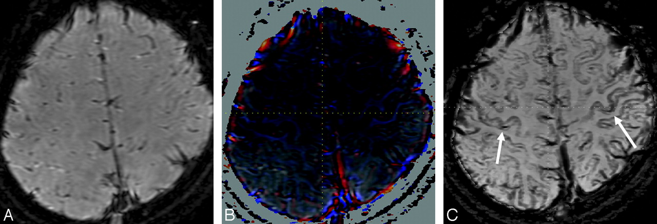
  • Fig 3.
    • Download figure
    • Open in new tab
    • Download powerpoint
    Fig 3.

    Magnitude (A), phase (B), and phase-weighted images (C) obtained in a 14-year-old girl. The signal intensity of the bilateral motor cortices (arrows) on the phase-weighted image (C) is definitely hypointense (grade III) in comparison with that in other cortices.

PreviousNext
Back to top

In this issue

American Journal of Neuroradiology: 29 (6)
American Journal of Neuroradiology
Vol. 29, Issue 6
June 2008
  • Table of Contents
  • Index by author
Advertisement
Print
Download PDF
Email Article

Thank you for your interest in spreading the word on American Journal of Neuroradiology.

NOTE: We only request your email address so that the person you are recommending the page to knows that you wanted them to see it, and that it is not junk mail. We do not capture any email address.

Enter multiple addresses on separate lines or separate them with commas.
Signal Intensity of the Motor Cortex on Phase-Weighted Imaging at 3T
(Your Name) has sent you a message from American Journal of Neuroradiology
(Your Name) thought you would like to see the American Journal of Neuroradiology web site.
CAPTCHA
This question is for testing whether or not you are a human visitor and to prevent automated spam submissions.
Cite this article
S. Kakeda, Y. Korogi, K. Kamada, N. Ohnari, J. Moriya, T. Sato, M. Kitajima, H. Hasnine, N. Hirata
Signal Intensity of the Motor Cortex on Phase-Weighted Imaging at 3T
American Journal of Neuroradiology Jun 2008, 29 (6) 1171-1175; DOI: 10.3174/ajnr.A1002

Citation Manager Formats

  • BibTeX
  • Bookends
  • EasyBib
  • EndNote (tagged)
  • EndNote 8 (xml)
  • Medlars
  • Mendeley
  • Papers
  • RefWorks Tagged
  • Ref Manager
  • RIS
  • Zotero
0 Responses
Respond to this article
Share
Bookmark this article
Signal Intensity of the Motor Cortex on Phase-Weighted Imaging at 3T
S. Kakeda, Y. Korogi, K. Kamada, N. Ohnari, J. Moriya, T. Sato, M. Kitajima, H. Hasnine, N. Hirata
American Journal of Neuroradiology Jun 2008, 29 (6) 1171-1175; DOI: 10.3174/ajnr.A1002
del.icio.us logo Twitter logo Facebook logo Mendeley logo
  • Tweet Widget
  • Facebook Like
  • Google Plus One
Purchase

Jump to section

  • Article
    • Abstract
    • Materials and Methods
    • Results
    • Discussion
    • Conclusion
    • References
  • Figures & Data
  • Info & Metrics
  • Responses
  • References
  • PDF

Related Articles

  • No related articles found.
  • PubMed
  • Google Scholar

Cited By...

  • The "White Gray Sign" Identifies the Central Sulcus on 3T High-Resolution T1-Weighted Images
  • Diffusion-weighted MRI hyperintensity patterns differentiate CJD from other rapid dementias
  • Demonstration of Cerebral Venous Variations in the Region of the Third Ventricle on Phase-Sensitive Imaging
  • Crossref (21)
  • Google Scholar

This article has been cited by the following articles in journals that are participating in Crossref Cited-by Linking.

  • Diffusion-weighted MRI hyperintensity patterns differentiate CJD from other rapid dementias
    P. Vitali, E. Maccagnano, E. Caverzasi, R.G. Henry, A. Haman, C. Torres-Chae, D.Y. Johnson, B.L. Miller, M.D. Geschwind
    Neurology 2011 76 20
  • In vivo assessment of iron content of the cerebral cortex in healthy aging using 7-Tesla T2*-weighted phase imaging
    Mathijs Buijs, Nhat Trung Doan, Sanneke van Rooden, Maarten J. Versluis, Baldur van Lew, Julien Milles, Jeroen van der Grond, Mark A. van Buchem
    Neurobiology of Aging 2017 53
  • Tensor‐valued diffusion MRI differentiates cortex and white matter in malformations of cortical development associated with epilepsy
    Björn Lampinen, Ariadne Zampeli, Isabella M. Björkman‐Burtscher, Filip Szczepankiewicz, Kristina Källén, Maria Compagno Strandberg, Markus Nilsson
    Epilepsia 2020 61 8
  • A novel tract imaging technique of the brainstem using phase difference enhanced imaging: normal anatomy and initial experience in multiple system atrophy
    Shingo Kakeda, Yukunori Korogi, Tetsuya Yoneda, Johji Nishimura, Toru Sato, Yasuhiro Hiai, Norihiro Ohnari, Kazumasa Okada, Haruki Hayashi, Eiji Matsusue, Takenori Uozumi, Sadatoshi Tsuji
    European Radiology 2011 21 10
  • Demonstration of Cerebral Venous Variations in the Region of the Third Ventricle on Phase-Sensitive Imaging
    S. Fujii, Y. Kanasaki, E. Matsusue, S. Kakite, T. Kminou, T. Ogawa
    American Journal of Neuroradiology 2010 31 1
  • The “White Gray Sign” Identifies the Central Sulcus on 3T High-Resolution T1-Weighted Images
    O.F. Kaneko, N.J. Fischbein, J. Rosenberg, M. Wintermark, M.M. Zeineh
    American Journal of Neuroradiology 2017 38 2
  • Zebra sign of precentral gyri in amyotrophic lateral sclerosis: A novel finding using phase difference enhanced (PADRE) imaging-initial results
    Shingo Kakeda, Tetsuya Yoneda, Satoru Ide, Mari Miyata, Tomoyo Hashimoto, Koichiro Futatsuya, Keita Watanabe, Atsushi Ogasawara, Junji Moriya, Toru Sato, Kazumasa Okada, Takenori Uozumi, Hiroaki Adachi, Yukunori Korogi
    European Radiology 2016 26 11
  • Comparison of increased venous contrast in ischemic stroke using phase-sensitive MR imaging with perfusion changes on flow-sensitive alternating inversion recovery at 3 Tesla
    Eijiro Yamashita, Yoshiko Kanasaki, Shinya Fujii, Takuro Tanaka, Yoshiharu Hirata, Toshihide Ogawa
    Acta Radiologica 2011 52 8
  • Parkinson’s disease: diagnostic potential of high-resolution phase difference enhanced MR imaging at 3 T
    Shingo Kakeda, Yukunori Korogi, Tetsuya Yoneda, Keita Watanabe, Junji Moriya, Yu Murakami, Toru Sato, Yasuhiro Hiai, Norihiro Ohnari, Satoru Ide, Kazumasa Okada, Takenori Uozumi, Sadatoshi Tsuji, Toshinori Hirai, Yasuyuki Yamashita
    European Radiology 2013 23 4
  • Superficial Siderosis Associated With a Chronic Subdural Hematoma
    Shingo Kakeda, Yukunori Korogi, Norihiro Ohnari, Johji Nishimura, Junji Moriya, Junkoh Yamamoto, Shigeru Nishizawa
    Academic Radiology 2010 17 7

More in this TOC Section

  • Quiet PROPELLER MRI Techniques Match the Quality of Conventional PROPELLER Brain Imaging Techniques
  • Predictors of Reperfusion in Patients with Acute Ischemic Stroke
  • Enhanced Axonal Metabolism during Early Natalizumab Treatment in Relapsing-Remitting Multiple Sclerosis
Show more BRAIN

Similar Articles

Advertisement

Indexed Content

  • Current Issue
  • Accepted Manuscripts
  • Article Preview
  • Past Issues
  • Editorials
  • Editor's Choice
  • Fellows' Journal Club
  • Letters to the Editor
  • Video Articles

Cases

  • Case Collection
  • Archive - Case of the Week
  • Archive - Case of the Month
  • Archive - Classic Case

More from AJNR

  • Trainee Corner
  • Imaging Protocols
  • MRI Safety Corner
  • Book Reviews

Multimedia

  • AJNR Podcasts
  • AJNR Scantastics

Resources

  • Turnaround Time
  • Submit a Manuscript
  • Submit a Video Article
  • Submit an eLetter to the Editor/Response
  • Manuscript Submission Guidelines
  • Statistical Tips
  • Fast Publishing of Accepted Manuscripts
  • Graphical Abstract Preparation
  • Imaging Protocol Submission
  • Evidence-Based Medicine Level Guide
  • Publishing Checklists
  • Author Policies
  • Become a Reviewer/Academy of Reviewers
  • News and Updates

About Us

  • About AJNR
  • Editorial Board
  • Editorial Board Alumni
  • Alerts
  • Permissions
  • Not an AJNR Subscriber? Join Now
  • Advertise with Us
  • Librarian Resources
  • Feedback
  • Terms and Conditions
  • AJNR Editorial Board Alumni

American Society of Neuroradiology

  • Not an ASNR Member? Join Now

© 2025 by the American Society of Neuroradiology All rights, including for text and data mining, AI training, and similar technologies, are reserved.
Print ISSN: 0195-6108 Online ISSN: 1936-959X

Powered by HighWire