Advertisement

Main menu

  • Home
  • Content
    • Current Issue
    • Accepted Manuscripts
    • Article Preview
    • Past Issue Archive
    • Video Articles
    • AJNR Case Collection
    • Case of the Week Archive
    • Case of the Month Archive
    • Classic Case Archive
  • Special Collections
    • AJNR Awards
    • Low-Field MRI
    • Alzheimer Disease
    • ASNR Foundation Special Collection
    • Photon-Counting CT
    • View All
  • Multimedia
    • AJNR Podcasts
    • AJNR SCANtastic
    • Trainee Corner
    • MRI Safety Corner
    • Imaging Protocols
  • For Authors
    • Submit a Manuscript
    • Submit a Video Article
    • Submit an eLetter to the Editor/Response
    • Manuscript Submission Guidelines
    • Statistical Tips
    • Fast Publishing of Accepted Manuscripts
    • Graphical Abstract Preparation
    • Imaging Protocol Submission
    • Author Policies
  • About Us
    • About AJNR
    • Editorial Board
    • Editorial Board Alumni
  • More
    • Become a Reviewer/Academy of Reviewers
    • Subscribers
    • Permissions
    • Alerts
    • Feedback
    • Advertisers
    • ASNR Home

User menu

  • Alerts
  • Log in

Search

  • Advanced search
American Journal of Neuroradiology

American Journal of Neuroradiology

ASHNR American Society of Functional Neuroradiology ASHNR American Society of Pediatric Neuroradiology ASSR
  • Alerts
  • Log in

Advanced Search

  • Home
  • Content
    • Current Issue
    • Accepted Manuscripts
    • Article Preview
    • Past Issue Archive
    • Video Articles
    • AJNR Case Collection
    • Case of the Week Archive
    • Case of the Month Archive
    • Classic Case Archive
  • Special Collections
    • AJNR Awards
    • Low-Field MRI
    • Alzheimer Disease
    • ASNR Foundation Special Collection
    • Photon-Counting CT
    • View All
  • Multimedia
    • AJNR Podcasts
    • AJNR SCANtastic
    • Trainee Corner
    • MRI Safety Corner
    • Imaging Protocols
  • For Authors
    • Submit a Manuscript
    • Submit a Video Article
    • Submit an eLetter to the Editor/Response
    • Manuscript Submission Guidelines
    • Statistical Tips
    • Fast Publishing of Accepted Manuscripts
    • Graphical Abstract Preparation
    • Imaging Protocol Submission
    • Author Policies
  • About Us
    • About AJNR
    • Editorial Board
    • Editorial Board Alumni
  • More
    • Become a Reviewer/Academy of Reviewers
    • Subscribers
    • Permissions
    • Alerts
    • Feedback
    • Advertisers
    • ASNR Home
  • Follow AJNR on Twitter
  • Visit AJNR on Facebook
  • Follow AJNR on Instagram
  • Join AJNR on LinkedIn
  • RSS Feeds

AJNR Awards, New Junior Editors, and more. Read the latest AJNR updates

Research ArticleBRAIN

An Acute Ischemic Stroke Classification Instrument That Includes CT or MR Angiography: The Boston Acute Stroke Imaging Scale

F. Torres-Mozqueda, J. He, I.B. Yeh, L.H. Schwamm, M.H. Lev, P.W. Schaefer and R.G. González
American Journal of Neuroradiology June 2008, 29 (6) 1111-1117; DOI: https://doi.org/10.3174/ajnr.A1000
F. Torres-Mozqueda
  • Find this author on Google Scholar
  • Find this author on PubMed
  • Search for this author on this site
J. He
  • Find this author on Google Scholar
  • Find this author on PubMed
  • Search for this author on this site
I.B. Yeh
  • Find this author on Google Scholar
  • Find this author on PubMed
  • Search for this author on this site
L.H. Schwamm
  • Find this author on Google Scholar
  • Find this author on PubMed
  • Search for this author on this site
M.H. Lev
  • Find this author on Google Scholar
  • Find this author on PubMed
  • Search for this author on this site
P.W. Schaefer
  • Find this author on Google Scholar
  • Find this author on PubMed
  • Search for this author on this site
R.G. González
  • Find this author on Google Scholar
  • Find this author on PubMed
  • Search for this author on this site
  • Article
  • Figures & Data
  • Info & Metrics
  • Responses
  • References
  • PDF
Loading

Article Figures & Data

Figures

  • Tables
  • Fig 1.
    • Download figure
    • Open in new tab
    • Download powerpoint
    Fig 1.

    Classification algorithm. Proximal cerebral artery occlusions are depicted in the drawing on the left and are defined as including the following arteries: distal (intracranial) ICA, proximal (M1 or M2) MCA, and/or basilar artery (BA). As shown in the algorithm on the right, the first step was evaluation of CTA or MRA data to identify apparent proximal cerebral artery occlusions. If no proximal cerebral artery occlusion was found, the noncontrast CT or diffusion MR imaging data were reviewed for evidence of a large acute ischemic infarct as defined in the “Materials and Methods” section. If a large CT or DWI abnormality was detected, the patient was classified as having a major stroke. All other circumstances resulted in classification as a minor stroke by imaging.
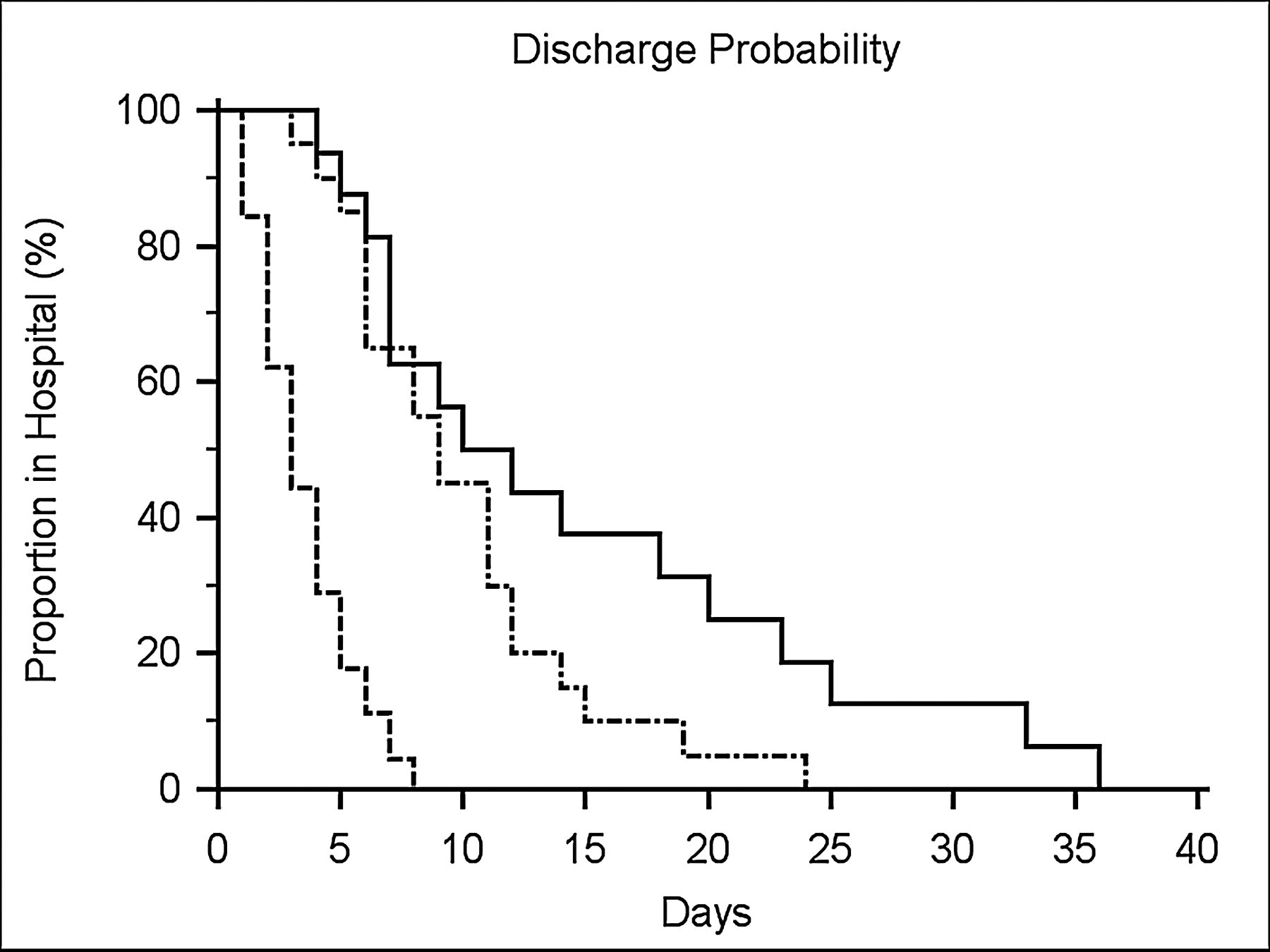
  • Fig 2.
    • Download figure
    • Open in new tab
    • Download powerpoint
    Fig 2.

    Kaplan-Meier curve of time to discharge. The Kaplan-Meier graph depicts the probability of discharge from hospital in days for patients classified as having major strokes by BASIS and ASPECT (solid line), major by BASIS but minor by ASPECT (dot and dash line), and minor by both classification instruments (dashed line). Overall, a highly significant difference (P < .0001) between the groups was found. In isolating the differences, both the BASIS major/ASPECT major and the BASIS major/ASPECT minor were significantly different from the BASIS and ASPECT minor group (P < .0001). However, there was no significant difference between the BASIS major/ASPECT major (solid line) and the BASIS major/ASPECT minor groups (P = .077).

Tables

  • Figures
    • View popup
    Table 1:

    Outcomes of 87 patients with ischemic stroke imaged by NCCT and CTA

    VariableMajor Stroke by Imaging (n = 43)Minor Stroke by Imaging (n = 44)P
    Deaths, n60<.0001
    Discharge to rehabilitation facility, n (%)32 (74)4 (9)<.0001
    Discharge to home, n (%)5 (12)34 (77)<.0001
    Length of stay, days (SE)12.1 (1.3)3.7 (0.4)<.0001
    • Note:—NCCT indicates noncontrast CT; CTA, CT angiography. Significant differences in outcomes between patients with major and minor stroke were assessed using Fisher exact test (deaths, discharge to a rehabilitation facility, or discharge home) and t test (length of stay). Highly significant differences in all outcome measures were found between patients with major and minor stroke who were initially imaged with NCCT and included CTA. Not included in the table are 11 patients who had NCCT without CTA.

    • View popup
    Table 2:

    Outcomes of 118 patients with ischemic stroke imaged by MRI and MRA

    VariableMajor Stroke by Imaging (n = 13)Minor Stroke by Imaging (n = 105)P
    Deaths, n20<.05
    Discharge to rehabilitation facility, n (%)8 (72)19 (18)<.001
    Discharge to home, n (%)3 (27)84 (80)<.001
    Length of stay, days (SE)12.9 (3.1)3.1 (0.2)<.005
    • Note:—MRI indicates MR imaging; MRA, MR angiography. Significant differences in all outcome measures were found between patients with major and minor stroke who had MRA as the first angiographic study. Statistical tests for each outcome measure are the same as in Table 1. Not included in this table are 14 patients who had MRI including diffusion MRI but not MRA.

    • View popup
    Table 3:

    Outcomes of 87 patients with ischemic stroke imaged by NCCT and CTA and classified by BASIS and ASPECTS

    VariableMajor Stroke by BASIS and ASPECTS (n = 22)Major by BASIS Minor by ASPECTS (n = 21)Minor Stroke by BASIS and ASPECTS (n = 44)
    Deaths, n5*10*
    Discharge to rehabilitation facility, n (%)16 (94)†‡16 (80)†‡4 (9)†
    Discharge to home, n (%)1 (6)†‡4 (20)†‡34 (77)†
    Length of stay, days (SE)14.8 (2.4)§‖9.9 (1.1)§‖3.7 (0.4)§
    • Note:—NCCT indicates noncontrast CT; CTA, CT angiography; BASIS, Boston Acute Stroke Imaging Scale; ASPECTS, Alberta Stroke Program Early CT Score. There were significant differences in deaths between the BASIS and ASPECTS major stroke group and the minor stroke group classified by BASIS and ASPECTS. Highly significant differences in discharge to rehabilitation and discharge to home were found between the 2 groups that had major stroke classification and the patients classified as minor strokes by BASIS and ASPECTS.

    • * P < .003.

    • † All P < .0001.

    • ‡ No significant differences in these outcomes were found between the BASIS and ASPECTS major stroke and BASIS major/ASPECTS minor stroke groups.

    • § P < .0001.

    • ‖ Significant differences were found in length of stay between the 2 groups that had major stroke classification and the patients classified as having minor strokes by BASIS and ASPECTS but not between the BASIS and ASPECTS major stroke and BASIS major/ASPECTS minor stroke groups.

PreviousNext
Back to top

In this issue

American Journal of Neuroradiology: 29 (6)
American Journal of Neuroradiology
Vol. 29, Issue 6
June 2008
  • Table of Contents
  • Index by author
Advertisement
Print
Download PDF
Email Article

Thank you for your interest in spreading the word on American Journal of Neuroradiology.

NOTE: We only request your email address so that the person you are recommending the page to knows that you wanted them to see it, and that it is not junk mail. We do not capture any email address.

Enter multiple addresses on separate lines or separate them with commas.
An Acute Ischemic Stroke Classification Instrument That Includes CT or MR Angiography: The Boston Acute Stroke Imaging Scale
(Your Name) has sent you a message from American Journal of Neuroradiology
(Your Name) thought you would like to see the American Journal of Neuroradiology web site.
CAPTCHA
This question is for testing whether or not you are a human visitor and to prevent automated spam submissions.
Cite this article
F. Torres-Mozqueda, J. He, I.B. Yeh, L.H. Schwamm, M.H. Lev, P.W. Schaefer, R.G. González
An Acute Ischemic Stroke Classification Instrument That Includes CT or MR Angiography: The Boston Acute Stroke Imaging Scale
American Journal of Neuroradiology Jun 2008, 29 (6) 1111-1117; DOI: 10.3174/ajnr.A1000

Citation Manager Formats

  • BibTeX
  • Bookends
  • EasyBib
  • EndNote (tagged)
  • EndNote 8 (xml)
  • Medlars
  • Mendeley
  • Papers
  • RefWorks Tagged
  • Ref Manager
  • RIS
  • Zotero
0 Responses
Respond to this article
Share
Bookmark this article
An Acute Ischemic Stroke Classification Instrument That Includes CT or MR Angiography: The Boston Acute Stroke Imaging Scale
F. Torres-Mozqueda, J. He, I.B. Yeh, L.H. Schwamm, M.H. Lev, P.W. Schaefer, R.G. González
American Journal of Neuroradiology Jun 2008, 29 (6) 1111-1117; DOI: 10.3174/ajnr.A1000
del.icio.us logo Twitter logo Facebook logo Mendeley logo
  • Tweet Widget
  • Facebook Like
  • Google Plus One
Purchase

Jump to section

  • Article
    • Abstract
    • Materials and Methods
    • Results
    • Discussion
    • Footnotes
    • References
  • Figures & Data
  • Info & Metrics
  • Responses
  • References
  • PDF

Related Articles

  • No related articles found.
  • PubMed
  • Google Scholar

Cited By...

  • Effect of definition and methods on estimates of prevalence of large vessel occlusion in acute ischemic stroke: a systematic review and meta-analysis
  • Prevalence of large vessel occlusion in patients presenting with acute ischemic stroke: a 10-year systematic review of the literature
  • Good Intracranial Collaterals Trump Poor ASPECTS (Alberta Stroke Program Early CT Score) for Intravenous Thrombolysis in Anterior Circulation Acute Ischemic Stroke
  • Percentage Insula Ribbon Infarction of >50% Identifies Patients Likely to Have Poor Clinical Outcome Despite Small DWI Infarct Volume
  • External Validation of the Boston Acute Stroke Imaging Scale and M1-BASIS in Thrombolyzed Patients
  • Current State of Acute Stroke Imaging
  • Computed Tomography Angiography in Hyperacute Ischemic Stroke: Prognostic Implications and Role in Decision-Making
  • The Massachusetts General Hospital acute stroke imaging algorithm: an experience and evidence based approach
  • Guidelines for the Early Management of Patients With Acute Ischemic Stroke: A Guideline for Healthcare Professionals From the American Heart Association/American Stroke Association
  • Location-weighted CTP analysis predicts early motor improvement in stroke: A preliminary study
  • Revised and Updated Recommendations for the Establishment of Primary Stroke Centers: A Summary Statement From the Brain Attack Coalition
  • Predicting Language Improvement in Acute Stroke Patients Presenting with Aphasia: A Multivariate Logistic Model Using Location-Weighted Atlas-Based Analysis of Admission CT Perfusion Scans
  • Hyperacute stent placement in acute cervical internal carotid artery occlusions: the potential role of magnetic resonance imaging
  • MRI-Based Selection for Intra-Arterial Stroke Therapy: Value of Pretreatment Diffusion-Weighted Imaging Lesion Volume in Selecting Patients With Acute Stroke Who Will Benefit From Early Recanalization
  • Arterial Wall Enhancement Overlying Carotid Plaque on CT Angiography Correlates With Symptoms in Patients With High Grade Stenosis
  • Comparing and Predicting the Costs and Outcomes of Patients with Major and Minor Stroke Using the Boston Acute Stroke Imaging Scale Neuroimaging Classification System
  • CT Angiography Clot Burden Score and Collateral Score: Correlation with Clinical and Radiologic Outcomes in Acute Middle Cerebral Artery Infarct
  • Response to Letter by Gonzalez-Hernandez et al
  • Crossref (61)
  • Google Scholar

This article has been cited by the following articles in journals that are participating in Crossref Cited-by Linking.

  • Guidelines for the Early Management of Patients With Acute Ischemic Stroke
    Edward C. Jauch, Jeffrey L. Saver, Harold P. Adams, Askiel Bruno, J.J. (Buddy) Connors, Bart M. Demaerschalk, Pooja Khatri, Paul W. McMullan, Adnan I. Qureshi, Kenneth Rosenfield, Phillip A. Scott, Debbie R. Summers, David Z. Wang, Max Wintermark, Howard Yonas
    Stroke 2013 44 3
  • CT Angiography Clot Burden Score and Collateral Score: Correlation with Clinical and Radiologic Outcomes in Acute Middle Cerebral Artery Infarct
    I.Y.L. Tan, A.M. Demchuk, J. Hopyan, L. Zhang, D. Gladstone, K. Wong, M. Martin, S.P. Symons, A.J. Fox, R.I. Aviv
    American Journal of Neuroradiology 2009 30 3
  • MRI-Based Selection for Intra-Arterial Stroke Therapy
    Albert J. Yoo, Luis A. Verduzco, Pamela W. Schaefer, Joshua A. Hirsch, James D. Rabinov, R. Gilberto González
    Stroke 2009 40 6
  • Revised and Updated Recommendations for the Establishment of Primary Stroke Centers
    Mark J. Alberts, Richard E. Latchaw, Andy Jagoda, Lawrence R. Wechsler, Todd Crocco, Mary G. George, E.S. Connolly, Barbara Mancini, Stephen Prudhomme, Daryl Gress, Mary E. Jensen, Robert Bass, Robert Ruff, Kathy Foell, Rocco A. Armonda, Marian Emr, Margo Warren, Jim Baranski, Michael D. Walker
    Stroke 2011 42 9
  • The Alberta Stroke Program Early CT Score in Clinical Practice: What have We Learned?
    V. Puetz, I. Dzialowski, M. D. Hill, A. M. Demchuk
    International Journal of Stroke 2009 4 5
  • Prevalence of large vessel occlusion in patients presenting with acute ischemic stroke: a 10-year systematic review of the literature
    Nikita Lakomkin, Mandip Dhamoon, Kirsten Carroll, Inder Paul Singh, Stanley Tuhrim, Joyce Lee, Johanna T Fifi, J Mocco
    Journal of NeuroInterventional Surgery 2019 11 3
  • Towards effective classification of brain hemorrhagic and ischemic stroke using CNN
    Anjali Gautam, Balasubramanian Raman
    Biomedical Signal Processing and Control 2021 63
  • Selection for Inpatient Rehabilitation After Acute Stroke: A Systematic Review of the Literature
    Sharon J. Hakkennes, Kim Brock, Keith D. Hill
    Archives of Physical Medicine and Rehabilitation 2011 92 12
  • Incidence of Acute Ischemic Stroke With Visible Arterial Occlusion
    Gauthier Duloquin, Mathilde Graber, Lucie Garnier, Valentin Crespy, Pierre-Olivier Comby, Laura Baptiste, Sophie Mohr, Benoit Delpont, Julien Guéniat, Christelle Blanc-Labarre, Marie Hervieu-Bègue, Guy-Victor Osseby, Maurice Giroud, Yannick Béjot
    Stroke 2020 51 7
  • Clinical MRI of acute ischemic stroke
    R. Gilberto González
    Journal of Magnetic Resonance Imaging 2012 36 2

More in this TOC Section

  • Quiet PROPELLER MRI Techniques Match the Quality of Conventional PROPELLER Brain Imaging Techniques
  • Predictors of Reperfusion in Patients with Acute Ischemic Stroke
  • Enhanced Axonal Metabolism during Early Natalizumab Treatment in Relapsing-Remitting Multiple Sclerosis
Show more BRAIN

Similar Articles

Advertisement

Indexed Content

  • Current Issue
  • Accepted Manuscripts
  • Article Preview
  • Past Issues
  • Editorials
  • Editor's Choice
  • Fellows' Journal Club
  • Letters to the Editor
  • Video Articles

Cases

  • Case Collection
  • Archive - Case of the Week
  • Archive - Case of the Month
  • Archive - Classic Case

More from AJNR

  • Trainee Corner
  • Imaging Protocols
  • MRI Safety Corner
  • Book Reviews

Multimedia

  • AJNR Podcasts
  • AJNR Scantastics

Resources

  • Turnaround Time
  • Submit a Manuscript
  • Submit a Video Article
  • Submit an eLetter to the Editor/Response
  • Manuscript Submission Guidelines
  • Statistical Tips
  • Fast Publishing of Accepted Manuscripts
  • Graphical Abstract Preparation
  • Imaging Protocol Submission
  • Evidence-Based Medicine Level Guide
  • Publishing Checklists
  • Author Policies
  • Become a Reviewer/Academy of Reviewers
  • News and Updates

About Us

  • About AJNR
  • Editorial Board
  • Editorial Board Alumni
  • Alerts
  • Permissions
  • Not an AJNR Subscriber? Join Now
  • Advertise with Us
  • Librarian Resources
  • Feedback
  • Terms and Conditions
  • AJNR Editorial Board Alumni

American Society of Neuroradiology

  • Not an ASNR Member? Join Now

© 2025 by the American Society of Neuroradiology All rights, including for text and data mining, AI training, and similar technologies, are reserved.
Print ISSN: 0195-6108 Online ISSN: 1936-959X

Powered by HighWire