Advertisement

Main menu

  • Home
  • Content
    • Current Issue
    • Accepted Manuscripts
    • Article Preview
    • Past Issue Archive
    • Video Articles
    • AJNR Case Collection
    • Case of the Week Archive
    • Case of the Month Archive
    • Classic Case Archive
  • Special Collections
    • Advancing NeuroMRI with High-Relaxivity Contrast Agents
    • AJNR Awards
    • Low-Field MRI
    • Alzheimer Disease
    • ASNR Foundation Special Collection
    • Photon-Counting CT
    • View All
  • Multimedia
    • AJNR Podcasts
    • AJNR SCANtastic
    • Trainee Corner
    • MRI Safety Corner
    • Imaging Protocols
  • For Authors
    • Submit a Manuscript
    • Submit a Video Article
    • Submit an eLetter to the Editor/Response
    • Manuscript Submission Guidelines
    • Statistical Tips
    • Fast Publishing of Accepted Manuscripts
    • Graphical Abstract Preparation
    • Imaging Protocol Submission
    • Author Policies
  • About Us
    • About AJNR
    • Editorial Board
    • Editorial Board Alumni
  • More
    • Become a Reviewer/Academy of Reviewers
    • Subscribers
    • Permissions
    • Alerts
    • Feedback
    • Advertisers
    • ASNR Home

User menu

  • Alerts
  • Log in

Search

  • Advanced search
American Journal of Neuroradiology

American Journal of Neuroradiology

ASHNR American Society of Functional Neuroradiology ASHNR American Society of Pediatric Neuroradiology ASSR
  • Alerts
  • Log in

Advanced Search

  • Home
  • Content
    • Current Issue
    • Accepted Manuscripts
    • Article Preview
    • Past Issue Archive
    • Video Articles
    • AJNR Case Collection
    • Case of the Week Archive
    • Case of the Month Archive
    • Classic Case Archive
  • Special Collections
    • Advancing NeuroMRI with High-Relaxivity Contrast Agents
    • AJNR Awards
    • Low-Field MRI
    • Alzheimer Disease
    • ASNR Foundation Special Collection
    • Photon-Counting CT
    • View All
  • Multimedia
    • AJNR Podcasts
    • AJNR SCANtastic
    • Trainee Corner
    • MRI Safety Corner
    • Imaging Protocols
  • For Authors
    • Submit a Manuscript
    • Submit a Video Article
    • Submit an eLetter to the Editor/Response
    • Manuscript Submission Guidelines
    • Statistical Tips
    • Fast Publishing of Accepted Manuscripts
    • Graphical Abstract Preparation
    • Imaging Protocol Submission
    • Author Policies
  • About Us
    • About AJNR
    • Editorial Board
    • Editorial Board Alumni
  • More
    • Become a Reviewer/Academy of Reviewers
    • Subscribers
    • Permissions
    • Alerts
    • Feedback
    • Advertisers
    • ASNR Home
  • Follow AJNR on Twitter
  • Visit AJNR on Facebook
  • Follow AJNR on Instagram
  • Join AJNR on LinkedIn
  • RSS Feeds

AJNR Awards, New Junior Editors, and more. Read the latest AJNR updates


Improved Turnaround Times | Median time to first decision: 12 days

Research ArticleNeurointervention

Patient Skin Dose during Neuroembolization by Multiple-Point Measurement Using a Radiosensitive Indicator

S. Suzuki, S. Furui, Y. Matsumaru, S. Nobuyuki, M. Ebara, T. Abe and D. Itoh
American Journal of Neuroradiology June 2008, 29 (6) 1076-1081; DOI: https://doi.org/10.3174/ajnr.A1045
S. Suzuki
  • Find this author on Google Scholar
  • Find this author on PubMed
  • Search for this author on this site
S. Furui
  • Find this author on Google Scholar
  • Find this author on PubMed
  • Search for this author on this site
Y. Matsumaru
  • Find this author on Google Scholar
  • Find this author on PubMed
  • Search for this author on this site
S. Nobuyuki
  • Find this author on Google Scholar
  • Find this author on PubMed
  • Search for this author on this site
M. Ebara
  • Find this author on Google Scholar
  • Find this author on PubMed
  • Search for this author on this site
T. Abe
  • Find this author on Google Scholar
  • Find this author on PubMed
  • Search for this author on this site
D. Itoh
  • Find this author on Google Scholar
  • Find this author on PubMed
  • Search for this author on this site
  • Article
  • Figures & Data
  • Info & Metrics
  • Responses
  • References
  • PDF
Loading

Article Figures & Data

Figures

  • Tables
  • Fig 1.
    • Download figure
    • Open in new tab
    • Download powerpoint
    Fig 1.

    Photograph of indicators. A total of 44 indicators were adhered to a cap (right aspect, n = 19; left aspect, n = 19; and parietal aspect, n = 6) at intervals of 5 cm.

  • Fig 2.
    • Download figure
    • Open in new tab
    • Download powerpoint
    Fig 2.

    Effects of tube voltage, filtration, and dose rate on the color difference of the indicator. A, Indicators were irradiated at the dose of 1 Gy by using 4 tube voltages (50, 75, 100, and 125 kV) and 4 types of filtration (2.2 mm of aluminum, 5.2 mm of aluminum, 3.7 mm of aluminum + 0.1 mm of copper; and 3.7 mm of aluminum + 0.4 mm of copper) with a 20-mA tube current. The difference in the color difference was within 2.20. B, The effect of the dose rate was also evaluated by using 2 tube currents (5 and 20 mA) for the 4 types of filtration at the dose of 1 Gy with tube voltage of 75 kVp. For each filtration, the difference in the color between the 2 dose rates was within 0.79. Al indicates aluminum; Cu, copper.

  • Fig 3.
    • Download figure
    • Open in new tab
    • Download powerpoint
    Fig 3.

    Maximum ESD distribution of each procedure. The maximum ESD of the patients exceeded 1 Gy in 84 procedures, 3 Gy in 20 procedures, and 5 Gy in 2 procedures.

  • Fig 4.
    • Download figure
    • Open in new tab
    • Download powerpoint
    Fig 4.

    Scatterplot of DAP and maximum ESD with regression line.
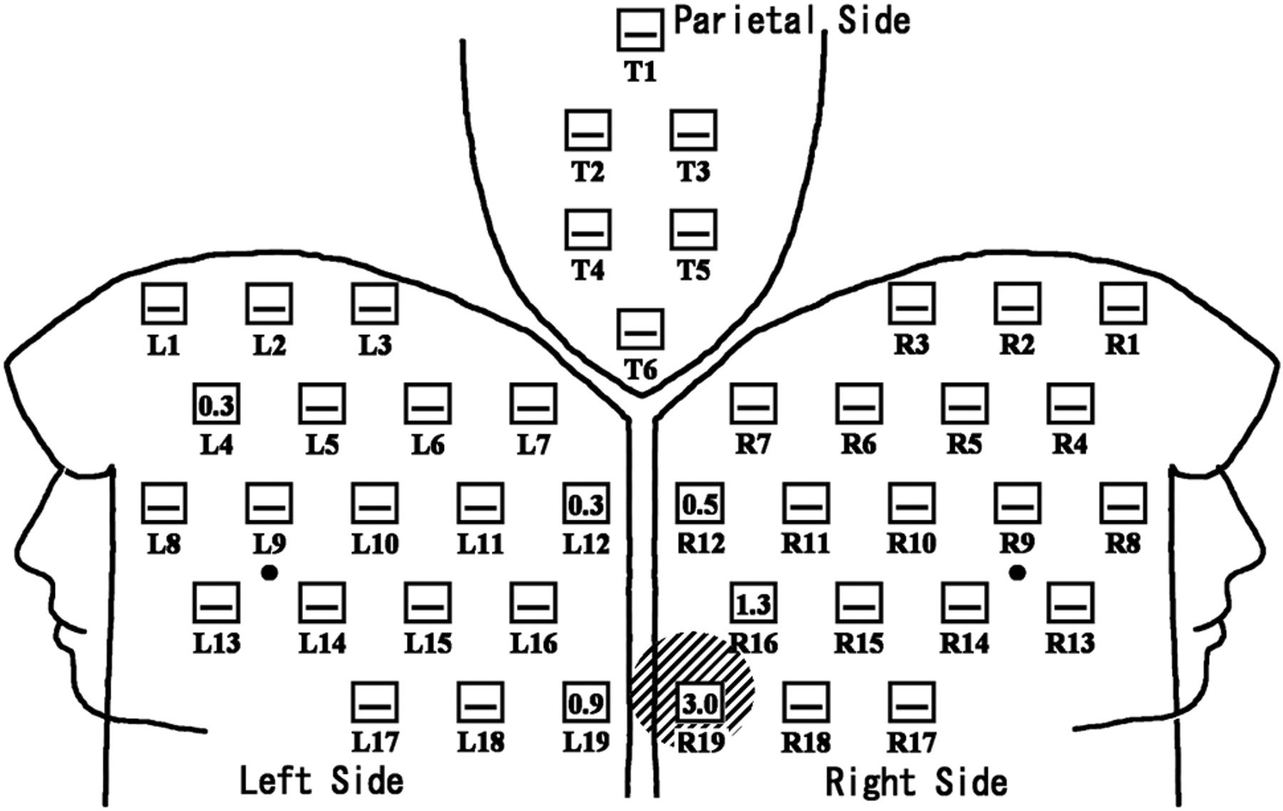
  • Fig 5.
    • Download figure
    • Open in new tab
    • Download powerpoint
    Fig 5.

    Distribution of ESDs in a patient with radiation skin injury. Neuroembolization for internal carotid-posterior communicating aneurysm was performed for a 43-year-old woman. The FOV mainly used was 14 cm. Total fluoroscopic time and DAP were 75.0 minutes and 182 Gy × cm2, respectively; 4° left anterior oblique with 17° cranial angulation view was mostly used. Exposed skin area was localized to the occipital region with the maximum ESD of 3.0 Gy. Temporary epilation was detected at the maximum exposure part (slanted lines) at 2 weeks and 3 months of follow-up. The number in the open square represents the ESD (Gy) at the point. — in the open square means the ESD at the point was less than 0.3 Gy.

Tables

  • Figures
    • View popup
    Table 1:

    Target lesions in each institution

    InstitutionNo. of Target Lesions
    ANAVFCCFAVM
    Institution 139243
    Institution 219010
    Institution 312300
    Institution 48110
    Institution 55000
    Institution 63101
    Overall86764
    • Note:—AN indicates cerebral aneurysm; AVF, dural arteriovenous fistula; CCF, internal carotid artery cavernous sinus fistula; AVM, arteriovenous malformation.

    • View popup
    Table 2:

    Data of angiographic units

    DataInstitution 1Institution 2Institution 3Institution 4Institution 5Institution 6
    Angiographic unit (No. of procedures)AXIOM Artis FA (48)AXIOM Artis dBA (20)AXIOM Artis dBA (15)Allura Xper FD20/10 (10)Advantx LCN PLUS (5)Advantx-E LCA (4)OEC9800 (1)
    DetectorI.I. (single plane)FD (Biplane)FD (Biplane)FD (Biplane)I.I. (Biplane)I.I. (Single plane)I.I. (Single plane)
    Period of use of the unit, y5111774
    Period of use of the detector, y5111314
    Dose area product meterAvailableAvailableAvailableAvailableNot availableNot available4
    Mainly used FOV, cm28, 20, 1448, 32, 22, 16*22, 16, 11*43, 40, 25, 20*15, 112323
    Minimal total filtration2.5-mm Al2.5-mm Al2.5-mm Al2.7-mm Al2.9-mm Al2.9-mm Al5.5-mm Al
    Fluoroscopy
        Removable filter0.1-, 0.2-, 0.3-mm Cu0.2-mm Cu0.1-mm Cu0.4-mm Cu + 1.0-mm Al0.1-mm Cu + 1.0-mm Al
        Pulse rate15 pulse/s15 pulse/s15, 10 pulse/s30, 15 pulse/s30 pulse/s25 pulse/s8 pulse/s
    Digital subtraction angiography
        Removable filter0.1-, 0.2-, 0.3-mm Cu0.1-mm Cu0.1-mm Cu0.1-mm Cu + 1.0-mm Al0.1-mm Cu +1.0-mm Al
        Frame rate2 frame/s1 frame/s6, 4, 3 frame/s2 frame/s1.9 frame/s3.1 frame/s30 frame/s
    • Note:—I.I. indicates image intensifier; FD, digital flat panel detector; Cu, copper; Al, aluminum.

    • * The FOV of the digital flat panel detector was specified by the diagonal length.

    • View popup
    Table 3:

    Averages of total fluoroscopic time, number of DSA frames, DAP, and maximum ESD in each institution

    VariableInstitutionOverall (n = 103)
    1 (n = 48)2 (n = 20)3 (n = 15)4 (n = 10)5 (n = 5)6 (n = 5)
    Total fluoroscopic time, min62.3 ± 35.150.6 ± 30.0114.8 ± 40.232.8 ± 24.056.4 ± 14.7107.8 ± 53.867.1 ± 41.6
    Total no. of DSA frames654 ± 376960 ± 3561447 ± 368604 ± 2311756 ± 2024770 ± 191883 ± 626
    DAP, Gy × cm2218 ± 141327 ± 162326 ± 98215 ± 162**257 ± 150
    Maximum ESD, Gy2.1 ± 1.11.6 ± 0.82.4 ± 1.31.5 ± 1.21.3 ± 0.71.0 ± 0.21.9 ± 1.1
    • Note:—Data are given as average ± SD. DSA indicates digital subtraction angiography; DAP, dose area product; ESD, entrance skin dose.

    • * DAP meters were not available.

    • View popup
    Table 4:

    Averages of total fluoroscopic time, number of DSA frames, DAP, and maximum ESD for each target lesion

    VariableTarget Lesions
    AN (n = 86)AVF (n = 7)CCF (n = 6)AVM (n = 4)
    Total fluoroscopic time, min59.8 ± 33.7120.3 ± 67.3125.9 ± 27.340.0 ± 8.7
    Total no. of DSA frames812 ± 221405 ± 7581125 ± 2511141 ± 320
    DAP, Gy × cm2233 ± 132380 ± 166428 ± 233307 ± 36
    Maximum ESD, Gy1.8 ± 1.12.0 ± 1.02.6 ± 0.91.6 ± 0.5
    • Note:—Data are given as average ± SD. AN indicates cerebral aneurysm; AVF, dural arteriovenous fistula; CCF, internal carotid artery cavernous sinus fistula; AVM, arteriovenous malformation; DSA, digital subtraction angiography; DAP, dose area product; ESD, entrance skin dose.

    • View popup
    Table 5:

    Six cases of radiation-induced epilation at the most exposed area

    Case No.InstitutionTarget LesionsMaximum ESD, GyTotal Fluoroscopic Time, minMost Exposed AreaTime of OnsetType of Epilation
    Case 11AN2.770.5Left occipital3 moTemporary
    Case 21AN5.696.5Right temporal2 wkTemporary
    Case 31AN3.075.0Right occipital2 wkTemporary
    Case 41AN3.968.0Right occipital2 wkTemporary
    Case 53AN5.2111.4Right occipital2 wkTemporary
    Case 63AN3.2128.7Right occipital2 wkTemporary
    • Note:—AN indicates cerebral aneurysm.

    • View popup
    Table 6:

    Data of patient's maximum skin dose (ESD) during neuroembolization in the literature

    Target Lesions or ProcedureNo. of PatientsAverage Maximum ESD, GyAverage Total Fluoroscopic Time, minDosimetryAuthorsPublished Year
    AN861.8 ± 1.159.8 ± 33.744 points by radiosensitive indicatorThis studyThis study
    AVF72.0 ± 1.0120.3 ± 67.3Same as aboveThis studyThis study
    CCF62.6 ± 0.9125.9 ± 27.3Same as aboveThis studyThis study
    AVM41.6 ± 0.540.0 ± 8.7Same as aboveThis studyThis study
    GDC embolization with an old angiography unit122.2565 points by TLDsMooney et al182006
    GDC embolization with a new angiography unit120.5315 points by TLDsMooney et al182006
    AN1431.973.8CareGraphMiller et al192003
    AN40.3 ± 0.144.3 ± 20.010 points by TLDsBergeron et al201994
    AVF93.1 ± 1.6*33.9 ± 33.55 points by TLDsKuwayama et al21994
    AVM1692.091.5CareGraphMiller et al192003
    AVM50.7 ± 0.5*59.8 ± 26.82 points by TLDsBerthelsen et al211991
    AVM31.9 ± 2.7*24.7 ± 9.65 points by TLDsKuwayama et al21994
    AVM20.8 ± 0.744.5 ± 10.610 points by TLDsBergeron et al201994
    AN, AVF, AVM942.1NAPEMNETO'Dea et al51999
    AN, AVM310.9 ± 0.534.8 ± 12.623 points by TLDsKemerink et al62002
    • Note:—AN indicates cerebral aneurysm; AVF, dural arteriovenous fistula; CCF, internal carotid artery cavernous sinus fistula; AVM, arteriovenous malformation; GDC, Guglielmi detachable coil; TLD, thermoluminescent dosimeter; NA, not available; ESD, entrance skin dose.

    • * Dose in Sv.

PreviousNext
Back to top

In this issue

American Journal of Neuroradiology: 29 (6)
American Journal of Neuroradiology
Vol. 29, Issue 6
June 2008
  • Table of Contents
  • Index by author
Advertisement
Print
Download PDF
Email Article

Thank you for your interest in spreading the word on American Journal of Neuroradiology.

NOTE: We only request your email address so that the person you are recommending the page to knows that you wanted them to see it, and that it is not junk mail. We do not capture any email address.

Enter multiple addresses on separate lines or separate them with commas.
Patient Skin Dose during Neuroembolization by Multiple-Point Measurement Using a Radiosensitive Indicator
(Your Name) has sent you a message from American Journal of Neuroradiology
(Your Name) thought you would like to see the American Journal of Neuroradiology web site.
CAPTCHA
This question is for testing whether or not you are a human visitor and to prevent automated spam submissions.
Cite this article
S. Suzuki, S. Furui, Y. Matsumaru, S. Nobuyuki, M. Ebara, T. Abe, D. Itoh
Patient Skin Dose during Neuroembolization by Multiple-Point Measurement Using a Radiosensitive Indicator
American Journal of Neuroradiology Jun 2008, 29 (6) 1076-1081; DOI: 10.3174/ajnr.A1045

Citation Manager Formats

  • BibTeX
  • Bookends
  • EasyBib
  • EndNote (tagged)
  • EndNote 8 (xml)
  • Medlars
  • Mendeley
  • Papers
  • RefWorks Tagged
  • Ref Manager
  • RIS
  • Zotero
0 Responses
Respond to this article
Share
Bookmark this article
Patient Skin Dose during Neuroembolization by Multiple-Point Measurement Using a Radiosensitive Indicator
S. Suzuki, S. Furui, Y. Matsumaru, S. Nobuyuki, M. Ebara, T. Abe, D. Itoh
American Journal of Neuroradiology Jun 2008, 29 (6) 1076-1081; DOI: 10.3174/ajnr.A1045
del.icio.us logo Twitter logo Facebook logo Mendeley logo
  • Tweet Widget
  • Facebook Like
  • Google Plus One
Purchase

Jump to section

  • Article
    • Abstract
    • Methods
    • Results
    • Discussion
    • Footnotes
    • References
  • Figures & Data
  • Info & Metrics
  • Responses
  • References
  • PDF

Related Articles

  • No related articles found.
  • PubMed
  • Google Scholar

Cited By...

  • Image Quality of Low-Dose Cerebral Angiography and Effectiveness of Clinical Implementation on Diagnostic and Neurointerventional Procedures for Intracranial Aneurysms
  • Patient Radiation Dose Management in the Follow-Up of Potential Skin Injuries in Neuroradiology
  • Guideline for Radiation Safety
  • Cumulative Radiation Dose in Patients Admitted with Subarachnoid Hemorrhage: A Prospective Study Using a Self-Developing Film Badge
  • Advances in Interventional Neuroradiology
  • Crossref
  • Google Scholar

This article has not yet been cited by articles in journals that are participating in Crossref Cited-by Linking.

More in this TOC Section

  • Prophylactic Endovascular Treatments for CBS
  • Smoking & Aneurysm Outcomes: A Meta-Analysis
  • “Buddy-wire anchoring” technique for TVE
Show more NEUROINTERVENTION

Similar Articles

Advertisement

Indexed Content

  • Current Issue
  • Accepted Manuscripts
  • Article Preview
  • Past Issues
  • Editorials
  • Editor's Choice
  • Fellows' Journal Club
  • Letters to the Editor
  • Video Articles

Cases

  • Case Collection
  • Archive - Case of the Week
  • Archive - Case of the Month
  • Archive - Classic Case

More from AJNR

  • Trainee Corner
  • Imaging Protocols
  • MRI Safety Corner
  • Book Reviews

Multimedia

  • AJNR Podcasts
  • AJNR Scantastics

Resources

  • Turnaround Time
  • Submit a Manuscript
  • Submit a Video Article
  • Submit an eLetter to the Editor/Response
  • Manuscript Submission Guidelines
  • Statistical Tips
  • Fast Publishing of Accepted Manuscripts
  • Graphical Abstract Preparation
  • Imaging Protocol Submission
  • Evidence-Based Medicine Level Guide
  • Publishing Checklists
  • Author Policies
  • Become a Reviewer/Academy of Reviewers
  • News and Updates

About Us

  • About AJNR
  • Editorial Board
  • Editorial Board Alumni
  • Alerts
  • Permissions
  • Not an AJNR Subscriber? Join Now
  • Advertise with Us
  • Librarian Resources
  • Feedback
  • Terms and Conditions
  • AJNR Editorial Board Alumni

American Society of Neuroradiology

  • Not an ASNR Member? Join Now

© 2025 by the American Society of Neuroradiology All rights, including for text and data mining, AI training, and similar technologies, are reserved.
Print ISSN: 0195-6108 Online ISSN: 1936-959X

Powered by HighWire