Advertisement

Main menu

  • Home
  • Content
    • Current Issue
    • Accepted Manuscripts
    • Article Preview
    • Past Issue Archive
    • Video Articles
    • AJNR Case Collection
    • Case of the Week Archive
    • Case of the Month Archive
    • Classic Case Archive
  • Special Collections
    • AJNR Awards
    • Low-Field MRI
    • Alzheimer Disease
    • ASNR Foundation Special Collection
    • Photon-Counting CT
    • View All
  • Multimedia
    • AJNR Podcasts
    • AJNR SCANtastic
    • Trainee Corner
    • MRI Safety Corner
    • Imaging Protocols
  • For Authors
    • Submit a Manuscript
    • Submit a Video Article
    • Submit an eLetter to the Editor/Response
    • Manuscript Submission Guidelines
    • Statistical Tips
    • Fast Publishing of Accepted Manuscripts
    • Graphical Abstract Preparation
    • Imaging Protocol Submission
    • Author Policies
  • About Us
    • About AJNR
    • Editorial Board
    • Editorial Board Alumni
  • More
    • Become a Reviewer/Academy of Reviewers
    • Subscribers
    • Permissions
    • Alerts
    • Feedback
    • Advertisers
    • ASNR Home

User menu

  • Alerts
  • Log in

Search

  • Advanced search
American Journal of Neuroradiology

American Journal of Neuroradiology

ASHNR American Society of Functional Neuroradiology ASHNR American Society of Pediatric Neuroradiology ASSR
  • Alerts
  • Log in

Advanced Search

  • Home
  • Content
    • Current Issue
    • Accepted Manuscripts
    • Article Preview
    • Past Issue Archive
    • Video Articles
    • AJNR Case Collection
    • Case of the Week Archive
    • Case of the Month Archive
    • Classic Case Archive
  • Special Collections
    • AJNR Awards
    • Low-Field MRI
    • Alzheimer Disease
    • ASNR Foundation Special Collection
    • Photon-Counting CT
    • View All
  • Multimedia
    • AJNR Podcasts
    • AJNR SCANtastic
    • Trainee Corner
    • MRI Safety Corner
    • Imaging Protocols
  • For Authors
    • Submit a Manuscript
    • Submit a Video Article
    • Submit an eLetter to the Editor/Response
    • Manuscript Submission Guidelines
    • Statistical Tips
    • Fast Publishing of Accepted Manuscripts
    • Graphical Abstract Preparation
    • Imaging Protocol Submission
    • Author Policies
  • About Us
    • About AJNR
    • Editorial Board
    • Editorial Board Alumni
  • More
    • Become a Reviewer/Academy of Reviewers
    • Subscribers
    • Permissions
    • Alerts
    • Feedback
    • Advertisers
    • ASNR Home
  • Follow AJNR on Twitter
  • Visit AJNR on Facebook
  • Follow AJNR on Instagram
  • Join AJNR on LinkedIn
  • RSS Feeds

AJNR Awards, New Junior Editors, and more. Read the latest AJNR updates

Research ArticleFUNCTIONAL

Extent of Microstructural White Matter Injury in Postconcussive Syndrome Correlates with Impaired Cognitive Reaction Time: A 3T Diffusion Tensor Imaging Study of Mild Traumatic Brain Injury

S.N. Niogi, P. Mukherjee, J. Ghajar, C. Johnson, R.A. Kolster, R. Sarkar, H. Lee, M. Meeker, R.D. Zimmerman, G.T. Manley and B.D. McCandliss
American Journal of Neuroradiology May 2008, 29 (5) 967-973; DOI: https://doi.org/10.3174/ajnr.A0970
S.N. Niogi
  • Find this author on Google Scholar
  • Find this author on PubMed
  • Search for this author on this site
P. Mukherjee
  • Find this author on Google Scholar
  • Find this author on PubMed
  • Search for this author on this site
J. Ghajar
  • Find this author on Google Scholar
  • Find this author on PubMed
  • Search for this author on this site
C. Johnson
  • Find this author on Google Scholar
  • Find this author on PubMed
  • Search for this author on this site
R.A. Kolster
  • Find this author on Google Scholar
  • Find this author on PubMed
  • Search for this author on this site
R. Sarkar
  • Find this author on Google Scholar
  • Find this author on PubMed
  • Search for this author on this site
H. Lee
  • Find this author on Google Scholar
  • Find this author on PubMed
  • Search for this author on this site
M. Meeker
  • Find this author on Google Scholar
  • Find this author on PubMed
  • Search for this author on this site
R.D. Zimmerman
  • Find this author on Google Scholar
  • Find this author on PubMed
  • Search for this author on this site
G.T. Manley
  • Find this author on Google Scholar
  • Find this author on PubMed
  • Search for this author on this site
B.D. McCandliss
  • Find this author on Google Scholar
  • Find this author on PubMed
  • Search for this author on this site
  • Article
  • Figures & Data
  • Supplemental
  • Info & Metrics
  • Responses
  • References
  • PDF
Loading

Article Figures & Data

Figures

  • Fig 1.
    • Download figure
    • Open in new tab
    • Download powerpoint
    Fig 1.

    Region-of-interest placement for DTI analysis. Shown are corresponding regions of interest for the right hemisphere. The solid ellipse within the white outline indicates the location and size of the region of interest. A, Middle cerebral peduncle. B, Pontine crossing tract. C, Superior cerebellar peduncle. D, Decussation of the superior cerebellar peduncle. E, Cerebral peduncle. F, Anterior inferior longitudinal fasciculi. G, Posterior inferior longitudinal fasciculi. H, Uncinate fasciculus. I, Genu of the corpus callosum. J, Forceps minor. K, Forceps major. L, Posterior limb of the internal capsule. M, Anterior limb of internal capsule. N, Splenium of the corpus callosum. O, Body of the corpus callosum. P, Superior corona radiata. Q, Anterior corona radiata. R, Superior longitudinal fasciculus at level of the body of the corpus callosum. S, Superior longitudinal fasciculus at level of cingulum bundle. T, Cingulum bundle. U, Anterior centrum semiovale. V, Posterior centrum semiovale. Regions of interest were placed on both cerebral hemispheres when applicable, resulting in a total of 39 regions of interest per subject.
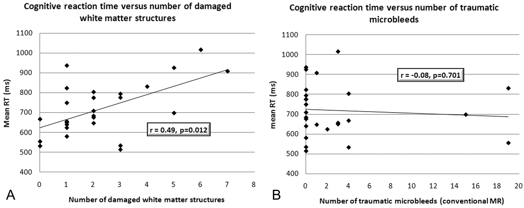
  • Fig 2.
    • Download figure
    • Open in new tab
    • Download powerpoint
    Fig 2.

    A, Correlation of the number of damaged white matter tracts and speed of processing. The correlation is statistically significant (P = .012) with r = 0.49. B, In contradistinction, the corresponding analysis, by using conventional MR imaging of the number of traumatic microhemorrhages correlated with speed of processing, is not statistically significant (r = −0.08, P = .70).

PreviousNext
Back to top

In this issue

American Journal of Neuroradiology: 29 (5)
American Journal of Neuroradiology
Vol. 29, Issue 5
May 2008
  • Table of Contents
  • Index by author
Advertisement
Print
Download PDF
Email Article

Thank you for your interest in spreading the word on American Journal of Neuroradiology.

NOTE: We only request your email address so that the person you are recommending the page to knows that you wanted them to see it, and that it is not junk mail. We do not capture any email address.

Enter multiple addresses on separate lines or separate them with commas.
Extent of Microstructural White Matter Injury in Postconcussive Syndrome Correlates with Impaired Cognitive Reaction Time: A 3T Diffusion Tensor Imaging Study of Mild Traumatic Brain Injury
(Your Name) has sent you a message from American Journal of Neuroradiology
(Your Name) thought you would like to see the American Journal of Neuroradiology web site.
CAPTCHA
This question is for testing whether or not you are a human visitor and to prevent automated spam submissions.
Cite this article
S.N. Niogi, P. Mukherjee, J. Ghajar, C. Johnson, R.A. Kolster, R. Sarkar, H. Lee, M. Meeker, R.D. Zimmerman, G.T. Manley, B.D. McCandliss
Extent of Microstructural White Matter Injury in Postconcussive Syndrome Correlates with Impaired Cognitive Reaction Time: A 3T Diffusion Tensor Imaging Study of Mild Traumatic Brain Injury
American Journal of Neuroradiology May 2008, 29 (5) 967-973; DOI: 10.3174/ajnr.A0970

Citation Manager Formats

  • BibTeX
  • Bookends
  • EasyBib
  • EndNote (tagged)
  • EndNote 8 (xml)
  • Medlars
  • Mendeley
  • Papers
  • RefWorks Tagged
  • Ref Manager
  • RIS
  • Zotero
0 Responses
Respond to this article
Share
Bookmark this article
Extent of Microstructural White Matter Injury in Postconcussive Syndrome Correlates with Impaired Cognitive Reaction Time: A 3T Diffusion Tensor Imaging Study of Mild Traumatic Brain Injury
S.N. Niogi, P. Mukherjee, J. Ghajar, C. Johnson, R.A. Kolster, R. Sarkar, H. Lee, M. Meeker, R.D. Zimmerman, G.T. Manley, B.D. McCandliss
American Journal of Neuroradiology May 2008, 29 (5) 967-973; DOI: 10.3174/ajnr.A0970
del.icio.us logo Twitter logo Facebook logo Mendeley logo
  • Tweet Widget
  • Facebook Like
  • Google Plus One
Purchase

Jump to section

  • Article
    • Abstract
    • Methods
    • Results
    • Discussion
    • Conclusion
    • Footnotes
    • References
  • Figures & Data
  • Supplemental
  • Info & Metrics
  • Responses
  • References
  • PDF

Related Articles

  • PubMed
  • Google Scholar

Cited By...

  • Volumetric and Diffusion Tensor Imaging biomarkers indicating long lasting post-concussion abnormalities in a youth pig model of mild Traumatic Brain Injury
  • Abnormal Neurite Density and Orientation Dispersion in Frontal Lobe Link to Elevated Hyperactive/Impulsive Behaviors in Young Adults with Traumatic Brain Injury
  • Connectomic Assessment of Injury Burden and Longitudinal Structural Network Alterations in Moderate-to-severe Traumatic Brain Injury
  • Assessment of fractional anisotropy outcomes in combat sport athletes with mild traumatic brain injury
  • Assessment of Maturational Changes in White Matter Anisotropy and Volume in Children: A DTI Study
  • Randomised controlled clinical trial of a structured cognitive rehabilitation in patients with attention deficit following mild traumatic brain injury: study protocol
  • Altered Relationship between Working Memory and Brain Microstructure after Mild Traumatic Brain Injury
  • Accuracy of different modalities of reaction time testing: Implications for online cognitive assessment tools
  • A common neural signature of brain injury in concussion and subconcussion
  • Assessing Postconcussive Reaction Time Using Transport-Based Morphometry of Diffusion Tensor Images
  • Longitudinal increases in structural connectome segregation and functional connectome integration are associated with better recovery after mild TBI
  • Experimental Traumatic Brain Injury Identifies Distinct Early and Late Phase Axonal Conduction Deficits of White Matter Pathophysiology, and Reveals Intervening Recovery
  • Toward Precision and Reproducibility of Diffusion Tensor Imaging: A Multicenter Diffusion Phantom and Traveling Volunteer Study
  • Bidirectional Changes in Anisotropy Are Associated with Outcomes in Mild Traumatic Brain Injury
  • Concussion is confusing us all
  • A Decade of DTI in Traumatic Brain Injury: 10 Years and 100 Articles Later
  • American Medical Society for Sports Medicine position statement: concussion in sport
  • Diffusion tensor imaging studies of mild traumatic brain injury: a meta-analysis
  • Acute Effects of Alcohol on the Human Brain: Diffusion Tensor Imaging Study
  • Salience network integrity predicts default mode network function after traumatic brain injury
  • Diffusion Tensor Imaging in Mild Traumatic Brain Injury Litigation
  • A prospective diffusion tensor imaging study in mild traumatic brain injury
  • Voxel-Based Analysis of Diffusion Tensor Imaging in Mild Traumatic Brain Injury in Adolescents
  • Contributions of neuroimaging, balance testing, electrophysiology and blood markers to the assessment of sport-related concussion
  • Crossref (470)
  • Google Scholar

This article has been cited by the following articles in journals that are participating in Crossref Cited-by Linking.

  • Animal models of traumatic brain injury
    Ye Xiong, Asim Mahmood, Michael Chopp
    Nature Reviews Neuroscience 2013 14 2
  • American Medical Society for Sports Medicine position statement: concussion in sport
    Kimberly G Harmon, Jonathan A Drezner, Matthew Gammons, Kevin M Guskiewicz, Mark Halstead, Stanley A Herring, Jeffrey S Kutcher, Andrea Pana, Margot Putukian, William O Roberts
    British Journal of Sports Medicine 2013 47 1
  • A review of magnetic resonance imaging and diffusion tensor imaging findings in mild traumatic brain injury
    M. E. Shenton, H. M. Hamoda, J. S. Schneiderman, S. Bouix, O. Pasternak, Y. Rathi, M.-A. Vu, M. P. Purohit, K. Helmer, I. Koerte, A. P. Lin, C.-F. Westin, R. Kikinis, M. Kubicki, R. A. Stern, R. Zafonte
    Brain Imaging and Behavior 2012 6 2
  • White matter damage and cognitive impairment after traumatic brain injury
    K. M. Kinnunen, R. Greenwood, J. H. Powell, R. Leech, P. C. Hawkins, V. Bonnelle, M. C. Patel, S. J. Counsell, D. J. Sharp
    Brain 2011 134 2
  • Cognitive Sequelae of Traumatic Brain Injury
    Amanda R. Rabinowitz, Harvey S. Levin
    Psychiatric Clinics of North America 2014 37 1
  • The Neuropathology and Neurobiology of Traumatic Brain Injury
    Kaj Blennow, John Hardy, Henrik Zetterberg
    Neuron 2012 76 5
  • Salience network integrity predicts default mode network function after traumatic brain injury
    Valerie Bonnelle, Timothy E. Ham, Robert Leech, Kirsi M. Kinnunen, Mitul A. Mehta, Richard J. Greenwood, David J. Sharp
    Proceedings of the National Academy of Sciences 2012 109 12
  • Detection of Blast-Related Traumatic Brain Injury in U.S. Military Personnel
    Christine L. Mac Donald, Ann M. Johnson, Dana Cooper, Elliot C. Nelson, Nicole J. Werner, Joshua S. Shimony, Abraham Z. Snyder, Marcus E. Raichle, John R. Witherow, Raymond Fang, Stephen F. Flaherty, David L. Brody
    New England Journal of Medicine 2011 364 22
  • Biomarkers of mild traumatic brain injury in cerebrospinal fluid and blood
    Henrik Zetterberg, Douglas H. Smith, Kaj Blennow
    Nature Reviews Neurology 2013 9 4
  • Neuroimaging after mild traumatic brain injury: Review and meta-analysis
    Cyrus Eierud, R. Cameron Craddock, Sean Fletcher, Manek Aulakh, Brooks King-Casas, Damon Kuehl, Stephen M. LaConte
    NeuroImage: Clinical 2014 4

More in this TOC Section

  • Kurtosis and Epileptogenic Tubers: A Pilot Study
  • Glutaric Aciduria Type 1: DK vs. Conventional MRI
  • Multiparametric MRI in PEDS Pontine Glioma
Show more FUNCTIONAL

Similar Articles

Advertisement

Indexed Content

  • Current Issue
  • Accepted Manuscripts
  • Article Preview
  • Past Issues
  • Editorials
  • Editor's Choice
  • Fellows' Journal Club
  • Letters to the Editor
  • Video Articles

Cases

  • Case Collection
  • Archive - Case of the Week
  • Archive - Case of the Month
  • Archive - Classic Case

More from AJNR

  • Trainee Corner
  • Imaging Protocols
  • MRI Safety Corner
  • Book Reviews

Multimedia

  • AJNR Podcasts
  • AJNR Scantastics

Resources

  • Turnaround Time
  • Submit a Manuscript
  • Submit a Video Article
  • Submit an eLetter to the Editor/Response
  • Manuscript Submission Guidelines
  • Statistical Tips
  • Fast Publishing of Accepted Manuscripts
  • Graphical Abstract Preparation
  • Imaging Protocol Submission
  • Evidence-Based Medicine Level Guide
  • Publishing Checklists
  • Author Policies
  • Become a Reviewer/Academy of Reviewers
  • News and Updates

About Us

  • About AJNR
  • Editorial Board
  • Editorial Board Alumni
  • Alerts
  • Permissions
  • Not an AJNR Subscriber? Join Now
  • Advertise with Us
  • Librarian Resources
  • Feedback
  • Terms and Conditions
  • AJNR Editorial Board Alumni

American Society of Neuroradiology

  • Not an ASNR Member? Join Now

© 2025 by the American Society of Neuroradiology All rights, including for text and data mining, AI training, and similar technologies, are reserved.
Print ISSN: 0195-6108 Online ISSN: 1936-959X

Powered by HighWire