
Article Figures & Data
Figures
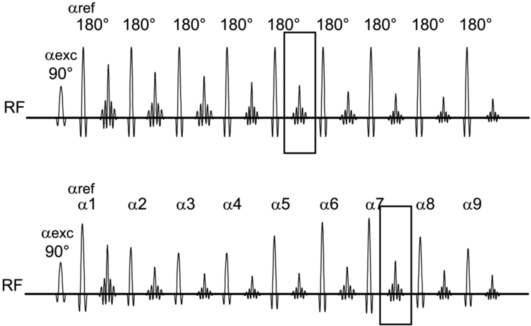
- Fig 1.
The basic principle of TSE180° and hyperTSE. A sketch of the radio-frequency (RF) of the refocusing pulses applying flip angles αref is shown with time in TSE180° (upper graph) and hyperTSE (lower graph). In TSE180°, a constant αref = 180° is used, whereas in hyperTSE, αref is varied along the echo train. The box indicates the echo written in the center of the k-space, which corresponds to the respective TE. Note that the hyperTSE uses a substantial later echo than the TSE180°. αexec indicates excitation pulse.
- Fig 2.
Representative regions of interest. A, Regions of interest for gray matter obtained from the frontal and temporal lobes (arrows). An average of the measurements was calculated. B, Region of interest for white matter obtained from the genu of the corpus callosum (arrow). C, Region of interest for CSF obtained from the frontal horn of the left ventricle (arrow). D, Region of interest for the SD of noise obtained from the noise in the upper left quadrant of the image.
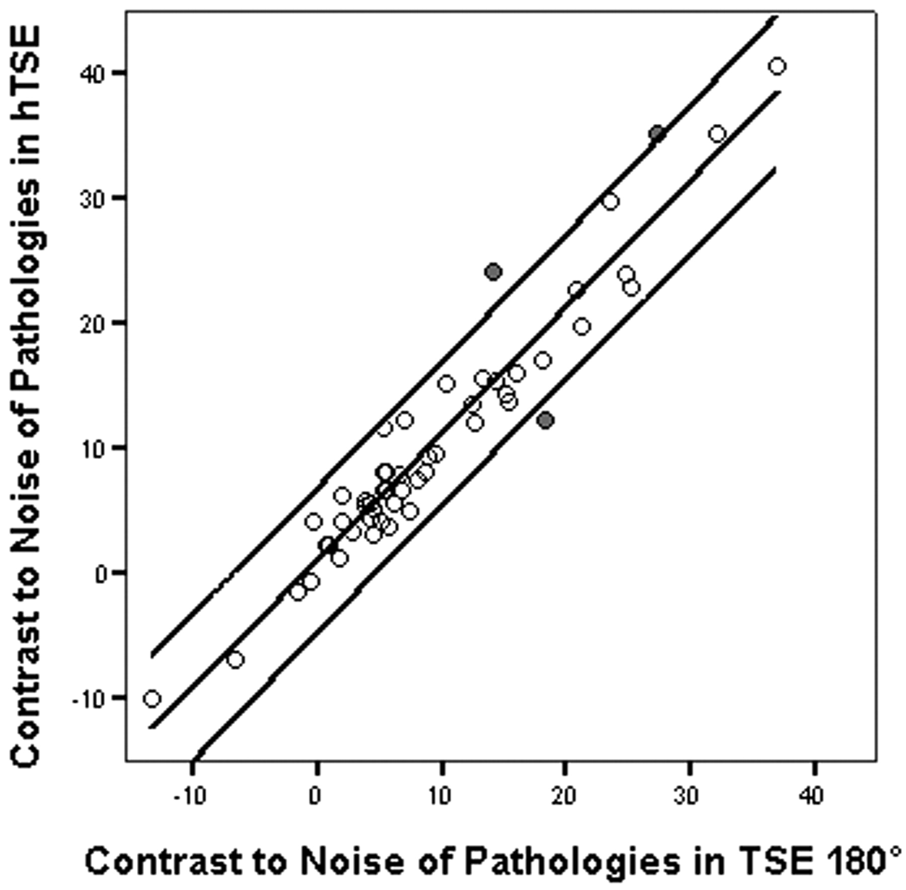
- Fig 3.
Linear regression analysis and 95% predictive interval of single values between contrast to noise of pathology in hyperTSE and TSE180° (r = 0.93, P = .001). Three outliers are marked in gray. hTSE indicates hyperTSE.
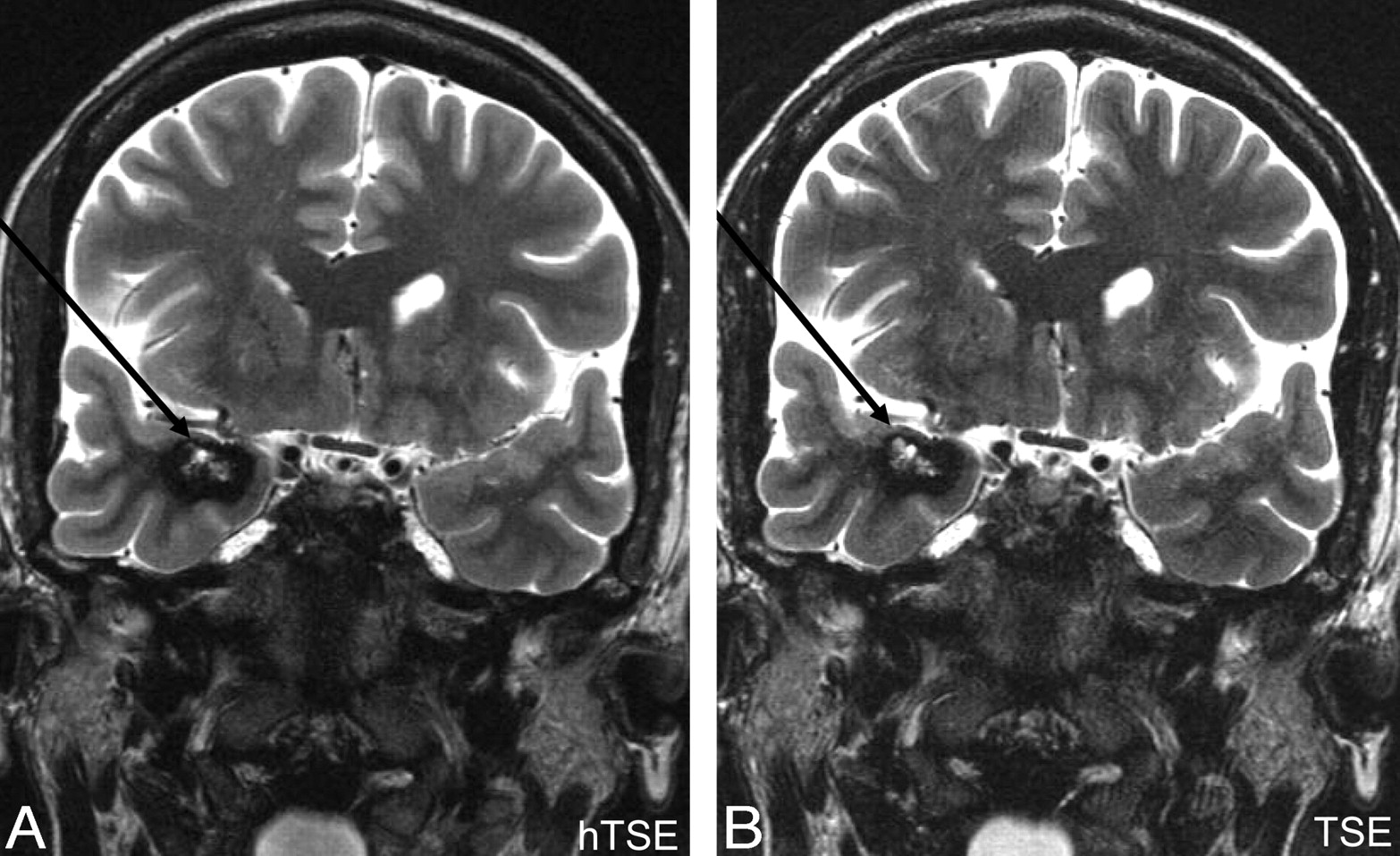
- Fig 4.
A, hyperTSE (hTSE). B, TSE180°. Note the gliosis in the white and gray matter of the left temporal lobe after trauma. In both sequences, the hyperintense findings are clearly visible.
- Fig 5.
A, hyperTSE (hTSE). B, TSE180°. The left hippocampus is atrophic and has increased signal intensity attributed to hippocampal sclerosis (arrows). This finding was confirmed by surgery. The right hippocampus shows the typical dentation, and the particular layers are visible on both sequences.
- Fig 6.
A, HyperTSE (hTSE). B, TSE180°. A right mesiotemporal cavernoma (arrows) consisting of a hypointense hemosiderin rim and a central hyperintense matrix containing methemoglobin is clearly depicted in both sequences. It is important that both parts of blood degradation products are depicted; this depiction was successful for both sequences. The diagnosis was confirmed by surgery.


Tables
Pathology Tumor Hippocampal Sclerosis FCD ED Neuropediatric Misc. No. of Pathologies 5 12 15 3 3 16 C/N of pathology in hyperTSE Mean 22.7 8.0 6.6 15.6 14.0 11.0 SD 13.6 4.3 7.1 17.1 8.2 11.0 C/N of pathology in TSE180° Mean 16.9 7.9 5.9 11.9 13.3 9.5 SD 17.4 4.5 5.8 13.4 11.4 11.7 Note:—C/N indicates contrast-to-noise ratio; SD, standard deviation; FCD, focal cortical dysplasia; ED, encephalomyelitis disseminata; Misc., miscellaneous.
- Table 2:
Visual ratings of 2 experienced neuroradiologists concerning motion and flow artifacts, lesion conspicuity, and subjective appraisal of image quality*
Rater Artifacts Lesion Conspicuity Quality hyperTSE TSE 180° hyperTSE TSE 180° hyperTSE TSE 180° 1 4.50/5/0.61 4.46/5/0.64 4.51/5/0.64 4.44/5/0.69 4.19/4/0.48 4.11/4/0.60 2 4.59/5/0.74 4.65/5/0.70 4.17/5/1.09 4.09/51.25 4.04/4/0.85 3.98/4/0.90 * Given are mean/median/SD of a 5-point scoring scale within a range of 1 (poor) through 5 (excellent).
In this issue


Jump to section
Related Articles
Cited By...
- No citing articles found.