
Article Figures & Data
Figures
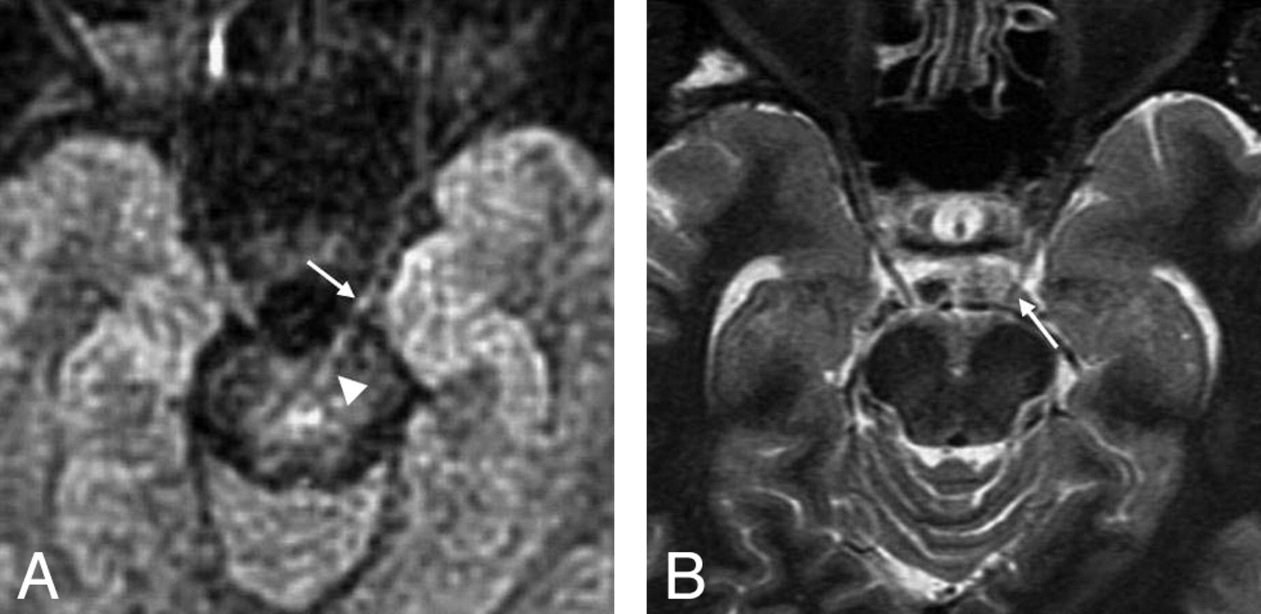
- Fig 1.
Images of a 44-year-old male volunteer. A, Multishot diffusion-weighted image with MPG applied in the SI direction shows the left ophthalmic nerve not only in the cistern (arrow) but also in the midbrain (arrowhead). The bright signals in the center of the midbrain are considered to be the decussation of the superior cerebellar peduncle running in the lateral direction. B, STIR image shows the cisternal portion of the left ophthalmic nerve (arrow); however, the nerve is not identified in the midbrain.
- Fig 2.
Images of a 30-year-old female volunteer. A,B, Multishot diffusion-weighted images with MPG applied in the SI direction show both trigeminal nerves as slightly curved high-intensity lines in the mid-pons (arrows). C,D, The trigeminal nerves are not identified in the mid-pons on the STIR images.
- Fig 3.
Images of a 34-year-old male volunteer. A, Multishot diffusion-weighted image with MPG applied in the SI direction shows the left acoustic nerve as a linear high-intensity line in the brain stem (black arrow) as well as in the cistern (white arrow). B, STIR image shows the left acoustic nerve in the cerebellar-pontine cistern (arrow); however, it is not identified in the brain stem.


