
Article Figures & Data
Figures
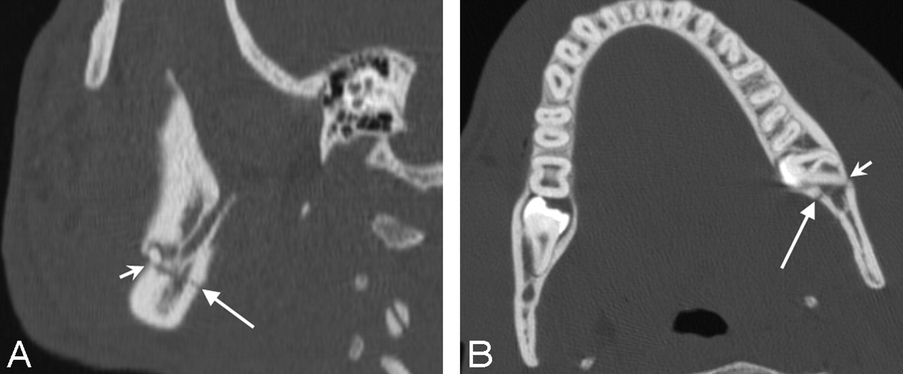
- Fig 1.
A, Sagittal reformatted image through the mandibular angle shows a nondisplaced very minimally distracted left mandibular angle fracture (long arrow), extending mildly obliquely anteriorly. Note that the fracture just enters the socket of the left third molar (short arrow). B, Axial CT scan shows the left mandibular angle fracture (long arrow) entering the socket of the third molar (short arrow).
- Fig 2.
A, Axial CT scan at the level of the glenoid fossa shows a right condylar fracture, with typical displacement. Note that the condylar head (solid arrow) is dislocated and displaced anteriorly and inferiorly from the glenoid fossa (asterisk) and that the ramus/neck component of the fracture (dashed arrow) is “telescoped” with respect to the condylar head component and displaced superiorly toward the glenoid fossa. Note the overlapping of the fracture fragments, with the condylar head component lying medial to the ramus/neck component. B and C, Coronal reformatted images through the mandibular condyle show the “telescoping,” with upward retraction of the ramus/neck component (dashed arrow) and anterior inferior medial displacement of the condylar head component (solid arrow) and resultant overlap of fracture fragments. This is the typical pattern of dislocation/displacement seen in this type of fracture and was present in all but 1 of the fractures that involved the condylar head or condylar neck. (The asterisk in C indicates the glenoid fossa.)


Tables
Location and displacement/distraction of mandible fractures
Location No. Percentage Displ/Distr (none/mild [≤3 mm]) Displ/Distr Mod or Greater Comminuted* (% of all unifocal fractures in same location) Parasymphyseal 7 16% 2 5 4 (57) Body† 11 26% 4 6 8 (73) Angle 13 30% 12 1 1 (8) Condyle or neck 5 12% 0 5 3 (60) Ramus 2 5% 2 0 0 Coronoid process 1 2% 1 0 0 Subcondylar 2 5% 2 0 0 Alveolar ridge 2 5% 2 0 0 Total 43 100% 26/43 17/43 16 (37) Note:—Displ/Distr indicates displacement and/or distraction; Mod, moderate.
* Comminuted includes fractures that may or may not be associated with condylar subluxation; 5/7 fractures with condylar subluxations were comminuted.
† The films for 1 of the body fractures were not available, so Displ/Distr could not be quantified.


