Advertisement

Main menu

  • Home
  • Content
    • Current Issue
    • Accepted Manuscripts
    • Article Preview
    • Past Issue Archive
    • Video Articles
    • AJNR Case Collection
    • Case of the Week Archive
    • Case of the Month Archive
    • Classic Case Archive
  • Special Collections
    • Advancing NeuroMRI with High-Relaxivity Contrast Agents
    • AJNR Awards
    • Low-Field MRI
    • Alzheimer Disease
    • ASNR Foundation Special Collection
    • Photon-Counting CT
    • View All
  • Multimedia
    • AJNR Podcasts
    • AJNR SCANtastic
    • Trainee Corner
    • MRI Safety Corner
    • Imaging Protocols
  • For Authors
    • Submit a Manuscript
    • Submit a Video Article
    • Submit an eLetter to the Editor/Response
    • Manuscript Submission Guidelines
    • Statistical Tips
    • Fast Publishing of Accepted Manuscripts
    • Graphical Abstract Preparation
    • Imaging Protocol Submission
    • Author Policies
  • About Us
    • About AJNR
    • Editorial Board
    • Editorial Board Alumni
  • More
    • Become a Reviewer/Academy of Reviewers
    • Subscribers
    • Permissions
    • Alerts
    • Feedback
    • Advertisers
    • ASNR Home

User menu

  • Alerts
  • Log in

Search

  • Advanced search
American Journal of Neuroradiology

American Journal of Neuroradiology

ASHNR American Society of Functional Neuroradiology ASHNR American Society of Pediatric Neuroradiology ASSR
  • Alerts
  • Log in

Advanced Search

  • Home
  • Content
    • Current Issue
    • Accepted Manuscripts
    • Article Preview
    • Past Issue Archive
    • Video Articles
    • AJNR Case Collection
    • Case of the Week Archive
    • Case of the Month Archive
    • Classic Case Archive
  • Special Collections
    • Advancing NeuroMRI with High-Relaxivity Contrast Agents
    • AJNR Awards
    • Low-Field MRI
    • Alzheimer Disease
    • ASNR Foundation Special Collection
    • Photon-Counting CT
    • View All
  • Multimedia
    • AJNR Podcasts
    • AJNR SCANtastic
    • Trainee Corner
    • MRI Safety Corner
    • Imaging Protocols
  • For Authors
    • Submit a Manuscript
    • Submit a Video Article
    • Submit an eLetter to the Editor/Response
    • Manuscript Submission Guidelines
    • Statistical Tips
    • Fast Publishing of Accepted Manuscripts
    • Graphical Abstract Preparation
    • Imaging Protocol Submission
    • Author Policies
  • About Us
    • About AJNR
    • Editorial Board
    • Editorial Board Alumni
  • More
    • Become a Reviewer/Academy of Reviewers
    • Subscribers
    • Permissions
    • Alerts
    • Feedback
    • Advertisers
    • ASNR Home
  • Follow AJNR on Twitter
  • Visit AJNR on Facebook
  • Follow AJNR on Instagram
  • Join AJNR on LinkedIn
  • RSS Feeds

AJNR Awards, New Junior Editors, and more. Read the latest AJNR updates


Improved Turnaround Times | Median time to first decision: 12 days

OtherPHYSICS REVIEW

Diffusion Tensor MR Imaging and Fiber Tractography: Technical Considerations

P. Mukherjee, S.W. Chung, J.I. Berman, C.P. Hess and R.G. Henry
American Journal of Neuroradiology May 2008, 29 (5) 843-852; DOI: https://doi.org/10.3174/ajnr.A1052
P. Mukherjee
  • Find this author on Google Scholar
  • Find this author on PubMed
  • Search for this author on this site
S.W. Chung
  • Find this author on Google Scholar
  • Find this author on PubMed
  • Search for this author on this site
J.I. Berman
  • Find this author on Google Scholar
  • Find this author on PubMed
  • Search for this author on this site
C.P. Hess
  • Find this author on Google Scholar
  • Find this author on PubMed
  • Search for this author on this site
R.G. Henry
  • Find this author on Google Scholar
  • Find this author on PubMed
  • Search for this author on this site
  • Article
  • Figures & Data
  • Info & Metrics
  • Responses
  • References
  • PDF
Loading

Article Figures & Data

Figures

  • Tables
  • Fig 1.
    • Download figure
    • Open in new tab
    • Download powerpoint
    Fig 1.

    Shift of DWIs due to eddy currents. A, The b = 0 s/mm2 image with the brain-CSF interface outlined (yellow). B, Corresponding DWI with the same outline (red), unchanged in position compared with A, shows a shift of the brain anteriorly, most easily seen at the ventricular margins and at the occipital lobes. C, Another DWI with the diffusion gradient pointing in a different direction than in B shows a different degree of anterior shift.

  • Fig 2.
    • Download figure
    • Open in new tab
    • Download powerpoint
    Fig 2.

    Combined DWI image shows pronounced artifacts at the anterior temporal lobes and around the superior cerebellar vermis (black lines) due to mechanical vibration.

  • Fig 3.
    • Download figure
    • Open in new tab
    • Download powerpoint
    Fig 3.

    Pulse sequence diagrams show the benefits of parallel imaging for DWI. At an acceleration factor of R = 2, the echo-train length for the single-shot EPI acquisition is only half as long. This is reflected in a shorter readout time (tacq) and allows the echo train to be better centered at the peak of the spin-echo, improving SNR, decreasing T2 and T2* contrast blurring, and reducing off-resonance artifacts that cause geometric distortions. The shorter readout time also enables a reduction of TE, further improving SNR and reducing geometric distortion. However, the use of parallel imaging results in an intrinsic loss of SNR that may offset the aforementioned SNR gains. RF indicates radio-frequency.

  • Fig 4.
    • Download figure
    • Open in new tab
    • Download powerpoint
    Fig 4.

    Parallel imaging ameliorates susceptibility-induced geometric distortions and T2 and T2* contrast blurring in 3T DWI performed with a single-shot echo-planar sequence. A, The b = 1000 s/mm2 DWI image acquired at 3T without parallel imaging shows warping of the pons and anterior temporal lobes. There is also signal-intensity void with adjacent regions of signal-intensity pileup in the temporal lobes. These are typical artifacts encountered with 3T ssEPI DWI due to susceptibility effects from the adjacent air-filled mastoid sinuses and sphenoid sinus. B, The b = 1000 s/mm2 3T DWI image acquired at the same axial level with ASSET parallel imaging (R = 2) demonstrates reduced foreshortening of the pons and reduced warping and signal-intensity distortions in the temporal lobes. There is also mitigation of contrast blurring, seen as improved definition of the cerebellar fissures and folia as well as better gray-white matter differentiation in the occipital lobes.

  • Fig 5.
    • Download figure
    • Open in new tab
    • Download powerpoint
    Fig 5.

    3T-versus-7T DTI with 36 diffusion-encoding directions at b = 3000 s/mm2 and 2.0 × 2.0 × 2.0 mm isotropic voxel resolution. Directionally encoded color FA maps at the axial level of cingulum bundles and the callosal striations are shown for 3T (A) and for 7T (B) in a healthy adult volunteer. Both scanners were equipped with 40 mT/m gradients and 8-channel phased-array head coils, and ASSET parallel imaging was used with an acceleration factor of 2. The standard DTI color conventions are used, with red representing left-right fiber orientation, green representing anteroposterior, and blue representing craniocaudal. With this combination of high spatial resolution and very strong diffusion weighting, the 3T image appears grainy because of inadequate SNR. However, with identical scanning parameters, the additional SNR at 7T produces a higher quality image. Parallel imaging is essential for SS-EPI at ultra-high field to combat the increased susceptibility artifacts as well as the signal intensity loss and contrast blurring due to shorter T2 ad T2* relaxation times.
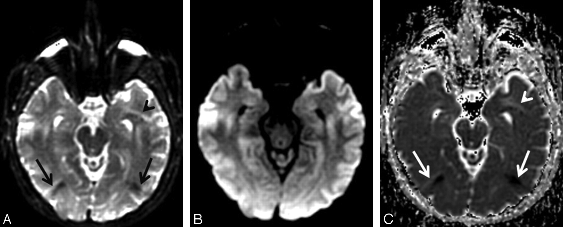
  • Fig 6.
    • Download figure
    • Open in new tab
    • Download powerpoint
    Fig 6.

    Unfolding artifacts from the globes in ASSET-accelerated DWI. A, The b = 0 s/mm2 image acquired at 3T with an ASSET acceleration factor of R = 2 shows unfolding artifacts from the distorted high-signal-intensity globes appearing as dark bands in the occipital regions (black arrows) as well as a bright band in the anterior left temporal lobe (black arrowhead). B, The unfolding artifacts are not apparent on the combined DWI image because the globes contain fluid with high diffusivity; therefore, the globes signal intensity is suppressed by the diffusion gradients. C, However, the unfolding artifacts are again apparent on the ADC map (white arrows and arrowhead) because the b = 0 s/mm2 image is required for ADC calculation (equation 5, Part I1).

Tables

  • Figures
  • Typical optimized whole-brain DTI acquisition parameters in a 1.5T or 3T MR imaging system*

    Acquisition Parameters3T1.5T
    Spatial resolution2.0 × 2.0 × 2.0 mm2.5 × 2.5 × 2.5 mm
    Acquisition matrix128 × 128 × 6096 × 96 × 50
    FOV256 mm240 mm
    No. DWIs3030
    No. minimally weighted images55
    No. repetitions (NEX)11
    b-value1000 s/mm21000 s/mm2
    TE/TR70 ms/<12 seconds70 ms/<10 seconds
    Total acquisition time<8 minutes<7 minutes
    • * The hardware is assumed to be equipped with an 8-channel head coil and gradients of 40 mT/m. It is also assumed that parallel acquisition is done with a SENSE reduction factor of 2, partial k-space acquisition of 62.5% (only 5/8 of the phase-encoding lines after a reduction to half by parallel acquisition is acquired), and an interleaved multisection SS-EPI sequence with no gap.

PreviousNext
Back to top

In this issue

American Journal of Neuroradiology: 29 (5)
American Journal of Neuroradiology
Vol. 29, Issue 5
May 2008
  • Table of Contents
  • Index by author
Advertisement
Print
Download PDF
Email Article

Thank you for your interest in spreading the word on American Journal of Neuroradiology.

NOTE: We only request your email address so that the person you are recommending the page to knows that you wanted them to see it, and that it is not junk mail. We do not capture any email address.

Enter multiple addresses on separate lines or separate them with commas.
Diffusion Tensor MR Imaging and Fiber Tractography: Technical Considerations
(Your Name) has sent you a message from American Journal of Neuroradiology
(Your Name) thought you would like to see the American Journal of Neuroradiology web site.
CAPTCHA
This question is for testing whether or not you are a human visitor and to prevent automated spam submissions.
Cite this article
P. Mukherjee, S.W. Chung, J.I. Berman, C.P. Hess, R.G. Henry
Diffusion Tensor MR Imaging and Fiber Tractography: Technical Considerations
American Journal of Neuroradiology May 2008, 29 (5) 843-852; DOI: 10.3174/ajnr.A1052

Citation Manager Formats

  • BibTeX
  • Bookends
  • EasyBib
  • EndNote (tagged)
  • EndNote 8 (xml)
  • Medlars
  • Mendeley
  • Papers
  • RefWorks Tagged
  • Ref Manager
  • RIS
  • Zotero
0 Responses
Respond to this article
Share
Bookmark this article
Diffusion Tensor MR Imaging and Fiber Tractography: Technical Considerations
P. Mukherjee, S.W. Chung, J.I. Berman, C.P. Hess, R.G. Henry
American Journal of Neuroradiology May 2008, 29 (5) 843-852; DOI: 10.3174/ajnr.A1052
del.icio.us logo Twitter logo Facebook logo Mendeley logo
  • Tweet Widget
  • Facebook Like
  • Google Plus One
Purchase

Jump to section

  • Article
    • Abstract
    • Technical Considerations for State-of-the-Art DWI
    • Technical Considerations for State-of-the-Art DTI
    • Conclusion
    • References
  • Figures & Data
  • Info & Metrics
  • Responses
  • References
  • PDF

Related Articles

  • No related articles found.
  • PubMed
  • Google Scholar

Cited By...

  • The Influence of Nonaerated Paranasal Sinuses on DTI Parameters of the Brain in 6- to 9-Year-Old Children
  • Dementia risk factors modify hubs but leave other connectivity measures unchanged in asymptomatic individuals: a graph theoretical analysis
  • Predicting MEG brain functional connectivity using microstructural information
  • White matter microstructural changes in short-term learning of a continuous visuomotor sequence
  • Myelin development in visual scene-network tracts beyond late childhood: A multimethod neuroimaging study
  • Tractography-Pathology Correlations in Traumatic Brain Injury: A TRACK-TBI Study
  • Individual structural features constrain the mouse functional connectome
  • Individual structural features constrain the mouse functional connectome
  • Connectivity Gradient in the Human Left Inferior Frontal Gyrus: Intraoperative Cortico-Cortical Evoked Potential Study
  • Toward Precision and Reproducibility of Diffusion Tensor Imaging: A Multicenter Diffusion Phantom and Traveling Volunteer Study
  • Cervical Spinal Cord DTI Is Improved by Reduced FOV with Specific Balance between the Number of Diffusion Gradient Directions and Averages
  • Choice of Diffusion Tensor Estimation Approach Affects Fiber Tractography of the Fornix in Preterm Brain
  • Assessment of Whole-Brain White Matter by DTI in Autosomal Recessive Spastic Ataxia of Charlevoix-Saguenay
  • Cerebral Diffusion Tensor MR Tractography in Tuberous Sclerosis Complex: Correlation with Neurologic Severity and Tract-Based Spatial Statistical Analysis
  • Acquisition Guidelines and Quality Assessment Tools for Analyzing Neonatal Diffusion Tensor MRI Data
  • Reduced-Distortion Diffusion MRI of the Craniovertebral Junction
  • Diffusion tensor imaging of normal prostate at 3 T: effect of number of diffusion-encoding directions on quantitation and image quality
  • Differentiation of Tumefactive Demyelinating Lesions from High-Grade Gliomas with the Use of Diffusion Tensor Imaging
  • Differential corticospinal tract degeneration in homozygous 'D90A' SOD-1 ALS and sporadic ALS
  • Correlation of Quantitative Diffusion Tensor Tractography with Clinical Grades of Subacute Sclerosing Panencephalitis
  • Principles and Limitations of Computational Algorithms in Clinical Diffusion Tensor MR Tractography
  • Wallerian Degeneration in the Corticospinal Tract Evaluated by Diffusion Tensor Imaging Correlates with Motor Deficit 30 Days after Middle Cerebral Artery Ischemic Stroke
  • The Evolution of Clinical Functional Imaging during the Past 2 Decades and Its Current Impact on Neurosurgical Planning
  • A new sensitive MRI marker for memory deficits in normal aging
  • Enhanced Detection of Diffusion Reductions in Creutzfeldt-Jakob Disease at a Higher B Factor
  • Crossref
  • Google Scholar

This article has not yet been cited by articles in journals that are participating in Crossref Cited-by Linking.

More in this TOC Section

  • Theoretic Basis and Technical Implementations of CT Perfusion in Acute Ischemic Stroke, Part 2: Technical Implementations
  • Theoretic Basis and Technical Implementations of CT Perfusion in Acute Ischemic Stroke, Part 1: Theoretic Basis
  • Susceptibility-Weighted Imaging: Technical Aspects and Clinical Applications, Part 2
Show more PHYSICS REVIEW

Similar Articles

Advertisement

Indexed Content

  • Current Issue
  • Accepted Manuscripts
  • Article Preview
  • Past Issues
  • Editorials
  • Editor's Choice
  • Fellows' Journal Club
  • Letters to the Editor
  • Video Articles

Cases

  • Case Collection
  • Archive - Case of the Week
  • Archive - Case of the Month
  • Archive - Classic Case

More from AJNR

  • Trainee Corner
  • Imaging Protocols
  • MRI Safety Corner
  • Book Reviews

Multimedia

  • AJNR Podcasts
  • AJNR Scantastics

Resources

  • Turnaround Time
  • Submit a Manuscript
  • Submit a Video Article
  • Submit an eLetter to the Editor/Response
  • Manuscript Submission Guidelines
  • Statistical Tips
  • Fast Publishing of Accepted Manuscripts
  • Graphical Abstract Preparation
  • Imaging Protocol Submission
  • Evidence-Based Medicine Level Guide
  • Publishing Checklists
  • Author Policies
  • Become a Reviewer/Academy of Reviewers
  • News and Updates

About Us

  • About AJNR
  • Editorial Board
  • Editorial Board Alumni
  • Alerts
  • Permissions
  • Not an AJNR Subscriber? Join Now
  • Advertise with Us
  • Librarian Resources
  • Feedback
  • Terms and Conditions
  • AJNR Editorial Board Alumni

American Society of Neuroradiology

  • Not an ASNR Member? Join Now

© 2025 by the American Society of Neuroradiology All rights, including for text and data mining, AI training, and similar technologies, are reserved.
Print ISSN: 0195-6108 Online ISSN: 1936-959X

Powered by HighWire