
Article Figures & Data
Figures
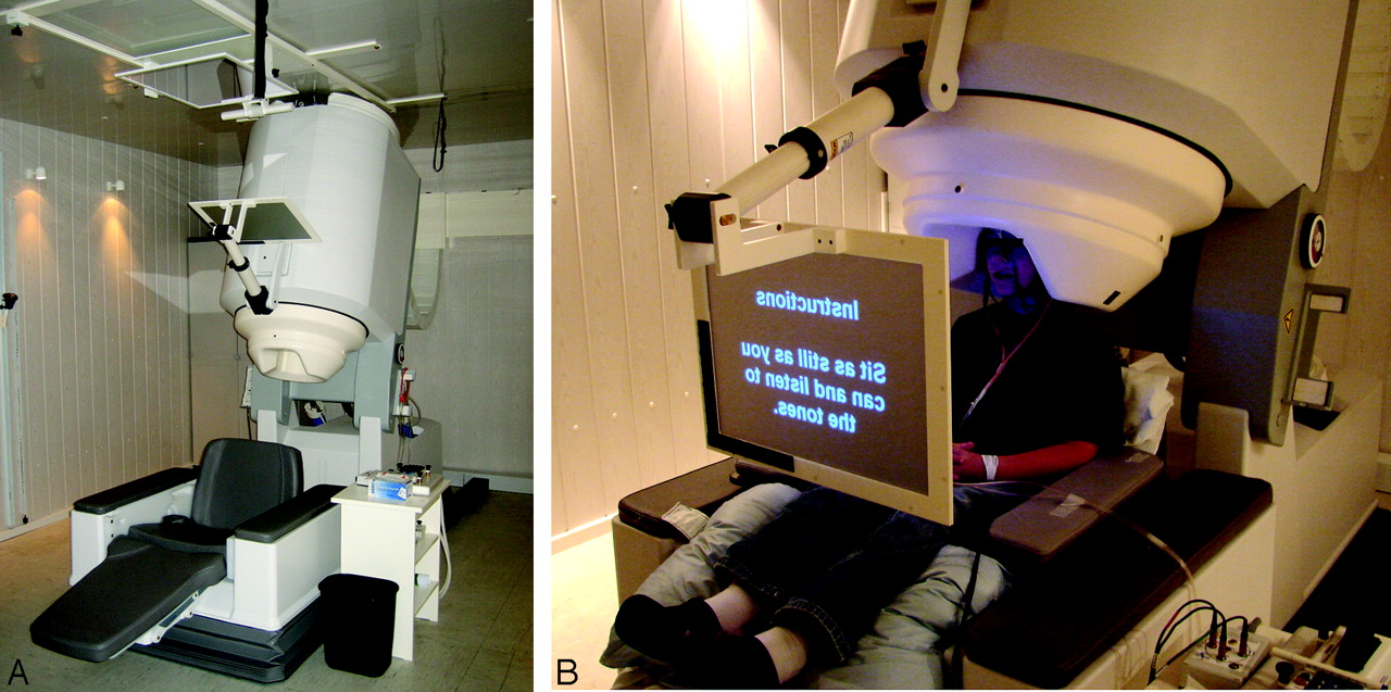
- Fig 1.
A, Biomagnetometer in an electrically shielded room. Patients can be comfortably studied in a seated or supine position. B, The video screen is easily viewed and headphones facilitate communication with the technologists and allow auditory stimulus delivery. The wires attached to the 3 electrically active fiducial coils are visible.
- Fig 2.
MEG SAMg2 data superimposed on coronal reformation (A), sagittal reformation (B), and axial MPRAGE MR imaging (C). Note the value of the t statistic indicated at the point of peak activity in the region. The statistical map overlay is thresholded to display only pixels with a t statistic value exceeding 2.0.
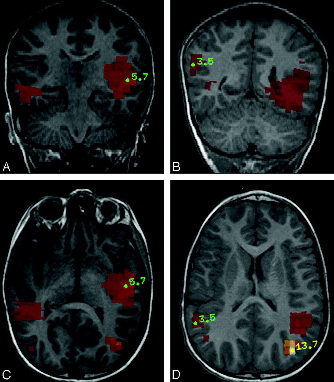
- Fig 3.
Multifocal Activity. MEG interictal activity is seen as regions of color superimposed over the midportion of the superior temporal gyri bilaterally and the posterior temporal-occipital lobe junctions bilaterally on coronal reformation (A and B) and axial (C and D) MPRAGE MR images.


In this issue


Jump to section
Related Articles
Cited By...
- No citing articles found.